CASE REPORT
새로운 MEN1 유전자 돌연변이가 증명된 위, 십이지장, 췌장의 신경내분비종양
양민아, 이웅기, 신홍식, 박성현, 김병선, 김지웅, 조진웅, 윤소희
예수병원 소화기내과
Neuroendocrine Tumors in the Stomach, Duodenum, and Pancreas Accompanied by Novel MEN1 Gene Mutation
Min A Yang, Woong Ki Lee, Hong Shik Shin, Sung Hyun Park, Byung Sun Kim, Ji Woong Kim, Jin Woong Cho and So Hee Yun Division of Gastroenterology, Department of Internal Medicine, Presbyterian Medical Center, Jeonju, Korea
Multiple endocrine neoplasia type 1 (MEN1) syndrome is a relatively rare disease, characterized by the occurrence of multiple endo- crine tumors in the parathyroid and pituitary glands as well as the pancreas. Here, we report a case of MEN1 with neuroendocrine tumors (NETs) in the stomach, duodenum, and pancreas. A 53-year-old man visited our hospital to manage gastric NET. Five years prior to his visit, he had undergone surgery for incidental meningioma. His brother had pancreatic nodules and a history of surgery for adrenal adenoma. His brother’s daughter also had pancreatic nodules, but had not undergone surgery. The lesion was treated by endoscopic submucosal dissection and diagnosed as a grade 1 NET. Another small NET was detected in the second duodenal portion, resected by endoscopic submucosal dissection, which was also diagnosed as a grade 1 NET. During evaluation, three nodules were detected in the pancreas, and no evidence of pituitary, parathyroid tumors, or metastasis was observed. After surgery, the pan- creatic lesions were diagnosed as NETs, with the same immunohistochemical patterns as those of the stomach and duodenum.
Genetic testing was performed, and a heterozygous mutation was detected in the MEN1 gene, which is located on 11q13.
(Korean J Gastroenterol 2017;69:181-186)
Key Words: Multiple endocrine neoplasia type 1; Neuroendocrine tumors; INDEL mutation; Endoscopy; Germ-line mutation
Received November 18, 2016. Revised November 18, 2016. Accepted January 17, 2017.
CC This is an open access article distributed under the terms of the Creative Commons Attribution Non-Commercial License (http://creativecommons.org/licenses/
by-nc/4.0) which permits unrestricted non-commercial use, distribution, and reproduction in any medium, provided the original work is properly cited.
Copyright © 2017. Korean Society of Gastroenterology.
교신저자: 윤소희, 54987, 전주시 완산구 서원로 365, 예수병원 소화기내과
Correspondence to: So Hee Yun, Division of Gastroenterology, Department of Internal Medicine, Presbyterian Medical Center, 365 Seowon-ro, Wansan-gu, Jeonju 54987, Korea. Tel: +82-63-230-1310, Fax: +82-63-230-1329, E-mail: [email protected]
Financial support: None. Conflict of interest: None.
INTRODUCTION
Neuroendocrine tumors (NETs) are rare tumors arising from enterochromaffin cells. NETs secrete biomarkers and hormones, including insulin, C-peptide, chromogranin A, pancreatic polypeptide, and 5-hydroxyindoleacetic acid, and are diagnosed based on expression of these neuroendocrine markers.1
Gastroenteropancreatic (GEP) NETs can present synchro-
nously with various genetic diseases, such as multiple endo- crine neoplasia type 1 (MEN1), von Hippel-Lindau disease, and neurofibromatosis.1 MEN1 is characterized by the pres- ence of tumors or hyperplasia of the endocrine glands, such as parathyroid glands, pituitary gland, and enteropancreatic system, and its incidence is very low.2-4 We report a case of a patient with NETs in the stomach, duodenum, and pan- creas, accompanied by a newly discovered MEN1 gene mutation.
Fig. 1. Abdominal computed tomography scan. (A) An arterial enhancing nodule in the pancreatic neck (arrow) and one thick-walled cystic nodule with a mural component and marginal calcification in the tail (arrowhead). (B) Another arterial enhancing nodule in the pancreatic tail (arrow).
Fig. 2. Endoscopic findings for the neuroendocrine tumors in the stomach. (A) A raised nodule with a whitish color and 8×8 mm in the area of the lesser curvature at the lower body of the stomach (arrow). (B) Endoscopic submucosal dissection of the gastric lesion using a soft trans- parent hood. (C) The completely resected gastric lesion.
CASE REPORT
A 53-year old man was diagnosed with NET in the lesser curvature of the lower body of the stomach using an endo- scopic forceps biopsy during a regular health check-up. He was taking medications for diabetes and hypertension. He had previously undergone surgery for meningothelial menin- gioma in the right frontal area, which had been incidentally discovered after an automobile accident 3 years before. He has a history of drinking (287 g/week) and smoking (20 packs/year). In his familial history, his brother had under- gone surgery for adrenal adenoma and had multiple pancre- atic tumors; his brother’s daughter also had multiple pancre-
atic tumors. The findings of physical examination were unremarkable. The laboratory results were as follows: total bilirubin, 0.4 mg/dL; amylase, 56 U/dL; serum carcinoem- bryonic antigen, 9.0 ng/mL; carbohydrate antigen 19-9, 5.09 U/mL; and gastrin, 133.7 pg/mL. The microscopic urine test results were normal. To evaluate metastasis and gastric wall invasion, abdominal computed tomography was performed;
we detected the following: Arterial enhancement of a 10×10 mm nodule in the neck of the pancreas, a 6×6 mm nodule in the tail, and a 30×30 mm thick-walled cyst adjacent to the en- hanced nodule in the tail (Fig. 1). Moreover, for an evaluation of potential distant metastasis, a positron emission tomog- raphy was performed; the results showed normal stand-
A B
A B C
Fig. 4. Pathologic findings after endoscopic submucosal dissection of a gastric neuroendocrine tumor invading the submucosa and mucosa.
(A) Tumor cells forming a mostly glandular and trabecular pattern (H&E, ×100). (B) Immunoreactivity for synaptophysin (synaptophysin, x100).
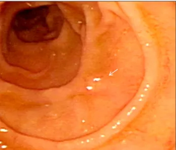
Fig. 3. Endoscopic findings for a 3×3 mm yellowish nodule in the second duodenal portion (arrow).
ardized uptake values.
Endoscopic submucosal dissection for gastric NET (GIF-Q260; Olympus, Tokyo, Japan) was performed using an IT knife (KD-611L; Olympus, Tokyo, Japan) and needle knife (KD-1L-1; Olympus, Tokyo, Japan) (Fig. 2A–C). During this pro- cedure, a raised 3×3 mm nodule was discovered incidentally in the second duodenal portion (Fig. 3). The final histologic analysis revealed an 8 × 8 mm grade 1 gastric NET (Ki-67
<2%, mitotic rate <1/10 high-power fields [HPF]) (Fig. 4). A NET was also revealed using a duodenal forceps biopsy.
Therefore, endoscopic mucosal resection was performed for
the duodenal lesion, which was finally diagnosed as a grade 1 NET (Ki-67 <2%, mitotic rate <1/10 HPF) (Fig. 5).
Endoscopic ultrasound-guided fine-needle aspiration bi- opsies of the pancreatic lesions were then performed.
Cytopathological examination of the neck nodule revealed clusters of small round cells that were not arranged in any pattern. The cystic lesion in the tail contained clusters of rela- tively small cells arranged in a ribbon-like pattern. Surgery was required to make a definitive diagnosis and to treat pan- creatic tumors. The post-operative pathologic findings for the pancreatic lesions revealed that they were grade 1 NETs, based on a Ki-67 labeling index <2% and mitoses <2/10 HPF, with positive staining for synaptophysin and chromogranin A.
Due to the simultaneous existence of NETs in the stomach, duodenum, and pancreas, we clinically suspected MEN1 and performed a diagnostic evaluation. An ultrasonography of the thyroid and parathyroid glands revealed normal findings.
The additional laboratory findings were as follows: Serum cal- cium, 9.6 mg/dL; phosphorus, 3.5 mg/dL; serum para- thyroid hormone, 33.3 pg/mL; thyroid-stimulating hormone, 1.37 μIU/mL; and calcitonin, <1.0 pg/mL. The 24-hour urine epinephrine, norepinephrine, metanephrine, vanillylmandelic acid (VMA), free cortisol, and 5-hydroxyindoleacetic acid levels were all within its respective normal range. Brain magnetic reso- nance imaging (MRI) showed no pituitary gland abnormalities.
Finally, DNA sequencing analysis was performed using the peripheral blood cells with an MEN1 cDNA reference se- quence (GenBank accession number NM_130799.1), and a
A B
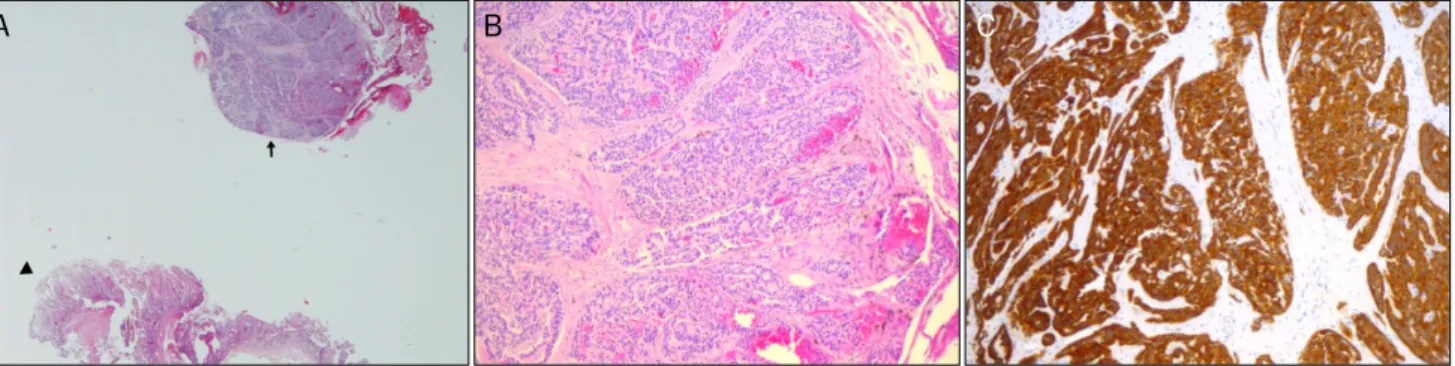
Fig. 5. Pathologic findings for an endoscopically-resected mucosal specimen containing a duodenal neuroendocrine tumor. (A) Neuroendocrine tumor (arrow) and normal duodenal epithelium (arrowhead) (H&E, ×100). (B) Monotonous proliferation of small round cells with hyperchromatic nuclei and scant cytoplasm forming nests, exhibiting a trabecular growth pattern (H&E, ×100). (C) Immunoreactivity for synaptophysin (synaptophysin, ×100).
Fig. 6. DNA sequencing analysis of exon 2 of the MEN1 gene, show- ing a heterozygous c.2_9delinsAGGGGGTT mutation. MEN1, multi- ple endocrine neoplasia type 1.
heterozygous mutation, c.2_9delinsAGGGGGTT, was newly detected in exon 2 (Fig. 6).
DISCUSSION
MEN1 is an autosomal dominant disorder. In 1988, the MEN1 gene was discovered in chromosome 11q13, by genet- ic linkage analysis based on a DNA single-nucleotide poly- morphism microarray.5 The MEN1 gene encodes menin, which is a putative tumor suppressor protein that is critical in the development and function of neuroendocrine cells, and it modulates gene transcription, as well as DNA repli- cation and repair.5,6 A germline mutation in this gene results in the production of an abnormal menin protein, causing MEN1.6
More than 1133 germline mutations and 203 somatic mu- tations in the MEN1 gene have been reported to date.
Although some potential mutational hot spots have been identified, most of the reported mutations are diffusely scat- tered throughout the 1830-bp coding region of the MEN1 gene.7
Clinical manifestations of MEN1 vary among families due
to the diversity of family-specific MEN1 gene mutations.6 Patients with the familial MEN1 Burin variant have been re- ported to exhibit an increased frequency of prolactinoma and low prevalence of gastrinoma, also possessing two common nonsense mutations (Tyr312Ter and Arg460Ter).8 Similar to our patient, 67 patients―out of 306 MEN1 patients―showed GEP-NETs as a first manifestation, as reported by Schaaf et al.9 These patients tended to have familial, rather than spora- dic, GEP-NETs, and they possessed truncating mutations.9 Another example is familial isolated primary hyperparathyroidism.
In contrast with the common MEN1 gene mutations, mis- sense mutations and in-frame deletions account for the ma- jority of familial isolated primary hyperparathyroidism gene mutations; however, the coding regions are also scattered.7 Therefore, various genotype-phenotype correlations in MEN1 are difficult to establish and must be verified in the future.7 To establish genotype-phenotype correlations be- tween the novel MEN1 gene mutation and presence of multi- ple NETs with meningioma in our case, further studies are necessary.
Our patient did not present with hyperparathyroidism;
however, it is an early symptom in most MEN1 patients, devel- oping sometime between the 2nd and 4th decades in life.2-4 Approximately 60% of MEN1 patients have Zollinger- Ellison syndrome, which is a characteristic syndrome of func- tioning NETs. In addition, many asymptomatic patients have an elevated serum gastrin level.3,10
The serum gastrin level of our patient was not high enough to suspect gastrinoma, and he did not show symptoms of Zollinger-Ellison syndrome. Moreover, the gastroscopy find- ings did not reveal peptic ulcer or thickened gastric folds.
A B C
VIPoma and insulinoma were also excluded, as the patient did not experience secretory diarrhea even during fasting.
The baseline insulin level and electrolytes were normal.
Pancreatic NETs are rare, comprising less than 2% of all pancreatic tumors.11 Non-functioning NETs are most com- monly found in the pancreatic/duodenal area in MEN1.3,12,13 At the time of diagnosis, approximately 60% of non-function- ing NETs have already metastasized.13 However, if the tumor size is 20 mm or smaller, the metastasis and mortality rates are low, and the life expectancy of patients with non-function- ing NETs is similar to that of those with functioning gastrinoma.12 A recent study reported that in MEN1 asso- ciated pancreaticoduodenal NET, the metastatic rate in- creased with age at surgery and metastatic disease was seen to be more common in gastrinoma. In addition, among pa- tients without metastasis at the time of surgery, 8% of pa- tients developed metastasis, and their mortality was 11%
during the follow-up; however, metastasis was not associated with age or tumor size. Therefore, regardless of age or tumor size, patients with MEN1 associated pancreaticoduodenal NETs should be monitored actively, and even be considered for treatment with surgery.14
As non-functioning duodenal NETs grow, obstruction, jaundice, pancreatitis, and bleeding may occur―albeit rarely
―in addition to carcinoid syndrome-like diarrhea and ab- dominal pain.15
Our patient did not exhibit any symptoms of functioning NETs; therefore, he was diagnosed as having non-functioning NETs in the duodenum and pancreas with an accompanied type 2 gastric NET.
In a previous study using brain MRI, meningioma was re- ported in 8% of 74 MEN1 patients compared with 0.9% of in the general population, and it was 11 times more frequent in patients with MEN1 than in those with pancreatic endocrine tumors alone.16,17 Most patients with meningioma are asymptomatic; therefore, if MEN1 is clinically suspected in a patient, brain MRI should be performed, especially if the pa- tient is in the 5th decade of life.17
Chang et al.18 reported that metastatic risk factors for NETs include a tumor size of 10 mm or larger, a central de- pression or ulcer at the tumor surface, extension beyond the muscularis propria, lymphatic or venous involvement, at least 3 mitoses per 10 HPF, and a Ki-67 labeling index of 3%
or higher. In general, NETs can be completely cured by endo-
scopic resection or by surgical removal; however, chemo- therapy is also recommended for patients with distant metastasis.19,20
Although typical MEN1 phenotypic features, such as hyper- parathyroidism and pituitary adenoma, were not present in our patient, we suspected MEN1 due to his family history, the pa- tient's history of brain surgery for meningioma and multiple gastric, duodenal and pancreatic NETs. DNA sequencing analy- sis of the MEN1 gene revealed an in-frame mutation, c.2_9delinsAGGGGGTT, in exon 2; such a mutation has not been previously described in the literature. Thus, we report a novel MEN1 gene mutation associated with specific pheno- typic features, including brain meningioma and three NETs in the stomach, pancreas and duodenum. These findings dem- onstrate novel genotype- phenotype correlations in MEN1.
REFERENCES
1. Kaltsas GA, Besser GM, Grossman AB. The diagnosis and medi- cal management of advanced neuroendocrine tumors. Endocr Rev 2004;25:458-511.
2. Marx S, Spiegel AM, Skarulis MC, Doppman JL, Collins FS, Liotta LA. Multiple endocrine neoplasia type 1: clinical and genetic topics. Ann Intern Med 1998;129:484-494.
3. Thakker RV, Newey PJ, Walls GV, et al. Clinical practice guidelines for multiple endocrine neoplasia type 1 (MEN1). J Clin Endocrinol Metab 2012;97:2990-3011.
4. Brandi ML, Gagel RF, Angeli A, et al. Guidelines for diagnosis and therapy of MEN type 1 and type 2. J Clin Endocrinol Metab 2001;86:5658-5671.
5. Larsson C, Skogseid B, Oberg K, Nakamura Y, Nordenskjold M.
Multiple endocrine neoplasia type 1 gene maps to chromosome 11 and is lost in insulinoma. Nature 1988;332:85-87.
6. Lips CJ, Dreijerink KM, Höppener JW. Variable clinical expression in patients with a germline MEN1 disease gene mutation: clues to a genotype-phenotype correlation. Clinics (Sao Paulo) 2012;
67 Suppl 1:49-56.
7. Lemos MC, Thakker RV. Multiple endocrine neoplasia type 1 (MEN1): analysis of 1336 mutations reported in the first decade following identification of the gene. Hum Mutat 2008;29:22-32.
8. Agarwal SK, Kester MB, Debelenko LV, et al. Germline mutations of the MEN1 gene in familial multiple endocrine neoplasia type 1 and related states. Hum Mol Genet 1997;6:1169-1175.
9. Schaaf L, Pickel J, Zinner K, et al. Developing effective screening strategies in multiple endocrine neoplasia type 1 (MEN 1) on the basis of clinical and sequencing data of German patients with MEN 1. Exp Clin Endocrinol Diabetes 2007;115:509-517.
10. Pipeleers-Marichal M, Somers G, Willems G, et al. Gastrinomas in the duodenums of patients with multiple endocrine neoplasia type 1 and the Zollinger-Ellison syndrome. N Engl J Med 1990;
322:723-727.
186 양민아 등. 새로운 MEN1 유전자 돌연변이가 증명된 신경내분비종양
11. Carriaga MT, Henson DE. Liver, gallbladder, extrahepatic bile ducts, and pancreas. Cancer 1995;75(1 Suppl):171-190.
12. Triponez F, Dosseh D, Goudet P, et al. Epidemiology data on 108 MEN 1 patients from the GTE with isolated nonfunctioning tu- mors of the pancreas. Ann Surg 2006;243:265-272.
13. Halfdanarson TR, Rabe KG, Rubin J, Petersen GM. Pancreatic neuroendocrine tumors (PNETs): incidence, prognosis and re- cent trend toward improved survival. Ann Oncol 2008;19:
1727-1733.
14. Donegan D, Singh Ospina N, Rodriguez-Gutierrez R, et al.
Long-term outcomes in patients with multiple endocrine neo- plasia type 1 and pancreaticoduodenal neuroendocrine tumours.
Clin Endocrinol (Oxf) 2017;86:199-206.
15. Soga J. Endocrinocarcinomas (carcinoids and their variants) of the duodenum. An evaluation of 927 cases. J Exp Clin Cancer Res
2003;22:349-363.
16. Vernooij MW, Ikram MA, Tanghe HL, et al. Incidental findings on brain MRI in the general population. N Engl J Med 2007;357:
1821-1828.
17. Asgharian B, Chen YJ, Patronas NJ, et al. Meningiomas may be a component tumor of multiple endocrine neoplasia type 1. Clin Cancer Res 2004;10:869-880.
18. Chang JH, Kim SW, Chung WC, et al. Clinical review of gastro- intestinal carcinoid tumor and analysis of the factors predicting metastasis. Korean J Gastroenterol 2007;50:19-25.
19. Sun JM, Jung HC. Gastrointestinal carcinoid tumor. Korean J Gastroenterol 2004;44:59-65.
20. Basu B, Sirohi B, Corrie P. Systemic therapy for neuroendocrine tumours of gastroenteropancreatic origin. Endocr Relat Cancer 2010;17:R75-90.