© 2013 The Korean Academy of Medical Sciences.
This is an Open Access article distributed under the terms of the Creative Commons Attribution Non-Commercial License (http://creativecommons.org/licenses/by-nc/3.0) which permits unrestricted non-commercial use, distribution, and reproduction in any medium, provided the original work is properly cited.
pISSN 1011-8934 eISSN 1598-6357
A Novel Mutation of the TAZ Gene in Barth Syndrome: Acute Exacerbation after Contrast-Dye Injection
A 14-month-old boy was transferred because of dilated and hypertrophied left ventricle, neutropenia, and developmental delay. After checking computed tomographic
angiography with contrast-dye, the patient showed acute exacerbation and finally died from multi-organ failure despite intensive cares. From genetic analysis, we revealed that the patient had Barth syndrome and found a novel hemizygous frame shift mutation in his TAZ gene, c.227delC (p.Pro76LeufsX7), which was inherited from his mother. Herein, we report a patient with Barth syndrome who had a novel mutation in TAZ gene and experienced unexpected acute exacerbation after contrast dye injection for computed tomographic angiography.
Key Words: Barth Syndrome; Tafazzin Gene; Cardiomyopathy; Cardiolipin; Neutropenia Gi Beom Kim,1 Bo Sang Kwon,1
Eun Jung Bae,1 Chung Il Noh,1
Moon-Woo Seong,2 and Sung Sup Park2 Departments of 1Pediatrics and 2Laboratory Medicine, Seoul National University Children’s Hospital, Seoul, Korea
Received: 11 October 2012 Accepted: 11 January 2013 Address for Correspondence:
Bo Sang Kwon, MD
Department of Pediatrics, Seoul National University Children’s Hospital, 101 Daehak-ro, Jongno-gu, Seoul 110-744, Korea Tel: +82.2-2072-3097, Fax: +82.2-743-3455 E-mail: [email protected]
http://dx.doi.org/10.3346/jkms.2013.28.5.784 • J Korean Med Sci 2013; 28: 784-787
CASE REPORT
Human Genetics & Genomics
INTRODUCTION
Barth syndrome is a rare X-linked recessive disorder presenting from infancy, with characteristic symptoms of cardiomyopathy, neutropenia, skeletal myopathy, growth retardation, and 3-me- thylglutaconic aciduria (1, 2). It is the first reported inborn error associated with cardiolipin dysfunction induced by mutations in the tafazzin (TAZ) gene at Xq28. Cardiolipin is an inner mito- chondrial membrane lipid with an essential role in the stability of the mitochondrial respiratory chain (3). Herein, we report a case of a novel mutation of the TAZ gene in Barth syndrome in a young child who showed acute exacerbation after contrast- dye injection and died from multiorgan failure.
CASE DESCRIPTION
A 14-month-old boy was referred to our hospital from another hospital because of dilated and hypertrophied left ventricle (LV), neutropenia, and developmental delay on 27 October 2011. He was born at full term, with a body weight of 3.2 kg. Six- teen days after birth, he was hospitalized owing to persistent ir- ritability. A chest radiograph showed cardiomegaly, and an echocardiogram revealed decreased LV contractility (ejection fraction: 24%). Under the impression of myocarditis, he had been managed for 1 yr before presentation in our hospital. More- over, the patient showed feeding difficulty and developmental delay from birth. Before referral, he had been admitted to other hospitals 7 times because of infection episodes. He was taking
furosemide, spironolactone, enalapril, and carvedilol before re- ferral. When he was referred to our hospital at 14 months old, his body weight was 6 kg (less than 3rd percentile) and his height was 71 cm (less than 3rd percentile). His overall motor develop- ment was delayed, and he could not sit alone. He could say
“mama” and “papa.” The recorded blood pressure and heart rate were 94/30 mmHg and 132 beats per minute, respectively.
On physical examination, no definite heart murmur was audi- ble and the liver was not palpable. He also showed persistent neutropenia, which started during his stay in the previous hos- pital. His WBC and neutrophil counts were 8,800/μL and only 2% (176/μL), respectively. The B-natriuretic peptide level was 1,045 pg/mL. A chest radiograph showed mild cardiomegaly (cardiothoracic ratio: 62.8%; Fig. 1A), and an electrocardiogram showed a low QRS voltage at the limb leads. An echocardio- gram revealed a dilated and hypertrophied globular LV with a hypertrophied papillary muscle and hyper-trabeculation, which did not meet the criteria of LV non-compaction. The other echo- cardiographic parameters were as follows: LV internal diameter at diastole, 37.7 mm (Z = 10.2); ejection fraction, 36.6%; and LV mass index, 75.6 g (Z = 6.3; Fig. 1B). To rule out the systemic cause of the dilated and hypertrophied LV, we performed a tho- raco-abdominal computed tomographic (CT) angiography with contrast dye. The CT findings showed no abnormality in the kidney and other organs and vessels. However, after under- going CT angiography, the patient showed abrupt high-grade spiking fever (Fig. 2) and developed secretory diarrhea (800 cc per day) associated with metabolic acidosis. At this time, his
Kim GB, et al. • TAZ Mutation in Barth Syndrome with Cardiomyopathy
http://jkms.org 785
http://dx.doi.org/10.3346/jkms.2013.28.5.784
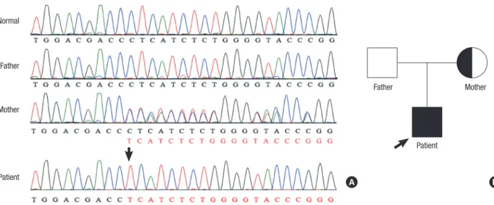
spite supportive care including intravenous fluid resuscitation and empirical antibiotics, the patient’s condition worsened, with aggravated metabolic acidosis and respiratory difficulty, requiring transfer to the intensive care unit (ICU). Just before the ICU transfer, the serum pH was 6.881; bicarbonate level, 7.8 mmol/L; and total CO2 was 41.4 mm Hg. Although the patient had been treated with intensive ventilator care, several inotro- pic agents, and other supportive care measures, he eventually died from the aggravated metabolic acidosis and acutely de- compensated heart failure 7 days after the ICU care. During the stay in ICU, we performed genetic analysis for Barth syndrome from the evidence of displayed cardiomyopathy, neutropenia, and developmental delay. The gene sequence analysis revealed that his TAZ gene harbored a novel hemizygous frameshift mu- tation, c.227delC (p.Pro76LeufsX7), which he inherited from his mother (Fig. 3).
DISCUSSION
Barth syndrome is a very rare X-linked recessive disorder with various types of cardiomyopathy as one of its characteristic symptoms. In this case, we found a novel mutation of the TAZ gene and experienced an unexpected acute exacerbation after contrast dye injection for CT angiography. Since the discovery of Barth syndrome by Barth in 1983, more than 100 different mutations of the TAZ gene have been reported in all exons of Xq28 (4). The level and composition of cardiolipin are affected by the TAZ gene mutation, resulting in the instability of the mi- Fig. 2. Vital signs after CT angiography. After undergoing CT angiography, the patient
spiked a high-grade fever and developed massive diarrhea with combined metabolic acidosis.
0 5 9 13 17 21 0
42.5 42.0 41.5 41.0 40.5 40.0 39.5 39.0 38.5 38.0 37.5 37.0 36.5 36.0 35.5 35.0 34.5 200 190 180 170 160 150 140 130 120 110 100 90 80 70 60 50 40 HR (BPM)
BT (˚C)
Timing of CT angiography
HR BT
A A
LV
Fig. 1. Chest radiograph and transthoracic echocardiogram at diagnosis. (A) Chest radiograph showing mild cardiomegaly (cardiothoracic ratio: 62.8%). (B) On echocardiogra- phy, the patient showed a dilated and hypertrophied globular left ventricle (LV) with hypertrophied papillary muscle and hyper-trabeculation (arrows).
WBC and neutrophil counts decreased to 3,290/μL and 1% (33/
μL), respectively. The B-natriuretic peptide level was greater than 4,901 pg/mL. He did not show associated respiratory symptoms.
The results of the respiratory and gastrointestinal viral studies were negative, and the blood and stool cultures were negative for pathogens. The C-reactive protein level was 7.08 mg/dL. De-
Kim GB, et al. • TAZ Mutation in Barth Syndrome with Cardiomyopathy
786 http://jkms.org http://dx.doi.org/10.3346/jkms.2013.28.5.784
Fig. 3. Results of the gene sequence (A) and pedigree analyses (B). The electropherogram (A) shows that the patient and his mother were hemizygous and heterozygous for the novel frameshift mutation, c.227delC (arrow), respectively.
Normal
Father
Father Mother
Mother
Patient
Patient
A B
tochondrial respiratory chain in the inner mitochondrial mem- brane lipid (3). This pathological mechanism provokes various expressions of cardiomyopathy, neutropenia, skeletal myopa- thy, and growth retardation (2). Various types of cardiomyopa- thy have been reported, including dilated cardiomyopathy, hy- pertrophic cardiomyopathy, endocardial fibroelastosis, and isolated non-compaction of the ventricular myocardium (5).
Spencer et al. (5) reported that approximately half of their pa- tients showed prominent LV trabeculations and 1 patient had hypertrophic CMP, although most patients had dilated CMP.
Our patient also showed a dilated and hypertrophied globular LV with prominent trabeculations.
From the abnormal shape of the LV, neutropenia, growth re- tardation, and developmental delay, we suspected Barth syn- drome, which was confirmed by genetic analysis after the pa- tient died. This is the first genetically confirmed case of Barth syndrome in Korea. There might have been many patients who had been treated and died under the impression of dilated CMP or non-compacted LV without definite diagnosis until now in Korea.
In our case, we experienced a sudden unexpected exacerba- tion of the patient’s condition with a high-grade fever, massive diarrhea, associated acute decompensated heart failure, and eventually death after the patient underwent CT angiography with iodinated contrast dye. Because the patient developed an acute high-grade fever and diarrhea and the blood and stool cul- tures were negative for pathogens during the treatment course, we assumed that the intravascular injection of iodinated con- trast dye induced cellular damage by neutrophil and cardiac muscle apoptosis and finally exacerbated the patient’s condi- tion. Fanning et al. (6) demonstrated that iodinated contrast media induce neutrophil apoptosis through a mitochondrial
and caspase-mediated pathway in vitro study using human neutrophil and provided insights of the pro-apoptotic mecha- nism of iodinated contrast dye in other cells. Our patient also showed decreased WBC and neutrophil counts 1 day after the contrast dye injection. Nevertheless, it is very difficult to con- clude the exact pathophysiological mechanism underlying the acute exacerbation of the patient’s condition.
It is interesting that some patients with Barth syndrome show improved cardiac and motor functions after meticulous sup- portive care as they grow older (7), although infantile Barth syn- drome shows a high mortality rate from severe heart failure and bacterial infections. To improve the prognoses of patients with Barth syndrome, physicians should be suspicious of Barth syn- drome in patients with dilated cardiomyopathy (especially as- sociated with increased LV hypertrophy including hyper-tra- beculations), neutropenia, growth retardation, and weakness in skeletal muscle.
In conclusion, we found a novel mutation of the TAZ gene and experienced unexpected acute exacerbation after contrast dye injection for CT angiography.
REFERENCES
1. Barth PG, Scholte HR, Berden JA, Van der Klei-Van Moorsel JM, Luyt- Houwen IE, Van ‘t Veer-Korthof ET, Van der Harten JJ, Sobotka-Plojhar MA. An X-linked mitochondrial disease affecting cardiac muscle, skele- tal muscle and neutrophil leucocytes. J Neurol Sci 1983; 62: 327-55.
2. Kelley RI, Cheatham JP, Clark BJ, Nigro MA, Powell BR, Sherwood GW, Sladky JT, Swisher WP. X-linked dilated cardiomyopathy with neutrope- nia, growth retardation, and 3-methylglutaconic aciduria. J Pediatr 1991;
119: 738-47.
3. Barth PG, Valianpour F, Bowen VM, Lam J, Duran M, Vaz FM, Wanders RJ. X-linked cardioskeletal myopathy and neutropenia (Barth syndrome):
Kim GB, et al. • TAZ Mutation in Barth Syndrome with Cardiomyopathy
http://jkms.org 787
http://dx.doi.org/10.3346/jkms.2013.28.5.784
an update. Am J Med Genet A 2004; 126A: 349-54.
4. Takeda A, Sudo A, Yamada M, Yamazawa H, Izumi G, Nishino I, Ariga T.
Eponym: Barth syndrome. Eur J Pediatr 2011; 170: 1365-7.
5. Spencer CT, Bryant RM, Day J, Gonzalez IL, Colan SD, Thompson WR, Berthy J, Redfearn SP, Byrne BJ. Cardiac and clinical phenotype in Barth syndrome. Pediatrics 2006; 118: e337-46.
6. Fanning NF, Manning BJ, Buckley J, Redmond HP. Iodinated contrast media induce neutrophil apoptosis through a mitochondrial and cas- pase mediated pathway. Br J Radiol 2002; 75: 861-73.
7. Schlame M, Ren M. Barth syndrome, a human disorder of cardiolipin metabolism. FEBS Lett 2006; 580: 5450-5.