© 2011 The Korean Academy of Medical Sciences.
This is an Open Access article distributed under the terms of the Creative Commons Attribution Non-Commercial License (http://creativecommons.org/licenses/by-nc/3.0) which permits unrestricted non-commercial use, distribution, and reproduction in any medium, provided the original work is properly cited.
pISSN 1011-8934 eISSN 1598-6357
Identification of a Novel Mutation in the ATP7A Gene in a Korean Patient with Menkes Disease
Menkes disease is an infantile-onset X-linked recessive neurodegenerative disorder caused by diverse mutations in a copper-transport gene, ATP7A. Affected patients are
characterized by progressive hypotonia, seizures, failure to thrive and death in early childhood. Here, we report a case of Menkes disease presented by intractable seizures and infantile spasms. A 3-month-old male infant had visited our pediatric clinic for lethargy, floppy muscle tone, poor oral intake and partial seizures. His hair was kinky, brown colored and fragile. Partial seizures became more frequent, generalized and intractable to
antiseizure medications. An EEG showed frequent posteriorly dominant generalized spikes that were consistent with a generalized seizure. From a genetic analysis, a c.2743C>T (p.Gln915X) mutation was detected and diagnosed as Menkes disease. The mutation is a novel one that has not been previously reported as a cause of Menkes disease.
Key Words: Menkes Disease; MNK Gene; ATP7A Mutation Yong Hyuk Kim1, Ran Lee1,
Han Wook Yoo2, Mi-Sun Yum2, Sun Hwan Bae1, So Chung Chung1, Yong Mean Park1 and Jae Sung Son1
1Department of Pediatrics, School of Medicine, Konkuk University; 2Department of Pediatrics, College of Medicine, University of Ulsan, Seoul, Korea Received: 28 February 2011
Accepted: 25 April 2011 Address for Correspondence:
Ran Lee, MD
Department of Pediatrics, Konkuk University Hospital, Konkuk University, School of Medicine, 255 Achasan-ro, Gwangjin-gu, Seoul 143-729, Korea
Tel: +82.2-2030-7557, Fax: +82.2-2030-7749 E-mail: [email protected]
DOI: 10.3346/jkms.2011.26.7.951 • J Korean Med Sci 2011; 26: 951-953
CASE REPORT
Human Genetics & Genomics
INTRODUCTION
Menkes disease (OMID 309400), also known as kinky hair dis- ease, is an infantile-onset X-linked recessive neurodegenerative disorder caused by diverse mutations in a copper-transport gene, ATP7A (1, 2). The ATP7A gene plays an important role in control- ling copper efflux from cells (3). Affected patients appear healthy at birth and develop normally for 6 to 8 weeks. Subsequently, hypotonia, seizures, failure to thrive and death in early child- hood are typical (4, 5). At present, a total of 170 different muta- tions have been identified worldwide (6).
CASE DESCRIPTION
A 3-month-old male infant had visited our pediatric clinic for lethargy, floppy muscle tone, poor oral intake and partial seizures on May 9, 2007. He was born at 38 weeks gestation by vaginal delivery. His birth weight was 3,180 g. He was healthy at birth and a neonatal metabolic screening test was negative. His hair was kinky, brown colored and fragile (Fig. 1). Symptoms of sei- zure were characterized by left facial twitching and left arm clonic movements. The initial EEG showed one episode of 2 Hz rhythmic spike and wave activity starting from the right central area evolving to the generalized slowings lasting about 100 sec- onds without clinical seizures, which was consistent with elec- trical partial seizures (Fig. 2A). There was no significant acidosis and ammonia was not increased. Serum lactate, tandem mass
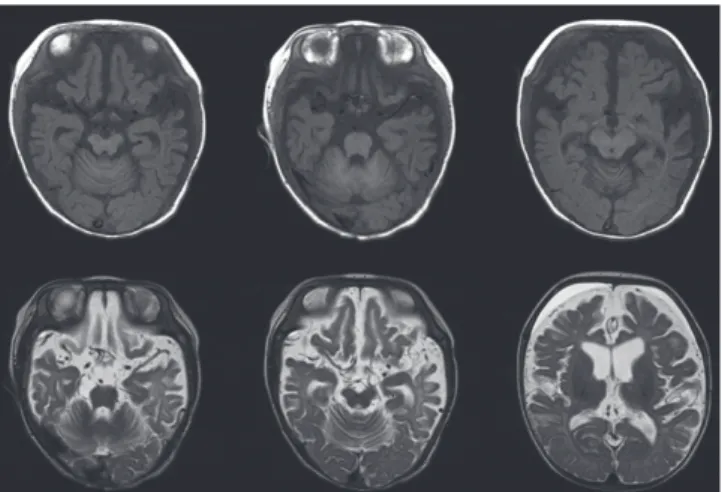
screening, serum amino acid and urine organic acids were all within the normal range. Pyruvate level (0.263 mM/L, reference range: 0.03-0.08 mM/L) was high. Partial seizures became more frequent and intractable to antiepileptic medications. About 1 month later, generalized flexor spasms developed. At this time, the EEG had changed to irregular high amplitude delta slowings on the background activities and frequent spikes from the right or left frontal or occipital areas that were consistent with hyp- sarrhythmia (Fig. 2B). Vascular tortuosity and diffuse brain at- rophy with callosal thinning were detected in an MRI scan (Fig.
3) at 3.5 months. Biochemical markers showed low serum cop- per (9.0 μg/dL, reference range: 70-130 μg/dL) and ceruloplas- min (5.6 mg/dL, reference range: 16-31.5 mg/dL) levels. From genetic analysis, a c.2743C>T (p.Gln915X) missense mutation (Fig. 4) in exon 13 of the ATP7A gene was detected, and the in- fant was diagnosed with Menkes disease (MD). The mutation was a novel one that has not been previously reported as a cause of MD. He died at 13 months of life.
DISCUSSION
We analyzed the ATP7A gene in a Korean patient with classical MD and identified one novel mutation. The ATP7A gene at Xq13.3 contains 23 exons and encodes a copper-transporting P-type ATPase of 1500 amino acids (3). To date, about 170 different mu- tations affecting ATP7A have been reported (6, 7). Approximate- ly 25% of the ATP7A mutations are gross deletions, ranging in
Kim YH, et al. • A Novel Mutation in the ATP7A Gene in Menkes Disease
952 http://jkms.org DOI: 10.3346/jkms.2011.26.7.951
size from a single exon to deletion of the whole gene, except for the first two exons (6). About 120 other different intragenic mu- tations of ATP7A have been reported: missense (33%), nonsense (16%), splice-site mutations (16%) and deletions/insertions/du- plications (33%) (Human Gene Mutation Database [HGMD];
www.hgmd.com) (7).
The biochemical result of low copper concentrations in MD is reduced activity of numerous copper-dependent enzymes such as ceruloplasmin, dopamine beta-hydroxylase, peptidylglycine alpha-amidating monooxygenase, cytochrome C oxidase, ascor- bate oxidase, lysyl oxidase, superoxide dismutase and tyrosi- nase, which leads to connective tissue abnormalities, tortuosity of blood vessels and peculiar hair (1, 8). The phenotypic features of Menkes disease can be divided into at least three categories:
classical MD with death in early childhood, mild MD with long survival and occipital horn syndrome (9). The majority of pa- tients suffer from classical MD, but milder forms are observed in 5%-10% of patients. There seems to be poor genotype–phe- notype correlation, and the clinical courses of MD patients may differ within a family, despite identical genetic changes (10).
Epilepsy is one of the main features of MD. Based on recent studies, the development of epilepsy can be divided into three phases: 1) an early stage characterized by focal clonic status, usually triggered by fever; 2) an intermediate stage with intrac- table infantile spasms, in which interictal EEG demonstrated modified hypsarrhythmia, with diffuse irregular slow waves, and spike waves; and 3) a late stage with multifocal seizures, tonic spasms and myoclonus (11, 12).
A B
Fig. 2. EEG records of the case. (A) At first admission, the pattern is consistent with electrical partial seizures from the right temporal area. (B) At 3 months later, the pattern is consistent with modified hypsarrhythmia.
Fig. 1. A photography of the patient’s head shows kinky, brown-colored and fragile hair.
Fig. 3. Brain MRI shows vascular tortuosity and diffuse brain atrophy.
Fig. 4. Gene analysis reveals c.2743C>T at exon 13 (p.Gln915X).
Normal
Patient
·Partial seq. of ATP7A gene.
Kim YH, et al. • A Novel Mutation in the ATP7A Gene in Menkes Disease
http://jkms.org 953
DOI: 10.3346/jkms.2011.26.7.951
Treatment in major cases is mainly symptomatic and sup- portive. However, neonatal diagnosis by plasma neurochemical measurement before symptoms appear and early parental cop- per–histidine supplement may modify the disease progression substantially (13-15). Prenatal diagnosis can be performed by biochemical analysis or DNA assay using chorionic villi sam- ples or amniocytes in the first trimester of an at-risk pregnancy (16, 17).
In summary, we report a case of Menkes disease presented by intractable seizures and infantile spasms because of a novel missense mutation (c.2743C>T) in the ATP7A gene.
REFERENCES
1. Kodama H, Murata Y. Molecular genetics and pathophysiology of menk- es disease. Pediatr Int 1999; 41: 430-5.
2. Mercer JF, Livingston J, Hall B, Paynter JA, Begy C, Chandrasekharappa S, Lockhart P, Grimes A, Bhave M, Siemieniak D. Isolation of a partial candidate gene for Menkes disease by positional cloning. Nat Genet 1993;
3: 20-5.
3. Voskoboinik I, Camakaris J. Menkes copper-translocating P-type ATPase (ATP7A): biochemical and cell biology properties, and role in Menkes dis- ease. J Bioenerg Biomembr 2002; 34: 363-71.
4. Bacopoulou F, Henderson L, Philip SG. Menkes disease mimicking non- accidental injury. Arch Dis Child 2006; 91: 919.
5. Vulpe C, Levinson B, Whitney S, Packman S, Gitschier J. Isolation of a candidate gene for Menkes disease and evidence that it encodes a copper- transporting ATPase. Nat Genet 1993; 3: 7-13.
6. Tümer Z, Birk Møller L, Horn N. Screening of 383 unrelated patients af- fected with Menkes disease and finding of 57 gross deletions in ATP7A.
Hum Mutat 2003; 22: 457-64.
7. Møller LB, Bukrinsky JT, Mølgaard A, Paulsen M, Lund C, Tümer Z, Larsen S, Horn N. Identification and analysis of 21 novel disease-caus- ing amino acid substitutions in the conserved part of ATP7A. Hum Mu- tat 2005; 26: 84-93.
8. Kim OH, Suh JH. Intracranial and extracranial MR angiography in Menk- es disease. Pediatr Radiol 1997; 27: 782-4.
9. Møller LB, Tümer Z, Lund C, Petersen C, Cole T, Hanusch R, Seidel J, Jensen LR, Horn N. Similar splice-site mutations of the ATP7A gene lead to different phenotypes: classical Menkes disease or occipital horn syn- drome. Am J Hum Genet 2000; 66: 1211-20.
10. Møller LB, Mogensen M, Horn N. Molecular diagnosis of Menkes disease:
genotype-phenotype correlation. Biochimie 2009; 91: 1273-7.
11. Bahi-Buisson N, Kaminska A, Nabbout R, Barnerias C, Desguerre I, De Lonlay P, Mayer M, Plouin P, Dulac O, Chiron C. Epilepsy in Menkes dis- ease: analysis of clinical stages. Epilepsia 2006; 47: 380-6.
12. White SR, Reese K, Sato S, Kaler SG. Spectrum of EEG findings in Menk- es disease. Electroencephalogr Clin Neurophysiol 1993; 87: 57-61.
13. George DH, Casey RE. Menkes disease after copper histidine replacement therapy: case report. Pediatr Dev Pathol 2001; 4: 281-8.
14. Choi JH, Yoo HW. Long term treatment of copper-histidine in a Menkes disease patient. J Korean Soc Inherit Metab Dis 2004; 4: 34-9.
15. Kaler SG, Holmes CS, Goldstein DS, Tang J, Godwin SC, Donsante A, Liew CJ, Sato S, Patronas N. Neonatal diagnosis and treatment of Menk- es disease. N Engl J Med 2008; 358: 605-14.
16. Poulsen L, Horn N, Heilstrup H, Lund C, Tümer Z, Møller LB. X-linked recessive Menkes disease: identification of partial gene deletions in affect- ed males. Clin Genet 2002; 62: 449-57.
17. Choi JH, Kim GH, Yoo HW. Identification of novel mutations of the ATP7A gene and prenatal diagnosis of Menkes disease by mutation analysis. J Genet Med 2007; 4: 38-44.