• 검색 결과가 없습니다.

Huge Intraabdominal and Thoracic Desmoid Tumor

N/A
N/A
Protected

Academic year: 2021

Share "Huge Intraabdominal and Thoracic Desmoid Tumor"

Copied!
4
0
0

로드 중.... (전체 텍스트 보기)

전체 글

(1)

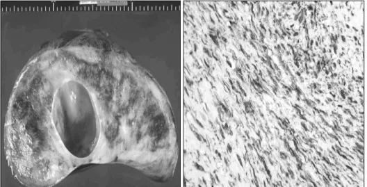
복강 및 흉강 내 거대 데스모이드 종양 수술 치험 1예

Huge Intraabdominal and Thoracic Desmoid Tumor

-Surgical experience in one case-

Desmoid tumors are benign neoplasms with high rates of recurrence. A case of huge desmoid tumor of the intrathoracic and intraabdominal space is presented. The patient was treated with resection, which involved hepatic left lobectomy and diaphragmatic resection and partial pericardiectomy and wedge resection of left lower lobe of lung. The resulting defect over the pericarium and diaphragm was reconstructed by bovine pericardium and Marlex mesh. Prevention of presumed local recurrence of desmoid tumors requires wide excision margin.

(Korean J Thorac Cardiovasc Surg 2003;36:623-626) Key words : 1. Diaphragm neoplasm

2. Desmoid

3. Thoracic neoplasms

(2)

Fig. 2. Gross finding of resected mass (29×22×15 in size, 5.9 kg in weight).

Fig. 1. Preoperative MRI shows

huge solid mass with cystic change

in upper abdominal cavity and

lower thoracic cavity.

(3)

Fig. 3. Cross section of mass re- vealed cystic formation (left) and positive vimentin staining on microscopic examination (×250, right).

Fig. 4. Postoperative chest PA revealed well reconstruction and

functioning of left diaphragm.

(4)

=국문 초록=

중심 단어

수치

Fig. 1. Preoperative MRI shows  huge solid mass with cystic change  in upper abdominal cavity and  lower thoracic cavity.
Fig. 3. Cross section of mass re- re-vealed cystic formation (left) and  positive vimentin staining on  microscopic examination (×250,  right).

참조

관련 문서

We report a case of 25 years old patient having recurrent giant cell tumor in the right distal femur which developed metastasis to lung. The primary bone lesion was treated with

Hence we report a patient with a papillary thyroid carcinoma metastasis to the right parietal lobe of brain, the lung, the left chest wall and right aceta- bulum, with a review of

Abdominal CT showed an exophytic, ovoid gastric mass having calcified components on the side of lesser curvature with huge, inhomogenous hepatic masses in the left lobe,

We report an unusual case of pulmonary cystic lymphangioma developed in the lingular segment of the left upper lobe which was diagnosed and treated by surgical resection.. (Korean

In our hospital, we have tried a less invasive method, the cryotherapy, to a patient who had a newly developed lung cancer at his right lower lobe after he had been treated with

We report a rare case of granular cell tumor arising in the left lower lobe (LLL) bronchus with secondary ob- structive change in a 60-year-old male.. The patient was found to have