J Korean Surg Soc 2012;82:128-133 http://dx.doi.org/10.4174/jkss.2012.82.2.128
CASE REPORT
Journal of the Korean Surgical Society
JKSS
pISSN 2233-7903ㆍeISSN 2093-0488
Received June 7, 2011, Revised July 18, 2011, Accepted August 16, 2011 Correspondence to: Chong Woo Chu
Division of Hepato-Biliary-Pancreatic Surgery and Transplantation, Department of Surgery, Pusan National University Yangsan Hospital, Pusan National University School of Medicine, Beomeo-ri, Mulgeum-eup, Yangsan 626-770, Korea
Tel: +82-55-360-2476, Fax: +82-55-360-2154, E-mail: [email protected]
cc Journal of the Korean Surgical Society is an Open Access Journal. All articles are distributed under the terms of the Creative Commons Attribution Non-Commercial License (http://creativecommons.org/licenses/by-nc/3.0/) which permits unrestricted non-commercial use, distribution, and reproduction in any medium, provided the original work is properly cited.
More than 7-year survival of a patient following repeat hepatectomy for total 20 colon cancer liver metastases
Kwang Ho Yang
1,2, Je Ho Ryu
1, Ki Myung Moon
1, Chong Woo Chu
1,21Division of Hepato-Biliary-Pancreatic Surgery and Transplantation, Department of Surgery, Pusan National University Yangsan Hospital, Pusan National University School of Medicine, 2Research Institute for Convergence of Biomedical Science and Technology, Pusan National University Yangsan Hospital, Yangsan, Korea
A 54-year-old man was transferred with sigmoid colon cancer combined with multiple bilobar liver metastases. Nine meta- stases were in the left lobe and 5 metastases were in the right lobe. After low anterior resection, all 9 lesions in the left lobe were completely removed by wedge resections. Because the remnant liver volume after multiple wedge resection of the left lobe was not sufficient to perform a right hepatectomy simultaneously, we planned a two-stage hepatectomy. Right portal vein embolization was performed one week after the first liver operation. A right hepatectomy was safely performed 22 days after the first hepatectomy. A recurrent mass developed in the segment III 18 months after the right hepatectomy.
Radiofrequency ablation (RFA) was performed to remove that lesion. Five other metastases developed 18 months after RFA whereby multiple wedge resections were performed. The patient has survived for more than 7 years after the first liver operation.
Key Words: Colorectal neoplasms, Neoplasm metastasis, Liver, Hepatectomy
INTRODUCTION
Curative hepatic resection is currently accepted as the most effective treatment for patients with colorectal can- cer liver metastases (CRLM). Recent reports showed that the mortality rate has decreased to less than 1%, and 5-year survival rates have increased after hepatectomy in these patients [1]. However, only 10 to 20% of the patients with CRLM are eligible for curative surgical procedures at the time of presentation [2]. The main cause of this low resect- ability is multiple bilobar liver metastases (MBLM). To
overcome this problem, multidisciplinary approaches were produced such as neoadjuvant chemotherapy, radio- frequency ablation (RFA), portal vein embolization (PVE), and two-stage hepatectomy [3]. Another problem is that the recurrent metastatic disease develops in most patients after liver resection, which mainly occurs in the liver. In this situation, repeat hepatectomy for recurrent liver metastasis can be performed safely and provide survival benefits [4].
In the case presented here, a patient initially had MBLM and recurrent lesions develop twice, yet, he was success-
7-year survival after resection of multiple CRLM
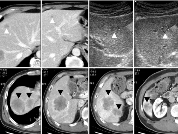
Fig. 1. Initial abdominal ultrasonography and computed tomography. Four lesions in left lobe and 5 lesions in right lobe were found (white arrow, metastases in left lobe; black arrow, metastases in right lobe).
fully managed with multidisciplinary approaches. The patient is now alive without disease, for more than 7 years after initial operation.
CASE REPORT
A 54-year-old man was referred to our department with sigmoid colon cancer combined with MBLM in November 2003. The sigmoid colon cancer infiltrated the pericolic fat tissue without lymph node invasion in the preoperative colon study. Nine metastatic lesions were preoperatively detected in the liver (5 lesions in the right lobe and 4 le- sions in the left lobe) (Fig. 1). Mean size of right side lesions was 4.3 ± 1.2 cm and mean size of left side lesions was 1.5
± 0.6 cm. We planned to resect the sigmoid colon cancer and perform staged hepatectomy; a simultaneous right hepatectomy with wedge resection of the left side meta- stases. The preoperative carcinoembryonic antigen (CEA)
level was 75.9 ng/mL.
The sigmoid colon cancer was completely removed by low anterior resection. The pathologic findings of the sig- moid colon cancer specimen were moderately differen- tiated adenocarcinoma, pericolic fat tissue infiltration, and no invasion of lymph nodes.
The staged hepatectomy was carried out 10 days after low anterior resection. Intraoperative ultrasonography (IOUS) was performed to detect the hepatic metastatic lesions. In contrast to the preoperative findings, a total of 9 lesions were detected in the left lobe. Therefore, we de- vised a new plan to perform non-anatomical resection of left side metastases followed by right hepatectomy after increasing the expected remnant liver volume. Mean size of left lobe lesions was 10 ± 2 mm. All lesions of the left lobe were completely removed with the cavitron ultrasonic surgical aspirator (CUSA; Integra LifeSciences Co., Plains- boro, NJ, USA). Abdominal computed tomography (CT) was checked the day after the first operation and the ex-
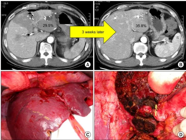
Fig. 2. (A) Abdominal computed tomography (CT) that was checked the day after first hepatectomy. Expected remnant liver volume was measured to be 29.5%. (B) Abdominal CT that was performed three weeks after first hepatectomy. Expected remnant liver volume increased to 35.8%. (C) Hypertrophied left lobe found in second hepatectomy. (D) Right hepatectomy was performed according to anatomical demarcation line.
pected remnant liver volume was measured at 29.5% (Fig.
2A). Embolization of the right portal vein was performed one week after the first hepatectomy. The patient recov- ered uneventfully. Abdominal CT was performed three weeks after the first operation and the expected remnant liver volume had increased to 35.8% (Fig. 2B). No meta- static lesions of the non-anatomically resected left lobe were found in the CT.
The second-stage operation was consequently per- formed 22 days after the first liver operation. Hypertro- phied left lobe was found (Fig. 2C). IOUS was rechecked to determine any metastatic lesions in the future remnant liv- er and there were no lesions. Therefore, the right hep- atectomy was safely performed (Fig. 2D). The CEA level was 2.67 ng/mL 10 days after the second liver operation.
The patient received 9 cycles of infusional 5-fluorouracil, leucovorin, and oxaliplatin (FOLFOX-4) chemotherapy af- ter two-stage hepatectomy.
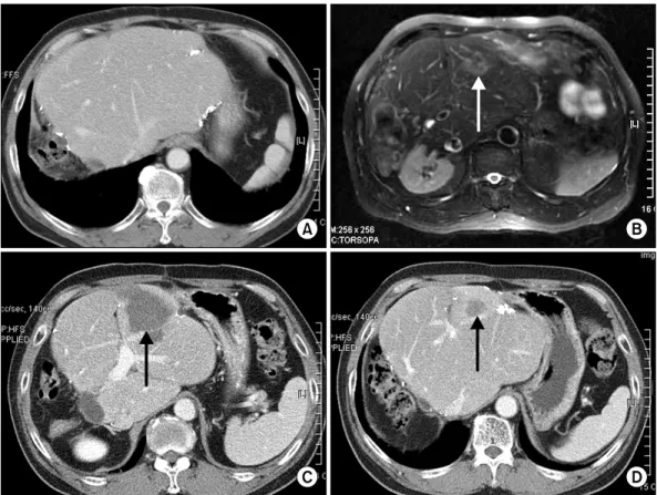
A recurred mass developed in segment III 18 months af- ter the second liver operation (Fig. 3B). Extracorporeal RFA was performed to remove a recurrent lesion. After RFA, the patient received 12 cycles of oral capecitabine.
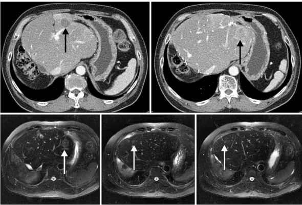
Unfortunately, 5 other metastases developed 18 months after RFA (Fig. 4). The third liver operation was performed and all metastatic lesions were completely removed by multiple wedge resections. The patient recovered un- eventfully and received 6 cycles of infusional 5-fluorour- acil, leucovorin, and irinotecan (FOLFIRI) chemotherapy after the third operation. He has survived for more than 7 years after the first operation (Fig. 5). The last CEA level was 3.35 ng/mL.
DISCUSSION
Over the last decade, the concept of treatment for CRLM
7-year survival after resection of multiple CRLM
Fig. 3. (A) Abdominal computed tomography (CT) taken at 15 months after second-stage hepatectomy showed no recurrent lesion. (B) Magnetic resonance image taken at 18 months after second-stage hepatectomy. Single recurrent mass (arrow) developed in remaining liver.
(C) Abdominal CT checked after radiofrequency ablation (arrow, post-radiofrequency ablation lesion). (D) Abdominal CT taken at 14 months after radiofrequency ablation showed no other recurrent lesion (arrow, post-radiofrequency ablation lesion).
has significantly changed. Many therapeutic options were introduced for curative hepatic resection, such as a novel regimen of chemotherapy, PVE, staged hepatic resection and RFA. As a result, more patients with CRLM could be- come candidates for curative surgical resections.
Traditionally, hepatectomy has been considered a con- troversial treatment strategy in cases with the presence of 4 or more colorectal metastatic lesions. However, recent studies have shown good results after hepatic resection in such cases [5]. Of particular interest, Imamura et al. [6] re- ported a case in which a patient had 22 metastatic nodules and underwent PVE and repeated hepatic resections. The author reported that disease-free survival time was 5 years 4 months after the repeat hepatic resection. Minagawa et al. [5] reported that 10-year survival rate of patients with 4 or more metastases after hepatic resection was 29%, which was almost equal to the long-term survival of patients with one solitary lesion. At present, it has been generally
accepted that the number of metastases does not sig- nificantly influence prognosis, if satisfactory resection could be achieved [5].
As one such innovative modality, two-stage hepatec- tomy with PVE was produced to increase curability in pa- tients with MBLM. PVE can be used to induce compensa- tory hypertrophy of the future remnant liver in patients who have inadequate liver volume. However, it also car- ries potential negative side effects. PVE can be associated with promotion of tumor growth in the future remnant liver [7], and enhancement of the disease recurrence [8].
Therefore, two-stage hepatectomy was developed. Meta- stases in the future remnant liver should ideally be re- sected before PVE. Then, major hepatic resection can be performed after PVE in patients with initially unresectable MBLM. Adam et al. [9] were the first to report a case un- dergoing this two-stage hepatectomy procedure com- bined with PVE. Unfortunately, the patient died of post-
Fig. 4. Abdominal computed tomography and magnetic resonance image taken at 36 months after second-stage hepatectomy (arrows, multiple recurrent lesions).
Fig. 5. Abdominal computed tomography taken at 7 years after initial hepatectomy. No recurrent lesion was found.
operative complications. Jaeck et al. [7] then reported that this strategy was successfully applied to patients with ini- tially unresectable MBLM. In our case, two-stage hep- atectomy with PVE was applied and the initial 14 lesions were completely resected. IOUS by a specialist also greatly contributed to removing all lesions.
Unfortunately, 60 to 70% of patients undergoing liver resection for colorectal metastases develop disease recur-
rence. Several articles have reported that one-third of pa- tients with recurrent liver metastases are suitable for re- peat liver resection [10]. And repeat hepatectomy for pati- ents with CRLM is a safe procedure that can provide long- term survival equal to that of a previous hepatectomy [4].
RFA may be an alternative therapeutic option for recur- rent lesions, but to date, no randomized, controlled trials of outcome of RFA for recurrence of CRLM compared with
7-year survival after resection of multiple CRLM
surgical resection exist; and the role of RFA is not yet well defined. In our case, the patient was successfully treated with RFA for one lesion and repeat wedge resections for five lesions in second recurrence.
In conclusion, we have reported that 20 accumulated metastatic lesions were successfully managed with multi- disciplinary and aggressive approaches in a patient with MBLM from colorectal origin. The patient is now alive without recurrence, more than 7 years after initial opera- tion.
CONFLICTS OF INTEREST
No potential conflict of interest relevant to this article was reported.
REFERENCES
1. Lochan R, White SA, Manas DM. Liver resection for color- ectal liver metastasis. Surg Oncol 2007;16:33-45.
2. Scheele J, Stang R, Altendorf-Hofmann A, Paul M. Resec- tion of colorectal liver metastases. World J Surg 1995;19:59- 71.
3. Tanaka K, Shimada H, Ueda M, Matsuo K, Endo I, Togo S.
Role of hepatectomy in treating multiple bilobar colorectal cancer metastases. Surgery 2008;143:259-70.
4. Antoniou A, Lovegrove RE, Tilney HS, Heriot AG, John TG, Rees M, et al. Meta-analysis of clinical outcome after first and second liver resection for colorectal metastases.
Surgery 2007;141:9-18.
5. Minagawa M, Makuuchi M, Torzilli G, Takayama T, Kawa- saki S, Kosuge T, et al. Extension of the frontiers of surgical indications in the treatment of liver metastases from color- ectal cancer: long-term results. Ann Surg 2000;231:487-99.
6. Imamura H, Sano K, Harihara Y, Noie T, Hasegawa K, Minagawa M, et al. Complete remission of disease for 5 years following initial and repeat resection of the liver for the removal of 22 metastases of colorectal origin. J Hepato- biliary Pancreat Surg 2003;10:321-4.
7. Jaeck D, Bachellier P, Nakano H, Oussoultzoglou E, Weber JC, Wolf P, et al. One or two-stage hepatectomy combined with portal vein embolization for initially nonresectable colorectal liver metastases. Am J Surg 2003;185:221-9.
8. Kokudo N, Tada K, Seki M, Ohta H, Azekura K, Ueno M, et al. Proliferative activity of intrahepatic colorectal meta- stases after preoperative hemihepatic portal vein emboli- zation. Hepatology 2001;34:267-72.
9. Adam R, Pascal G, Azoulay D, Tanaka K, Castaing D, Bismuth H. Liver resection for colorectal metastases: the third hepatectomy. Ann Surg 2003;238:871-83.
10. Pawlik TM, Scoggins CR, Zorzi D, Abdalla EK, Andres A, Eng C, et al. Effect of surgical margin status on survival and site of recurrence after hepatic resection for colorectal metastases. Ann Surg 2005;241:715-22.