간세포 암은 유병율이 전 세계적으로 약 백만 명으로 5번 째로 많은 암종이다. 따라서 고립성 간 종양을 가진 큰 동물 의 모형을 만드는 것은 유전자물질을 이용한 새로운 치료 약 제의 개발, 약의 독성 및 효능 연구를 위한 임상 전 시험 연 구를 하기 위해 꼭 필요하다고 하겠다. 이런 이유로, VX2 세 포를 이용한 적절한 토끼 모형을 만들기 위한 다양한 연구가 보고 되고 있다.
VX2 암종은 야생 토끼의 자궁 경부에서 바이러스 성 유두 종에서 기원한 역형성(anaplastic)의 편평 세포 암종이며, 집
토끼에서도 암종으로 발생할 수 있다(1-3). VX2 간 종양 모 형을 만들기 위해, 간 실질에 VX2 종양 세포 부유 액을 직접 주입하는 방법은 간편하고 쉽기 때문에 예전부터 널리 사용 되었다(1, 2, 4). 그러나 주입 경로를 통한 복강 내로의 종양 세포 누출이 빈번히 나타났으며, 복강 내 누출을 막기 위해 Lin 등(5)은 주입 후 천자 부위를 솜 거즈(cotton gauze)로 압박하는 방법을 고안하였으나, 만족스런 결과를 얻지는 못하 였다. 또한 이들은 녹인 정제우무(agarose)를 천자부위에 넣 고 굳혀서 VX2 종양 세포의 누출을 막고자 하였는데, 누출율 이 6.6%, 종양의 성공적 이식율이 86.6%로 비교적 높았으나, 시술 방법이 복잡하고 시간이 많이 걸리는 단점이 있다.
최근에는 VX2 세포 부유 액 대신에 종양 조직 절편을 이 식하는 방법이 시술되었는데, 이 방법은 종양의 조기 누출 및
바늘을 통한 VX2 입자의 간 내 이식 접종에 의한 고립성 간 종괴를 가진 가토 모형 생성
1조진한・최종철・신태범・박병호
목적: 바늘을 통한 VX2 입자를 접종하는 새로운 방법으로 가토 간 내에 적당한 크기의 단일 성 종괴를 가진 가토 모델을 구축하고, VX2 입자 접종 후 종괴의 성장 및 시간과 부피에 따 른 전이 형태를 알아보고자 하였다.
대상과 방법: 2.5-3 kg의 20마리의 가토를 대상으로 하였으며, 1 mm3 크기의 VX2 종괴를
21 G 바늘을 통해 주입될 정도의 입자로 잘라 투베르쿨린 주사기내에 식염수 0.1 ml와 함께 넣어 VX2 tumor를 준비하였다. 투베르쿨린 주사기내의 VX2 입자를 21 게이지 주사바늘 통 해 가토의 간 좌 내엽으로 주입한 후, VX2 입자의 복막 내 유출을 막기 위해 주입부 주위의 간 피막을 흑 견사로 봉합한 후‘Surgicel’Ⓡ 부착포(Oxidized regenerated cellulose, Johnson
& Johnson Medical Inc., Arlington, U.S.A.)로 주입구를 막았다. 접종 후 1주 간격으로 다중 채널 전산화 단층 촬영기를 이용하여 조영제 주입 후 10초, 30초, 60초에 간 내 종괴를 촬영 하였으며, 2주부터 매 주 3마리씩 부검을 하여 영상 소견과 비교하였다.
결과: 간 내 접종된 VX2 종괴는 첫 주에는 관찰되지 않았고, 둘째 주에 시행한 20마리 중 13 마리(65%)에서 종괴가 발견되었으며, 3주부터는 모든 가토(100 %)에서 종괴를 발견할 수 있었다. 3주 이내에 복강 내 종괴 유출을 보인 경우가 3마리(15%)였고, 나머지 17마리의 가 토(85 %)에서 성공적인 단일 종괴를 형성하였다. 성공적으로 접종된 17마리의 가토 중 3마 리(15%)는 성공적인 접종 후 크기가 증가하다 3-5주 후부터 크기가 줄어드는 모양을 보였 다. 접종된 부위 외 전이는 접종 후 4주 후부터 급격히 증가하는 소견을 보였고, 종괴의 부피 가 12×103 mm3 이상에서 전이가 증가하는 소견을 보였다.
결론: 저자들이 시행한 가토 내 단일성 VX2 종괴를 만드는 새로운 방법은 기존의 방법들에 비해 손쉽고 초기 복강 내 종괴 유출을 막는 데 유용한 방법으로 생각되며, 간 내 접종된 VX2 종괴는 접종 후 4주 이내, 그리고 부피가 12×103 mm3 이하에서 단일성 간 종괴를 유지 할 수 있었고, 향후 단일성 간 종괴를 이용한 실험 및 치료 효과 판정은 이 시기에 연구를 하는 것이 바람직하다고 생각한다.
1동아대학교병원 영상의학과학교실
This Paper was Supported by the Dong-A University Research Fund in 2004.
이 논문은 2005년 3월 30일 접수하여 2005년 6월 10일에 채택되었음.
양을 만들 수 있는 장점이 있다(6-8). 그러나, 외과적 시술 이 세포 부유 액 주입에 비해 보다 침습적이며, 출혈, 담즙 누 출, 농양과 같은 외과적 합병증도 유발하는 단점이 있다.
이에, 저자들은 적절한 고립성 간 내 종양을 가진 큰 동물 (토끼) 모형을 만들기 위한 누출율이 낮으며 침습적이지 않은 새로운 방법을 개발하고, 접종 후 시간의 경과와 부피 증가에 따라 간 내 종양이 전이를 일으키지 않은 고립성 종양으로서 유지할 수 있는 시기를 알고자 하였다.
대상과 방법
실험 동물 모형 실험 동물 준비
동아대학교 동물 실험 위원회의 승인 아래 체중 2.5-3 kg 의 20마리 뉴질랜드 흰 토끼를 실험대상으로 하였다. 실험 동 물의 사육은 본교 부설 동물 실험실에 의뢰하여 사육하였다.
Ketamine. Yuhan, Seoul, Korea), xylazinehydrochloride (5 mg/kg; Rumpun; Bayer Korea, Ansan, Korea)를 주입한 후 전신 마취 상태에서 시행하도록 하였다.
VX2 종양 유지
이식을 위해 필요한 VX2 종양 조직은 본 대학교 병리학 교 실에서 제공받았다. VX2 암종 균주는 보균 토끼의 후사지 (hindlimb)내에 연속적인 이식에 의해 보존하도록 하였다.
VX2 종양 입자 제작 및 VX2종양 접종 방법
저자들은 보균 토끼의 후사지(hindlimb)에 이식된 종양에 서 중심부 괴사 부위를 제거하고 살아있는 부위의 종양을 가 위로 적출하여 저온의 calcium and magnesium-free Hank’
s balanced salt solution(Grand Island Biological Co., Grand Island, New York, U.S.A.)과 혼합한 후 수술용 칼 & 가위를 이용하여 1 mm3의 VX2 종양 절편을 만들었다. 이 입자는 21
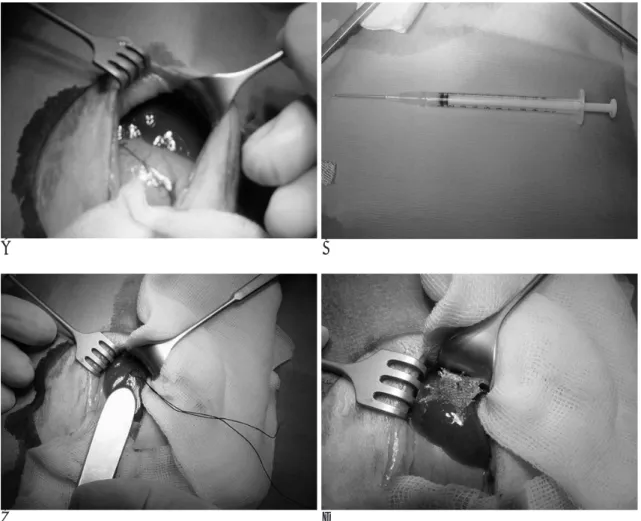
A B
C D
Fig. 1. Inoculation methods.
A. Median incision beneath the sternum is made to expose the left medial lobe of liver.
B. VX2 tumor is minced it to particles being able to pass through a 21 gauge needle and then mounted in a tuberculin syringe with 0.1 ml of normal saline.
C. First We make a purse-string suture using black silk 4-0, then inject tumor particles through the center of suture. During the re- moval of needle, I tighten the suture, and then cover the injection site with ‘Surgicel’Ⓡpatch (D) to prevent hemorrhage or leakage of VX2 tumor cells through the injection route.
게이지 바늘(gauge needle)을 통과할 수 있을 크기의 종양 입자로 만들어, 0.1 ml의 생리식염수와 함께 투베르쿨린 (tuberculin) 주사기에 넣었다. 흉골 아래로 정중 절개를 통해 간의 왼쪽 엽을 노출시킨 후 간 내부의 큰 혈관을 피하기 위 해 왼쪽 간엽의 간 피막 하 실질 내에 투베르쿨린 주사기에 담은 절개된 VX2 종양 입자를 21 게이지 바늘을 통해 주입 하였다(Fig. 1A, B). 주입 경로를 통한 VX2 종양 세포의 누 출 및 출혈을 막기 위해 흑견사 4-0를 이용한 purse-string 봉합을 하였으며(Fig. 1C), 봉합 중심을 통하여 바늘을 천자 하였다. 바늘을 제거하면서 봉합을 죄었으며, ‘SurgicelⓇ’부 착포(oxidized regenerated cellulose, Johnson & Johnson Medical Inc. Arlington, U.S.A.)로 주입 부위를 덮었다(Fig.
1D). 시술이 끝난 후 복벽은 각각의 층으로 모두 봉합을 하 였다.
CT 장비와 영상 분석
간 내에 주입된 VX2 종양은 16채널 다중 CT(Multidetec- tor CT, 이하 MDCT, Somaton Sensation 16; Siemens Medical Systems, Erlangen, Germany)를 이용하여 일주일
간격으로 폐 꼭대기에서 골반저부까지 0.75 mm collimation, 24 mm/s table speed, pitch factor=2, reconstruction thick- ness/interval=1/0.7 mm, 0.5sec rotation time, 80mA, 40KVP의 조건으로 촬영하였다. 조영제(UltravistⓇ; Schering, Berlin, Germany)는 가토 귓바퀴 정맥을 통해 0.7 ml/sec의 속도로 5 ml를 주입한 후 10, 30, 60초 간격으로 촬영을 시 행하였으며, 정보는 독자적인 다른 컴퓨터(Rapidia, Infinitt technology, Seoul, Korea)로 전송하여 분석하였다. 다중 면 재구성(Multiplanar reconstruction, MPR)은 종양의 장축에 대 해 비스듬히 1 mm 간격으로 재구성하여 측정하였다. 종양 용 적은 두 명의 방사선과의사의 협의 하에 측정하였으며, 용적 은 V=L×S2/2(L=종양의 가장 긴 직경, S=종양의 가장 짧은 직경)공식에 의해 계산하였다(9-11).
두 명의 방사선과 의사에 의해 종양 위치, 크기, 복강 내 파 종, 다른 장기 전이정도가 평가되어 졌으며, CT나 부검 소견 상 접종 후 3주내에 복강 내로의 누출되거나 간 내 종양이 보 이지 않을 시, 또는 간 내에 여러 개의 종양이 자라거나 다른 기관으로의 전이를 되었을 시는 이식 실패로 간주하고, 접종 후 3주내에 복강 내 누출이 없고 다른 기관으로의 전이 없이
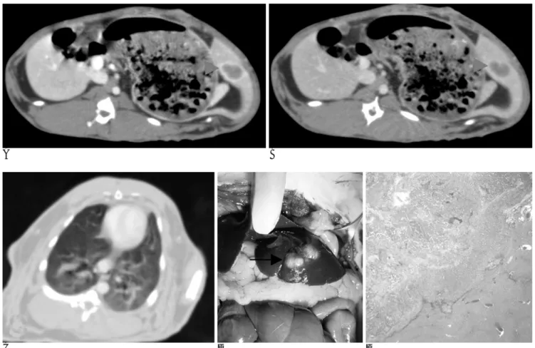
A B
C D E
Fig. 2. Successful inoculation of VX2 tumor particles into the hepatic parenchyma. Contrast enhanced CT of 10 seconds (A) and 60 seconds (B) delayed scans demonstrate typical VX2 tumor(arrowhead) appearance which shows peripheral enhancement with cen- tral low attenuation, at left lobe of rabbit liver on the third week after inoculation.
C. There is no visible distant metastasis on lung setting.
D. Autopsy findings show intrahepatic lobulated mass (arrow) growing at subcapsular area.
E. Microscopic finding reveals well-inoculated intrahepatic solitary VX2 tumor (H & E stain, ×100).
식으로 생각하였다.
조직 검사
계획서에 따라서, 종양 접종 후 2주째부터 4주간 무작위로 3마리씩의 토끼를 부검하여 CT소견과 병리학적 소견을 비교 하였다. 모든 동물은 가스 상자에서 이산화탄소를 흡입시켜 희 생시킨 후 부검을 시행토록 하였다. 복강 내 파종 전이는 육 안으로 확인한 후 모두 병리학 교실로 보내어 조직 검사를 하 였으며, 부검 후 나온 표본은 10% 완충 포르말린에 보관하였 으며, 파라핀으로 조직을 고정하여 5 mm 두께로 절개한 다 음 헤마토실린과 에오진 염색(H & E stain)처리를 통해 조 직 검사를 시행하였다.
결 과
종양 접종 후 20마리 모든 토끼에서 외과적 합병증 없이 VX2 종양의 간 내 증식이 이루어졌으며, 간 내 접종된 VX2
CT검사에서는 20마리 중 13마리(65%)에서 간 내 증식된 VX2종양을 확인할 수 있었다. 접종 후 3주째에는 모든 토끼 (100%)에서 종양을 확인할 수 있었다. 20마리 중 17마리 (85%)에서 접종 후 3주내에 종양의 복강 내 누출이나 다른 기관으로의 전이 없이 간 실질 내에 고립성 종양(solitary mass)으로 성공적으로 접종되었다(Fig. 2). 나머지 3마리 (15%)에서는 종양 증식이 간과 복강에서 나타났으며, 이것은 접종 후 주입 부위에서 종양세포가 누출되어 복강 내 파종에 의한 것으로 생각되어 진다(Fig. 3).
VX2 입자 접종 후 종양 증식은 지수적 증식 양상을 보였 다(Fig. 4). 간 내 또는 간 외 장기로의 종양 전이는 2주째에 는 관찰되지 않았으며, 시간이 지남에 따라 3주 이후에는 17 마리 중 1마리(5.9 %), 4주에는 14마리 중 7마리(50 %), 5 주에는 11마리 중 8마리(72.7%), 6주에는 8마리 중 7마리 (87.5%), 7주 이후부터는 모든 토끼에서 전이가 발견되어, VX2 접종 후 4 주부터 전이가 급격히 증가함을 볼 수 있었 다(Fig. 5). 종양 부피 증가에 따른 전이는 부피가 6×10³
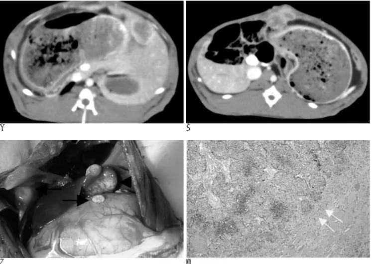
A B
C D
Fig. 3. Early peritoneal seeding due to VX2 tumor leakage.
A. Contrast enhanced CT scans demonstrate intrahepatic inoculated mass in left lobe of liver at the third week.
B. Early peritoneal seeding masses are noted at anterior peritoneum.
C. Autopsy reveals intrahepatic inoculated mass (arrowhead) and peritoneal seedings (arrow).
D. Microscopic finding shows peritoneal seeding (arrows) (H & E stain, ×200).
mm3이하에서 5.9%, 12×103 mm3이하에서 13.3%, 18×103 mm3이하에서 40%, 24×103 mm3이하에서 66.7%, 30×103 mm3이하에서 75%, 36×103 mm3이하에서 75%였으며 36×
103 mm3이상의 모든 토끼(100%)에서는 전이가 관찰되었고, 부피가 12×103 mm3이상에서 전이가 급격히 증가함을 볼 수 있었다(Fig. 6, 7). 전이 부위는 폐, 복강, 간, 임파선, 부신의 순서로 빈도가 높았다.
20마리 중 3마리(15%)에서 접종 후 3-5주후부터 간 내 VX2종양 퇴화를 CT 영상에서 관찰할 수 있었으며, 이것은 병 리학적 소견에서 접종된 부위가 괴사되고 섬유화 된 것을 관 찰하였다(Fig. 8).
복강 내 파종과 다른 기관으로의 전이 유무와 결과는 병리 학적 소견과 CT소견이 모두 일치하였다.
고 찰
간세포 암은 만성적인 간 질환을 앓고 있는 환자에서 발생 을 잘 하는 매우 악성의 종양으로 간세포 암 환자의 70-90%
가 진단되는 시점에 이미 간 경화를 앓고 있는 것으로 보고 되고 있다. 간세포 암이 잘 발생하는 간 경화는 만성 B, C형 간염, 술, aspergillus 곰팡이에서 오는 Aflatoxin과 관련이 있 으며, 간세포 암의 전 세계적인 분포는 B형 그리고 C형간염 보균자의 지역적인 유병율과 관계가 있다(12, 13). 수술 및 이식이 간세포 암을 완치시키는 유일한 치료법이나 간세포 암 환자의 근원적인 간경화에 따른 부작용으로 인해 그 대상이 되는 환자는 전체 중 5-10%로 제한되어 있다. 나머지 90%
에게는 완치를 위한 치료는 없으며 현재 사용되는 약물 치료 제는 15-20%정도로 만족스럽지 못한 반응율을 보이고 있어, 이로 인해 종양전문 내과 의사들은 간세포 암 환자의 약물치 료에 어려움을 겪고 있다. 이는 약물 치료를 하기에는 근원적 인 간경화로 인한 간의 제한된 간 용량 때문뿐 아니라, 간세 포 암이 독소로부터 다른 정상 세포를 보호하는 역할을 하는 간세포에서 생긴 아주 저항력이 강한 암이기 때문이다(14).
이러한 이유로 암의 진행을 막기 위해 알코올이나 고주파를 이용한 국소 소작 시술이나 유전자 분자 치료 같은 새로운 중 재 요법과 약의 개발이 이루어지고 있다.
VX2 암종은 토끼 자궁 경부에 생기는 매우 악성이며 역형 성(anaplastic)의 편평 세포 암이며, 일반적으로 토끼의 간에 이식할 수 있는 암종으로 받아들여지고 있다(1-3). VX2 암 종 세포는 사람의 간세포 암과 혈관신생 등 유사한 특징을 가 지므로, 임상적으로 국소적인 항암 치료 또는 방사선 치료의 적절한 실험 모델로 사용 가능하다(15-17). 따라서 간세포 암에 대한 효과적인 중재 요법 및 약의 개발이 이루어지기 위 해서는 효과적인 전 임상 시험을 위한 적절한 고립성 간 내 VX2 암종을 가진 큰 동물 모형이 필요하며, 접종 후의 자연 적인 종양 성장에 대한 정보는 적절한 치료 방법을 연구하는 데 있어서 매우 중요하다.
간 내 VX2 암종을 만들기 위해 가장 많이 사용되어 온 방 법인 VX2 세포 부유물을 직접 주사하는 방법은 시술이 간단 하지만 주사 경로를 통한 복강 내의 종양세포 누출이 빈번히 일어나는 단점이 있다(1, 2, 4). 따라서 초기 전이 및 복강 내 Fig. 4. Tumor volume according to the course of time after
VX2 tumor inoculation. On serial contrast enhanced CT scans, intrahepatic VX2 tumor shows a highly aggressive growth pat- tern and has grown exponentially.
Fig. 5. The tumor metastatic rates according to the course of time. Tumor metastasis to intrahepatic and/or extrahepatic or- gans is increased from the fourth week after inoculation of VX2 tumors.
Fig. 6. The tumor metastatic rates according to the inoculated tumor volume increment. Tumor metastasis to other sites in- cluding liver and peritoneal cavity, lung etc has been started to be observed at which tumor volume was reaching more than 12×103 mm3after inoculation of VX2 tumors.
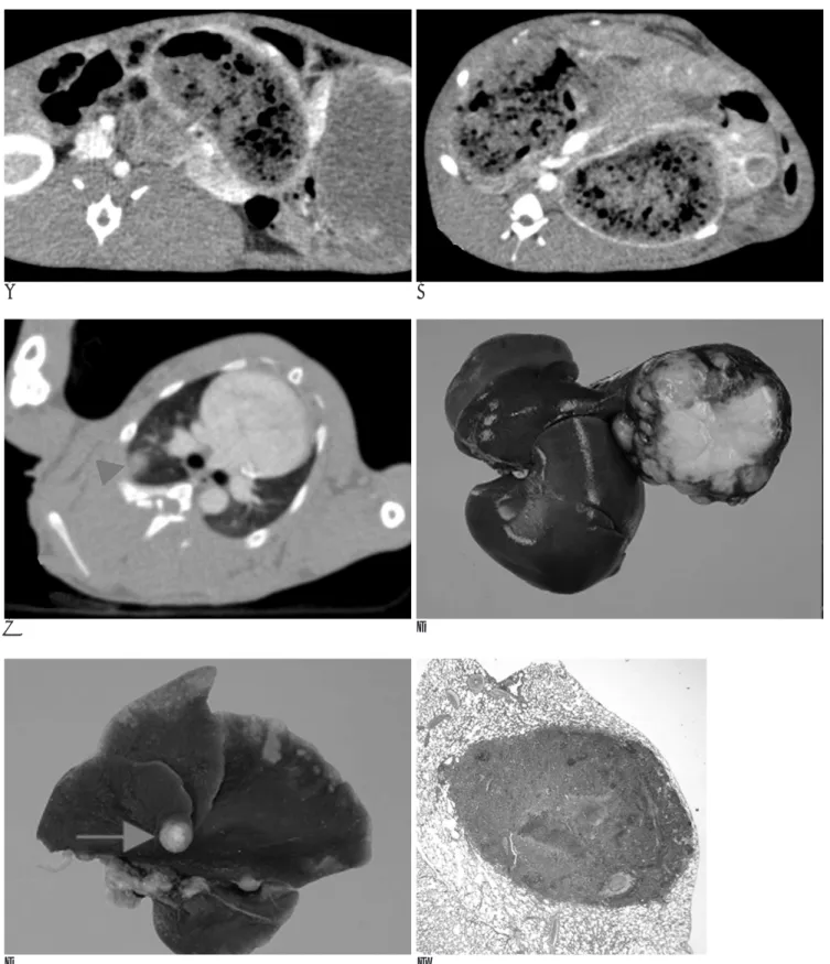
A B
C D
E F
Fig. 7. Tumor metastasis.
A. Contrast enhanced CT scans demonstrate huge VX2 tumor at left hepatic lobe of rabbit on the seventh week after inoculation.
B. Intrahepatic metastatic nodule is detected in another hepatic lobe of rabbit.
C. Pulmonary metastasis (arrowhead) is also noted at right upper lobe.
D. E. Gross specimen reveal exophytic grown large tumor with small intrahepatic metastatic nodule (arrow).
F. Microscpic finding shows lung metastasis (H & E stain, ×50).
누출을 막기 위해, 바늘 제거 후 천자 부위를 솜 거즈로 압박 하는 방법을 고안하였으나, 만족스런 결과를 얻지는 못하였다.
Lin 등(5)은 종양 부유물을 주입 하고, 같은 천자부위로 액상 으로 끓인 정제우무(agarose)를 0.2 ml 주입한 후 정제우무 가 고형으로 될 때 까지 2분간 유지하여 VX2 종양 세포의 누 출을 막고자 한 최근 연구는 낮은 누출율(6.6 %)을 보였으 나, 저자들의 시술보다 시간이 오래 걸리고 고형으로 변한 정 제우무에서 바늘을 제거해야 하는 등 시술이 복잡하였다. 또 한 간 실질에 종양부유물을 주입할 시에는 종양은 표적 위치 밖으로 퍼지게 되며, 간 실질 내에 수많은 작은 결절이 생기 게 된다(2, 4). 이것은 간 실질 내로 주입된 세포가 혈관 또 는 담관으로 들어가거나 아니면 천자부위로부터 종양세포의 누출이 일어나는 경우로 생각할 수 있다(5).
최근, 개복술 같은 외과적 시술을 이용한 VX2 종양 조각의 이식을 통한 간 내 암종을 만드는 방법이 사용되고 있으며, 이 시술은 정확한 이식 종양의 위치를 알 수 있으며, 종양의
누출 및 초기 확산을 줄일 수 있는 장점이 있다(3, 7, 8). 그 러나 이 외과적 종양 조각 이식 시술은 저자들의 방법 및 세 포 부유물 주입 방법보다 침습적이며 수술에 따른 출혈이나 담즙 누출, 농양 같은 외과적 합병증이 발생하는 단점이 있다.
저자들이 사용한 21 게이지 바늘을 통과할 정도의 종양 입 자를 사용하고, purse string을 이용한 주입 부위 주위의 간 피막 봉합 및‘SurgicelⓇ’조각(oxidized regenerated cellu- lose, Johnson & Johnson Medical Inc. Arlington, U.S.A.)을 이용한 새로운 VX2 종양 접종 방식은 주입 부위에서의 누출 율이 낮으며(15%), 우수한 이식 성공율(85%)을 보였다. 또 한 모든 토끼에서 접종된 VX2종양 입자는 시술과 관련된 어 떠한 합병증 없이 토끼 간 내에서 증식을 보였다.
간 내에 이식된 VX2종양을 관찰하기 위해 여러 가지 영상 기법이 이용되고 있으며, 초음파를 이용한 이전 연구의 민감 도는 단지 41-92%이며(18-20), CT가 간 내 이식 종양을 발견하고 치료 효과를 평가하는데 가장 우수한 영상기법으로
A B
C D
Fig. 8. Tumor regression after successful inoculation. VX2 tumor is detected at the second week after inoculation on contrast en- hanced CT scan (A, arrowhead), but regress after 2 weeks (B, arrow). Autopsy (C) and microscopic (D) finding reveals tumor fibro- sis (arrows) (H & E stain, ×100).
와 비교하여 신속한 영상을 얻을 수 있으므로 주위 조직과의 대조도를 높여 간세포 암과 같은 과 혈관성의 간암의 관찰에 좋기 때문이다(25-29). Mortele 등(30)은, 조영제를 사용한 이중 나선 CT의 간암의 발견 민감도와 특이도는 각각 82, 100%라고 하였다. 최근 MDCT는 여러 병원에서 널리 사용 되고 있으며, MDCT를 이용한 영상은 간의 강조 영상에서 필 요한 초기 및 후기 동맥기의 영상을 보다 자세하게 볼 수 있 게 되었고, 호흡에 의한 미영상을 줄였으며, Z-축 해상력을 개선시켰으며, 매우 미세한 조준이 가능하기 때문에 작은 병 소라도 높은 해상도의 영상을 가능하게 한다. 따라서, MDCT 는 간 종양의 발견 및 치료 효과를 평가하는데 있어 기존의 나선형 CT보다 높은 정확도를 보여준다. 저자들이 시행한 MDCT 소견 상, 접종 후 첫 주에 관찰한 MDCT, CT영상에 서는 토끼의 간에서 암종을 관찰할 수는 없었고, 2주째에 시 행한 CT소견상 20마리의 토끼 중 13마리에서 간 내에 종양 주위 조영 증강 양상을 보이는 종양을 관찰할 수가 있었다. 3 주째에 시행한 CT 소견에서는 모든 토끼에서 종양 주위 조 영 증강 양상을 보이는 고형 종괴를 관찰할 수 있었다. 복강 내 파종과 다른 기관으로의 전이 유무와 결과는 부검 소견과 MDCT결과가 모두 일치하는 소견을 보여 높은 정확도를 확 인하였다. 조영 증강 CT 영상에서 VX2종양은 매우 빨리 성 장하는 지수적 증식 양상을 볼 수 있었으며, 간을 포함한 간 외 기관으로의 전이 그리고 복강 내 파종은 VX2 종양 접종 후 4주 이상 지났거나 종양의 용적이 12×103 mm3 이상 되 었을 때 급격히 증가하였다.
20마리 중 3마리(15%)는 성공적인 접종 후 3-5주 사이에 종양이 퇴화되는 양상을 볼 수 있었다. 이에 대해 저자들은 야생 토끼의 면역력 또는 종양의 급속한 성장에 비해 상대적 으로 혈액 공급이 잘 공급되지 않아 종양이 괴사 및 섬유화 가 되면서 이러한 반응이 일어났을 것이라고 생각한다.
결론적으로 저자들이 시도한 바늘을 통한 간 내 VX2 종양 입자 주입법은 간 내 고립성 종양을 가진 동물 모형설계에 있 어 간단하고 효과적인 방법으로 생각된다. 성공적으로 간 내 접종된 VX2 종양은 접종 후 4주 이후 또는 종양 용적이 12
×103 mm3 이상으로 자라면 복강 내와 다른 기관으로의 전 이가 증가하므로, 고립성 간암을 이용한 중재 시술 개발 및 치료 효과에 대한 연구는 이 시기 이전에 시행하는 것이 좋 을 것으로 생각된다.
참 고 문 헌
1. Berkowitz DM, Alexander L, Hollenberg NK. A simple cell suspen- sion method for transplantation of VX2 carcinoma. J Natl Cancer Inst 1975;54:233-234
2. Izumi B, Tashiro S, Miyauchi Y. Anticancer effects of local admin- istration of mitomycin C via the hepatic artery or portal vein on implantation and growth of VX2 cancer injected into rabbit liver.
Cancer Res 1986;46:4167-4170
3. Ramirez LH, Zhao Z, Rougier P, Bognel C, Dzodic R, Vassal G. et al. Pharmacokinetics and antitumor effects of mitoxantrone after
Cancer Chemother Pharmacol 1996;37:371-376
4. Burgener FA, Violante MR. Comparison of hepatic VX2 carcino- mas after intraarterial, intraportal and intraparenchymal tumor cell injection: an angiographic and computed tomographic study in the rabbit. Invest Radiol 1979;14:410-414
5. Wan YL, Jeonhor C, Yungchang L, Kunwang H. Implantation of VX2 carcinoma into the liver of rabbits: a comparison of three di- rect-injection methods. J Vet Med Sci 2002;64:649-652
6. Ikeda Y, Matsumata T, Adachi E, Nishizaki T, Sugimachi K.
Ethanol injection therapy in RBT-1 carcinoma of the rat liver evokes enhancement of metastasis. J Surg Oncol 1993;54:9-12 7. Phillips JJ, Chang SL, Vargas HI, Dickman PS, Butler JA,
Lipcamon JD. MR and CT imaging of ethanol-treated liver tumors in an animal model. Magnet Reson Imag 1991;9:201-204
8. Thorstensen O, Isberg B, Svahn U, Jorulf H, Venizelos N, Jaremko G. Experimental tissue transplantation using a biopsy instrument and radiologic methods. Invest Radiol 1994;29:469-471
9. Okada M, Kudo S, Miyazaki O, Saino T, Ekimoto H, Iguchi H, et al. Antitumoral efficacy and pharmacokinetic properties of piraru- bicin upon hepatic intra-arterial injection in the rabbit VX2 tumor model. Br J Cancer 1995;71:518-524
10. Watanabe D, Ueo H, Inoue H, Matsuoka H, Honda M, Shinomiya Y, et al. Antitumor effects of intraarterial infusion of tumor necro- sis factor/lipiodol emulsion on hepatic tumor in rabbits. Oncology 1995;52:76-81
11. Yoon CJ, Chung JW, Park JH, Yoon YH, Lee JW, Jeong SY, et al.
Transcatheter arterial chemoembolization with paclitaxel-lipiodol solution in rabbit VX2 liver tumor. Radiology 2003; 229: 126-131 12. Shiu W, Dewar G, Leung N, Leung WT, Chan M, Tao M, et al.
Hepatocellular carcinoma in Hong Kong: clinical study on 340 cas- es. Oncology 1990;47:241-245
13. Macintosh EL, Minuk GY. Hepatic resection in patients with cir- rhosis and hepatocellular carcinoma. Surg Gynecol Obstet 1992;174:
245-254
14. Pergolizzi JV, Auster M, Conaway GL, Sardi A. Cryosurgery for unresectable primary hepatocellular carcinoma: a case report and review of literature. American Surgery 1999;65:402-405
15. Dickson JA, Shah SA. Technology for the hyperthermic treatment of large solid tumors at 50 degrees C. Clin Oncol 1997;3:301-318 16. Miller DL, O’Leary J, Girton M. Hepatic metastasis detection:
comparison of three CT contrast enhancement methods. Radiology 1987;162:849-852
17. Munck JN, Riggi M, Rougier P, Chabot GG, Ramirez LH, Zhao Z, et al. Pharmacokinetic and pharmacodynamic advantages of pi- rarubicin over adriamycin after intraarterial hepatic administra- tion in the rabbit VX2 tumor model. Cancer Res 1993;53:1550-15 18. Park SH, Kim TK, Lee KH, Kim AY, Choi JI, Han JK, et al.
Quantitative comparison of tumor vascularity of hepatocellular carcinoma after intravenous contrast agent: conventional versus harmonic power doppler US. Abdom Imaging 2001;26:178-183 19. Choi BI, Kim TK, Han JK, Chung JW, Park JH, Han MC. Power
versus conventional color doppler sonography: comparison in the depiction of vasculature in liver tumors. Radiology 1996;200:55-58 20. Kubota K, Hisa N, Fujiwara Y, Fukumoto M, Yoshida D, Yoshida
S. Evaluation of the intratumoral vasculature of hepatocellular car- cinoma by power doppler sonography: advantages and disadvan- tages versus conventional color doppler sonography. Abdom Imaging 2000;25:172-178
21. Lee FT, Chosy SG, Naidu SG, Goldfarb S, Weichert JP, Bakan DA, et al. CT depiction of experimental liver tumors: contrast enhance- ment with hepatocyte-selective iodinated triglyceride versus con-
ventional techniques. Radiology 1997;203:465-470
22. Gryspeerdt S, Hoe LV, Marchal G, Baert AL. Evaluation of hepatic perfusion disorders with double-phase spiral CT. RadioGraphics 1997;17:337-348
23. Heiken JP, Brink JA, Vannier MW. Spiral(helical) CT. Radiology 1993;189:647-656
24. Zeman RK, Fox SH, Silverman PM, Davros WJ, Carter LM, Griego D, et al. Helical(spiral) CT of abdomen. AJR Am J Roentgenol 1993;
160: 719-725
25. Lee HM, Lu DS, Krasny RM, Busuttil R, Kadell B, Lucas J. Hepatic lesion characterization in cirrhosis: significance of arterial hyper- vascularity on dual-phase helical CT. AJR Am J Roentgenol 1997;
169:125-130
26. Van Hoe L, Baert AL, Gryspeerdt S, Vandenbosh G, Nevens F, Van Steenbergen W, et al. Dual-phase helical CT of the liver: value of an early-phase acquisition in the differential diagnosis of non- cystic focal lesions. AJR Am J Roentgenol 1997;168:1185-1192
27. Oliver JH III, Baron RL, Federle MP, Rockette HE Jr. Detecting he- patocellular carcinoma: value of unenhanced or arterial phase CT imaging or both used in conjunction with conventional portal ve- nous phase contrast-enhanced CT imaging. AJR Am J Roentgenol 1996;167:71-77
28. Choi BI, Cho JM, Han JK, Choi DS, Han MC. Spiral CT for the de- tection of hepatocellular carcinomas: relative value of arterial and late-phase scanning. Abdom Imaging 1996;21:440-444
29. Murakami T, Kim T, Oi H, Nakamura H, Igarashi M, Matsushita M, et al. Detectability of hypervascular hepatocellular carcinoma by arterial phase images of MR and spiral CT. Acta Radiol 1995;
36:372-376
30. Mortele KJ, De Keukeleire K, Praet M, Van Vlierberghe H, De Hemptinne B, Ros PR. Malignant focal hepatic lesions complicat- ing underlying liver disease: dual-phase contrast-enhanced spiral CT sensitivity and specificity in orthotopic liver transplant pa- tients. Eur J Radiol 2001;11:1631-1638
J Korean Radiol Soc 2005;53:19-28
Address reprint requests to : Jong Cheol Choi, M.D., Department of Diagnostic Radiology, Dong-A University, School of Medicine 1-3ga, Dongdaesin-dong, Seo-gu, Busan 602-103, Korea.
Tel. 82-51-240-5367 Fax. 82-51-253-4931 E-mail: [email protected]
Intrahepatic Transneedle Inoculation of VX2 Particles for Obtaining a Solitary Hepatic Tumor in an Animal Model
1Jin Han Cho, M.D., Jong Cheol Choi, M.D., Tae Beom Shin, M.D., Byeong Ho Park, M.D.
1Department of Diagnostic Radiology, Dong-A University, School of Medicine
Purpose: The purpose of this study was to develop a large animal (rabbit) model which has a proper solitary intrahepatic tumor with lower leakage rates through less traumatic methods. Consequently, we evaluated tu- mor progression following the intrahepatic inoculation of VX2 cells into New Zealand white rabbits to acquire baseline data on the progression of a VX2 tumor.
Materials and Methods:Twenty New Zealand white rabbits, each weighting 2.5-3 kg, were selected for this study. A 1 mm3VX2 tumor fragment was created and then minced to enable the particles to pass through a 21 G needle mounting in a tuberculin syringe with 0.1 ml of normal saline. The minced VX2 tumor particles were injected into the subcapsular parenchyma of the left hepatic lobe. A 21 G needle was used to avoid pene- trating large hepatic vessels. In order to prevent hemorrhage or leakage of the VX2 tumor cells through the in- jection route, a purse-string suture around the puncture site was made using black silk 4-0. The tumor parti- cles were then injected through the center of the suture. While removing the needle, the suture was tightened to prevent hemorrhage or leakage of the VX2 tumor cells through the injection route. Finally, the injection site was covered with a SurgicelⓇpatch. The inoculated intrahepatic VX2 tumors were then imaged with a 16 channel multidetector CT every week for the duration of the study. The CT images covered from the lung apex to the pelvic floor. Two radiologists evaluated the size, location, and peritoneal seeding of the tumors as well as metastasis of other organs. Three rabbits were sacrificed at random beginning in the second week, and this process continued on a weekly basis for the duration of the study. The CT images and pathologic findings for the sacrificed rabbits were correlated.
Results: The inoculated intrahepatic VX2 tumors were not visible in the first week. By the second week 66.7%
were visible on CT images and by the third week all tumors were visible. Of the twenty rabbits, three (15%) had tumor growth both in the liver and the peritoneal cavity, suggesting tumor leakage from the injection site into the peritoneal cavity. The remaining rabbits (n=17) had successful inoculation in the liver parenchyma as a solitary mass. Three of twenty rabbits (15%) showed tumor regression after successful inoculation. Tumor metastasis in extratumoral regions, including the liver and peritoneal seeding, increased beginning in the fourth week and more than 12×103 mm3in volume after the initial inoculation of the VX2 tumors.
Conclusion: This new technique using innoculated intrahepatic VX2 tumor particles seems to be a simple and effective method for obtaining a solitary hepatic tumor in animal models. Results of this study suggest that a solitary intrahepatic tumor model without metastasis can be maintained. However, the evaluation of any ther- apeutic effects or any planned intervention should not occur until the fourth week following innoculation or less than 12×103 mm3in volume after the inoculation of the VX2 tumor. The second highlighted section does not seem to fir with the rest of the sentence. Consider rephrasing the last part of the sentence.
Index words :Liver
Liver, neoplasms
Computed tomography (CT)