432
술하였고, 소화기 결핵 중 빈도가 낮아, 위절제 검체의 0.04∼
0.9%로 발견된다고 하였고,(2-4) 국내에서는 1971년 서 등 (5)이 악성 위종양으로 오인되었던 위결핵 1예를 처음으로 보고한 이후 20여예(6,7)가 보고되었다. 간결핵은 Bristowe (8)가 1858년 처음으로 속립성 간결핵을 기술하였고, 국내 에선 1980년 박 등(9)이 속립성 간결핵 1예를 보고한 이후 10여 예 보고되었을 뿐이다.(10,11)
위나 간 결핵 환자는 대부분의 경우 특징적인 증상이 없 고, 검사실 및 방사선 소견 또한 특이사항이 없기 때문에 진단에 어려움이 있다. 특히 위결핵은 내시경하 조직검사 를 해도 만성 염증소견만 관찰되는 경우가 많고, 간결핵의 경우도 초음파나 전산화단층 촬영하 조직 생검을 해야만 확진이 가능하다. 이러한 진단 방법으로 확진된다면 항결 핵 약제의 복용으로 치료하면 되지만, 확진이 안 되거나 합 병증이 발생한 경우에는 수술적 진단 및 치료를 시행하는 경우가 빈번하다. 최근 저자들은 악성 위장관 간질종양의 간전이로 오인된 위와 간에 동시발생한 결핵을 치험하였기 에 문헌고찰과 함께 보고하는 바이다.
증 례
환 자: 49세 남자, 신○술
주 소: 2주간의 간헐적인 오심 및 식사 후 복부 충만감.
이학적 소견: 내원시 활력징후는 정상이고, 이학적 검사 에서 특이사항 없었다.
검사실 소견: 간기능검사를 포함한 전혈 및 생화학 검사 모두 정상범위였고, 적혈구침강속도가 25 mm/hr로 약간 상 승되었다.
방사선 소견: 흉부방사선상 결핵소견은 관찰되지 않았고 (Fig. 1), 단순복부 촬영상 특이 소견은 없었으며. 초음파상 격막을 지닌 저에코 종괴가 위-간 인대에서 관찰되었고, 또 다른 저에코성의 비균질성의 종괴들이 간 좌엽에서 다발성 으로 관찰되었다. 상부위장관조영상 위체부 중앙에서 위전 정부까지 소만부를 위부에서 압박하는 소견이 관찰되었고 Say June Kim, M.D. and Wook Kim, M.D.
Diagnosis of gastric and/or hepatic tuberculosis is often delayed or missed because of its non-specific symp- tomatology and rare occurrence. We present here a rare case of concomitant gastric and hepatic tuberculosis which was preoperatively mistaken for a malignant gastrointestinal stromal tumor (GIST) with hepatic metastasis in a 49- year-old male. The patient, with no past history of pulmonary tuberculosis, was admitted with indigestion and epigastric discomfort for 2 weeks. There were no abnormal findings on physical examination and chest radiology. Gastrofi- berscopic examination revealed a large, submucosal tumor with central ulceration on the middle third of the stomach and biopsy targeted on the ulceration site showed only chronic inflammation. Abdominal CT showed an exophytic, ovoid gastric mass having calcified components on the side of lesser curvature with huge, inhomogenous hepatic masses in the left lobe, requiring differentiation from possible he- matogenous metastasis of gastric lesion, most likely malig- nant GIST. The patient underwent distal gastrectomy and left lobectomy of the liver. The case was confirmed pathologi- cally as tuberculosis showing confluent epithelioid cell gra- nulomas and multinucleated giant cells with caseous ne- crosis. (J Korean Surg Soc 2002;63:432-436)
Key Words: Tuberculosis, Stomach, Liver 중심 단어: 위결핵, 간결핵
ꠏꠏꠏꠏꠏꠏꠏꠏꠏꠏꠏꠏꠏꠏꠏꠏꠏꠏꠏꠏꠏꠏꠏꠏꠏꠏꠏꠏꠏꠏꠏꠏꠏꠏꠏꠏꠏꠏꠏꠏꠏꠏꠏꠏꠏꠏꠏꠏꠏꠏ Department of Surgery, College of Medicine, The Catholoic University of Korea, Seoul, Korea
책임저자:김 욱, 경기도 부천시 소사동 2
ꂕ 420-717, 가톨릭대학교 의과대학 성가병원 외과 Tel: 032-340-7022, Fax: 032-340-2668
E-mail: [email protected]
접수일:2002년 7월 31일, 게재승인일:2002년 8월 6일
ꠏꠏꠏꠏꠏꠏꠏꠏꠏꠏꠏꠏꠏꠏꠏꠏꠏꠏꠏꠏꠏꠏꠏꠏꠏꠏꠏꠏꠏꠏꠏꠏꠏꠏꠏꠏꠏꠏꠏꠏꠏꠏꠏꠏꠏꠏꠏꠏꠏꠏꠏꠏꠏꠏꠏꠏꠏꠏꠏꠏꠏꠏꠏꠏꠏꠏꠏꠏꠏꠏꠏꠏꠏꠏꠏꠏꠏꠏꠏꠏꠏꠏꠏꠏꠏꠏꠏꠏꠏꠏꠏꠏꠏꠏꠏꠏꠏꠏꠏꠏꠏꠏꠏꠏꠏꠏꠏꠏꠏꠏꠏꠏꠏꠏꠏ
Fig. 1. Chest P-A shows no evidence of pulmonary tuberculosis. Fig. 2. UGIS shows extrinsic compression on lesser curvature of the gastric body.
Fig. 5. Operative finding shows multiple pale brown-colored lesions located mainly on the left lobe of the liver.
Fig. 3. (A) Abdominal CT shows exophytic tumor located on the gastric body (a) and heterogeneous tumors on the left lobe of the liver (b). (B) Abdominal CT shows multiple heterogenous tumors on the left lobe of the liver.
Fig. 4. Gastrofiberscopic finding shows submucosal protruding mass with a central ulceration.
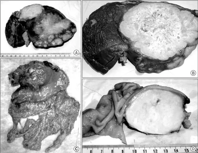
한 양상의 3 cm 크기의 종괴가 5번 분절에서 관찰되었다 (Fig. 5). 저자들은 악성 위장관 간질종양의 간전이로 판단 하여, 원위부 위절제, 간 좌엽절제 및 5번 분절의 쐐기 절제 를 시행하였다. 절제 검체의 육안소견상 간(Fig. 6 A, B) 및 위(Fig. 6C, D) 종괴의 절단면은 황색을 띠고, 균일한 괴사 성 병변이 관찰되었다.
발생하는데, 50% 이상에서 폐결핵이 병발되어 있다. 즉 폐 결핵 환자가 객담을 삼키거나 최근에는 드물지만 소 결핵 에 이환된 우유를 마심으로 인해 위장관의 어느 부위에서 나 발생가능한데, 특히 회맹장판막부가 가장 높은 빈도를 차지하고 상행결장, 공장, 충수돌기, 십이지장, 위, S자결장, 직장의 순서로 발생된다.(12)
Fig. 6. (A) The left lobe of the liver contains multiple non-capsulated necrotic tumors. (B) The cross section of the hepatic lesion shows well-demarcated, necrotic cut surface. (C) The specimen shows en-bloc resection of gastric distal portion including extrinsic tumors located on the lesser curvature. (D) The cross section of the gastric submucosal tumor shows heterogenous yellowish surface.
ꠏꠏꠏꠏꠏꠏꠏꠏꠏꠏꠏꠏꠏꠏꠏꠏꠏꠏꠏꠏꠏꠏꠏꠏꠏꠏꠏꠏꠏꠏꠏꠏꠏꠏꠏꠏꠏꠏꠏꠏꠏꠏꠏꠏꠏꠏꠏꠏꠏꠏꠏꠏꠏꠏꠏꠏꠏꠏꠏꠏꠏꠏꠏꠏꠏꠏꠏꠏꠏꠏꠏꠏꠏꠏꠏꠏꠏꠏꠏꠏꠏꠏꠏꠏꠏꠏꠏꠏꠏꠏꠏꠏꠏꠏꠏꠏꠏꠏꠏꠏꠏꠏꠏꠏꠏꠏꠏꠏꠏꠏꠏꠏꠏꠏꠏ
원발성 위결핵은 위산과 정상 위점막의 저항, 결핵균의 빠른 위내 통과 및 위벽의 적은 림프조직 때문으로 발생빈 도가 낮다고 생각되고 있고,(13) 발생부위는 주로 전정부와 체부위 소만측이며 병변은 결절형, 궤양형, 비후형으로 구 분되고, 가장 흔한 병변은 궤양형이다.(14) 저자의 증례는 궤양형으로 내시경 소견만으로는 점막하 종양, 즉 위장관 간질종양과의 감별이 어려웠다. 증상은 소화성궤양에서 나 타나는 간헐적인 상복부 동통, 체중감소, 전신쇠약, 발열, 식후 팽만감 등의 비특이적인 경우가 대부분이다. 위 내시 경 소견에서는 육안적 구분에 따라 위벽의 비후에 의한 전 정부 협착, 결절성 병변, 궤양 등이 관찰될 수 있다. 진단은 위내시경하 조직검사로 항산성 간균이나 랑거한 거대세포 및 건락성 육아종이 발견되야만 가능하지만 다른 육아종성 질환과도 감별되어야 한다. 또한 많은 예에서 만성 염증소 견만 관찰될 수 있어, 저자의 예와 같이 위 점막하 종양으로 오인되어 술 후 병리 조직검사로 확진된 예도 드물지 않다.
치료는 확진이 되면 12∼14개월간의 항결핵제 치료가 필수 적이나,(15) 폐색, 농양, 누공, 출혈 등의 합병증이 발생한 경우에는 수술의 적응이 되며, 본 예와 같이 술전 진단이 불확실하거나 병소가 매우 크고 다발성인 경우 수술을 고 려할 수 있다.(16)
간결핵은 주로 폐 및 장결핵에 의해 발생하는 이차성 병 변으로 간동맥과 문맥을 통한 혈행성 파급으로 이환되며 드물게는 림프관을 통해서도 병소를 유발하기도 한다. 간 동맥을 통한 혈행성 파급은 주로 속립성으로 발생되고, 균 은 간정맥 주위에 존재하지만, 문맥을 통한 파급은, 결핵균 의 일차병소인 폐나 소화관의 균은 성공적으로 제거되는 반면, 결핵균의 문맥을 통해 혈행성전이로 간의 망상내피 계에 지속적인 병변이 남아있어 고립성 병변을 남기는 경 우가 많고, 결핵결절은 문맥을 따라 위치하게 된다.(17) 증 상은 위결핵과 마찬가지로 비특이적이어서 기면, 오심, 체
중감소 및 복통 등이 있고, 이학적 검사상 발열, 상복부 압 통 및 간종대 소견이 관찰될 수 있다. 간기능 검사소견에서 는 저 알부민증, AST와 ALT, 알칼라인 포스파타제, 감마글 로불린 등이 상승되며 혈액학적 소견으로는 적혈구침강속 도의 증가, 범혈구 감소증, 비기능 항진 등의 소견이 나타날 수 있다.(18) 본 예에서는 적혈구침강속도가 25 mm/hr로 증 가된 것 외에는 다른 검사는 모두 정상범위였다. 방사선학 적으로는 단순 복부촬영상 우상복부에 석회화로 인해 무에 코성 음영을 동반한 다발성의 고에코를 유발하는 부위로 관찰된다. 컴퓨터 단층촬영에서도 저음영의 다발성 종괴로 보이는데 초기에는 격막이 있으며 경계가 불분명하기 때문 에 농양과 같은 양상을 보이지만, 진행되면 중심부가 균일 해지면서 석회화 소견이 나타날 수 있다.(17,18) 이러한 소 견들은 간혹 악성종양과의 감별이 요구되지만, 항상 결핵 의 가능성을 유념해야 하며, 최종진단은 조직학적으로 이 루어짐을 기억해야 한다. 조직학적 진단은 일차적으로 컴 퓨터 촬영 및 초음파하에서의 경피적 간생검으로 이루어지 며, 적절한 조직이 얻어지지 못할 경우 복강경검사를 통한 조직생검을 시도할 수 있다. 치료는 항결핵제 투여가 원칙 이며, 18∼24개월간의 치료로 85%에서 완치된다고 한다.
그러나 항결핵제에 반응이 없거나 병변이 거대성 결핵종일 경우 및 합병증이 병발될 때 수술의 적응증이 된다.
결 론
경제적인 성장과 더불어 국민들의 보건 위생 상태가 호 전되어 결핵의 이환율이 감소되고는 있지만 국내에서는 아 직 발생률이 높은 실정이다. 위나 간 결핵의 발병은 비교적 드물고 증상이나 검사소견이 비특이적이어서 위 내시경이 나 초음파 또는 단층촬영을 통한 생검이 필수적이다. 또한 생검을 하더라도 확진되지 않는 경우도 많아 진단에 어려 Fig. 7. (A) Gastric submucosal lesion shows multiple epitheloid granuloma (H&E ×40). (B) Hepatic lesion shows multi-nucleated giant
cells with epitheloid cells (H&E ×100).
acute miliary tuberculosis of gastric mucosa. Br J Surg 1928;15:626-30.
4) Good RW. Tuberculosis of the stomach; Analysis of cases recently reviewed. Arch Surg 1931;22:415-25.
5) Seo SI, Song YT, Kim KT, Kim IC. Gastric tuberculosis. J Korean Surg Soc 1971;13:83-7.
6) Lee DJ, Shon SH, Chin YJ, Lim CY, Song IH, Kim JW. A case of primary gastric tuberculosis diagnosed as a submucosal tumor. Korean J Gastrointest Endosc 1998;18:567-72.
7) Park JK, Lee S, Yun SS, Choi SH, Min KO, Lim KW. Gastric tuberculosis: a case report. J Korean Surg Soc 2000;59:548-53.
8) Bristowe JS. On the connection between abscess of the liver and gastrointestinal ulceration. Trans Pathol Coc 1858;9:241- 52.
tuberculosis: A review with particular reference to gross pathology and gastroscopic diagnosis. Am Rev Tuber 1960;61:
116-30.
14) Bhansali SK. Abdominal tuberculosis experience with 300 cases. Am J Gastroenterol 1977;67:324-37.
15) Gupta B, Mathew S, Bhalla S. Pyloric obstruction due to gastric tuberculosis. An endoscopic diagnosis. Post Grad Med J 1990;66:63-5.
16) Marshall JB. Tuberculosis of the gastrointestinal tract and peritoneum. Am J Gastroenterol 1993;88:996-1011.
17) Goodman PC, Jinkins JR. Imaging of tuberculosis and cra- niospinal tuberculosis. Radiol Clin North Am 1995;33:701-5.
18) Denath FM. Abdominal tuberculosis in children. CT findings Gastrointest Radiol 1990;15:303-5.