pISSN 1738-3544 eISSN 2288-1662
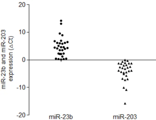
Comparison of the miR-23b and miR-203 Expressions in Endometrial Cancer
Kyung Eun Lee 1,2
1
Department of Clinical Laboratory Science, College of Health Sciences, Catholic University of Pusan, Busan, Korea
2
Brain Busan 21 Project for Catholic University of Pusan, Busan, Korea
자궁내막암종에서 miR-23b와 miR-203 발현 비교
이경은 1,2
1