• 검색 결과가 없습니다.

Diagnostic Value of miR-1260b in Cervical Cancer: A Pilot Study

N/A
N/A
Protected

Academic year: 2021

Share "Diagnostic Value of miR-1260b in Cervical Cancer: A Pilot Study"

Copied!
6
0
0

로드 중.... (전체 텍스트 보기)

전체 글

(1)

Biomedical Science Letters 2020, 26(1): 8~13 https://doi.org/10.15616/BSL.2020.26.1.8 eISSN : 2288-7415

Diagnostic Value of miR-1260b in Cervical Cancer: A Pilot Study

Jungho Kim

1,§,*

, Sunyoung Park

2,§,**

and Hyeyoung Lee

2,,*

1

Department of Clinical Laboratory Science, College of Health Sciences, Catholic University of Pusan, Busan 46252, Korea

2

Department of Biomedical Laboratory Science, College of Health Sciences, Yonsei University, Wonju, Gangwon 26493, Korea

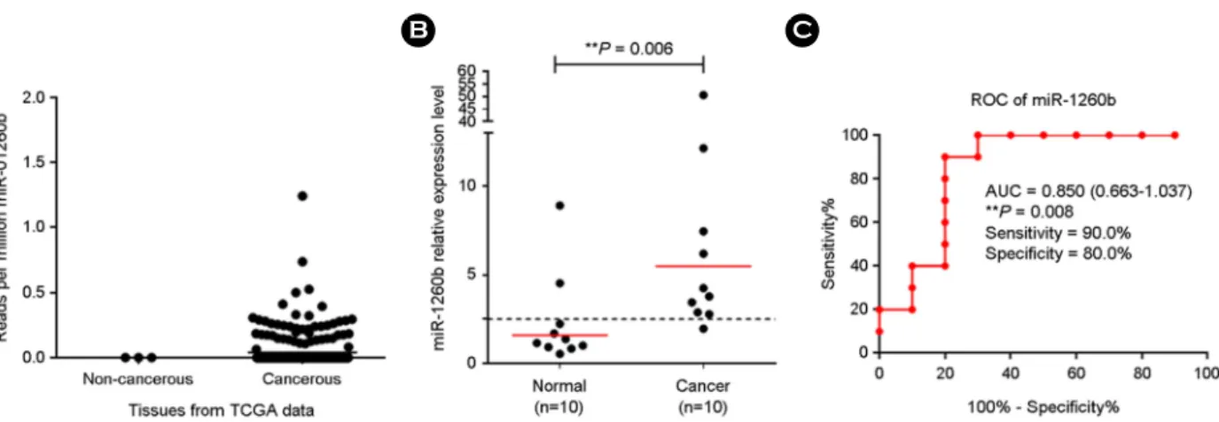
Cervical cancer is the fourth most common cancer in women, with approximately 528,000 new cases and 266,000 women dying of it per year in the world. MicroRNAs have recently been in the spotlight as potential biomarkers that regulate gene expression and are involved in tumorigenesis. In the present study, we evaluated miR-1260b as a potential biomarker for screening of cervical cancer by quantitative reverse transcription PCR. We profiled the TCGA data of miR-1260b in 307 cervical cancer tissues. Then, miR-1260b expression levels in 10 cervical cancer tissues and 10 non- cancerous tissues were investigated in a pilot study. miR-1260b was found to be significantly up-regulated in cervical cancer FFPE tissues as compared to those in cervical normal FFPE tissues (P = 0.006). The mean expression level of miR-1260b in late-stage (IIB-IVB) was higher than in those with early-stage (IA-IIA). Furthermore, high miR-1260b was found to be associated with high hTERT and Ki-67 mRNA expression, which are representative of tumor prognosis.

The results of the pilot study suggest that miR-1260b may be used as a novel biomarker for improving the diagnosis of cervical cancer.

Key Words: Cervical cancer, miR-1260b, RT-qPCR

INTRODUCTION

Cervical cancer is the fourth leading cause of death world- wide in women after breast cancer, colon cancer, and lung cancer (Torre et al., 2015). According to the World Health Organization (WHO) report, about 528,000 new cases of cervical cancer are recorded globally, and 266,000 women die of it each year (McGuire, 2016). Risk factors of cervical cancer include human papillomavirus (HPV) infection, pro- miscuous sex, long-term use of hormonal contraceptives,

sexually transmitted disease (STD) infection, and smoking (Burd, 2003). Current methods used to diagnose cervical cancer such as cytological and histological examinations are hampered by the examiner's subjectivity, lack of necessary expertise, and low sensitivity (Committee on Practice, 2012).

Recently, various molecular diagnostic methods including HPV genotype have been developed and have come into a wide use (Boone et al., 2012; Kim et al., 2015).

microRNAs are small (18-25 nucleotides) non-coding RNAs regulating gene expression by binding the 3'-UTR of the target messenger RNAs (mRNAs) (Bartel, 2004).

Original Article

Received: March 2, 2020 / Revised: March 24, 2020 / Accepted: March 25, 2020

*Professor, **Post-Doctor.

§Jungho Kim and Sunyoung Park contributed equally.

Corresponding author: Hyeyoung Lee. Department of Biomedical Laboratory Science, College of Health Sciences, Yonsei University, 1 yonseidae-gil, Wonju-si, Gangwon-do 26493, Korea.

Tel: +82-33-760-2740, Fax: +82-33-760-2561, e-mail: [email protected]

CThe Korean Society for Biomedical Laboratory Sciences. All rights reserved.

CCThis is an Open Access article distributed under the terms of the Creative Commons Attribution Non-Commercial License (http://creativecommons.org/licenses/by-nc/3.0/) which permits unrestricted non-commercial use, distribution, and reproduction in any medium, provided the original work is properly cited.

(2)

Numerous previous studies have reported that microRNAs involved in tumorigenesis are potential diagnosis, prognosis and therapeutic biomarkers in various types of cancer.

miRNAs have been proposed to function as tumor genes or tumor suppressors based on the inhibition of expression of tumor suppressor or oncogene, respectively (Lui et al., 2007;

Hebert and De Strooper, 2009; Bouyssou et al., 2014). In recent studies, several miRNAs have been considered to be diagnostic markers of cervical cancer. The Cancer Genome Atlas (TCGA) data were provided by clinical information and genomic sequencing data such as mRNA, microRNA, and methylation. However, since there are over 10,000 to 100,000 various genomic targets, there is a lack of clinical validation of specific targets (Banister et al., 2017).

Among these several miRNAs, miR-1260b has been re- ported to play an important role in the tumorigenesis of

renal, prostate, lung, and colon cancer cells. miR-1260b is located in human 11q21 and was first identified in human renal cancers using miRNA microarray (Hirata et al., 2013).

However, the clinical relevance of miR-1260b in cervical cancer is still poorly understood. In our pilot study, miR- 1260b as potential biomarkers for cervical cancer were inves- tigated using 20 formalin-fixed paraffin-embedded (FFPE) cervical tissues. In addition, we analyzed the association be- tween the expression levels of miR-1260b and FIGO stage, the immortalization marker hTERT, and the proliferation marker Ki-67 expression.

MATERIALS AND METHODS

Clinical samples

A total of 10 FFPE cervical tissues were obtained from cervical cancer patients diagnosed with cervical cancer at Yonsei University Wonju Severance Christian Hospital, Wonju, Republic of Korea from 2010 to 2014. A total of 10 FFPE normal cervical tissues were obtained from the patients with non-cervical, benign, uterine disease (Table 1).

All study participants provided written consent, and the study was approved by the institutional ethics committee at Yonsei severance hospital (approval no. CR315052).

Deparaffinization of FFPE tissue and total RNA ex- traction

For RNA extraction of FFPE cervical tissues, three to four 10-μm-thick sections were transferred to 1.5 mL tube each.

Before extracting RNA, the paraffin was removed from the tissue sections by adding 160 μL of deparaffinization solu- tion (Qiagen, Hilden, Germany), followed by incubation for 3 min at 56℃. Total RNA extraction was extracted accord- ing to the manufacturer's protocol (Qiagen RNeasy FFPE kit, Qiagen). Next, the concentration of the total RNA was measured with an Infinite 200 spectrophotometer (Tecan, Salzburg, Austria). Total RNA was stored at -80℃ until further use.

miRNA Reverse-Transcriptase (RT)- Quantitative (q) PCR analysis

TaqMan miRNA Reverse Transcription kit (Applied Bio- Table 1. Clinical characteristics of 20 FFPE cervical tissues

Specimen HPV infection status Histology FIGO

stage

d

Age

C01 16 SCC

a

ⅠB 65

C02 16 nkSCC

b

ⅡB 30

C03 16 SCC ⅠA 39

C04 16 SCC ⅡA 50

C05 16 nkSCC ⅡB 55

C06 45 nkSCC ⅡB 55

C07 16 nkSCC ⅡB 80

C08 16 SCC ⅣB 70

C09 16 nkSCC ⅡB 77

C10 16 kSCC

c

ⅡA 41

N01 52

N02 50

N03 31

N04 57

N05 30

N06 36

N07 67

N08 49

N09 44

N10 40

a

SCC, Squamous Cell Carcinoma

b

nkSCC, Nonkeratinizing Squamous Cell Carcinoma

c

kSCC, Keratinizing Squamous Cell Carcinoma

d

FIGO, International Federation of Gynecology and Obstetrics

(3)

systems, Foster City, CA, USA) was used to synthesize cDNA according to the manufacturer's instructions. Reverse transcription reactions were performed using 10 ng of total RNA and specific reverse transcription primers (Life Tech- nologies) for Homo sapiens (hsa)-miR-1260b and RNU6B.

Reverse transcription was performed at 16℃ for 30 min, 42℃ for 30 min, and 85 for 5 min.

After the reverse transcription, miRNA expression was quantified using the TaqMan small RNA assays (Applied Biosystems by Life Technologies) with miRNA specific primers (miR-1260b) measuring the cycle threshold (C

T

), the number of PCR cycles in which fluorescence exceeds the background fluorescence value. Briefly, the initiation step of PCR cycling conditions was set at 95℃ for 10 min, fol- lowed by an amplification step of 40 cycles at 95℃ for 15 s and 60℃ for 60 s. The samples were repeated twice, and the data were analyzed using the comparative ΔC

T

method (2-

ΔCT

) with RNU6B as an endogenous control (Pfaffl et al., 2002).

RT-qPCR for hTERT and Ki67 mRNA

M-MLV reverse transcriptase kit (Invitrogen, Carlsbad, CA, USA) was used for a complementary DNA (cDNA) synthesis. Briefly, 0.25 μg random hexamers, 1 μL 10 Mm dNTP mix, and 5 μL DEPC-treated water, and 10 μL total RNA were mixed. Then the mixture was incubated at 65℃

for 5 min and quickly cooled on ice. Next, 4 μL of 5 × buf- fer, 2 μL of 0.1 M dithiothreitol (DTT) and 1 μL of 200 U Murine Molony Leukemia Virus Reverse Transcriptase (MMLV-RT) were added to the first mix. cDNA synthesis was performed at 25℃ for 10 min, at 37℃ for 50 min, and at 70℃ for 15 min.

As described by Kim et al., hTERT and Ki67 mRNA expression in FFPE cervical tissues was assessed via a RT- qPCR TaqMan assay and the CFX-96 real-time PCR system (Bio-Rad, Hercules, CA, USA) for thermal cycling and fluorescence detection. Real-time PCR amplification for hTERT and Ki67 mRNA was performed in a reaction mix containing 10 μL of 2 × Thunderbird probe qPCR mix (Toyobo, Osaka, Japan), 3 μL of primer and TaqMan probe mixture, 2 μL of template cDNA, and distilled water (D.W) to the final volume of 20 μL per sample. Positive and nega-

tive controls were included throughout the procedure. The used PCR cycling conditions were 95℃ for 3 min, followed by 41 cycles of 95℃ for 3 s, and 55℃ for 30 s. The sam- ples were repeated twice, and the data were analyzed using the comparative ΔC

T

method (2-

ΔCT

) with glyceraldehyde- 3-phosphate dehydrogenase (GAPDH) as an endogenous control.

Statistical analyses

Statistical analyses were performed using GraphPad Prism software version 6 (GraphPad, LaJolla, CA, USA). Student's t-test, 95% confidence interval (CI), and ROC curve were used to evaluate the statistical significance of the results.

RESULTS

miR-1260b expression levels in cervical cancer and normal tissues

First, we investigated TCGA data of miR-1260b in 307 cervical cancer tissues and paired 3 normal tissues. The results showed the up-regulation in cervical cancer tissues.

However, the normal tissue was rare in the TCGA data, and we profiled the expression levels of miR-1260b in cervical cancer, ten cervical cancer tissues and ten normal tissues by RT-qPCR (Fig. 1). The results showed that the expression levels of miR-1260b were significantly higher in cervical cancer tissues as compared to those in cervical normal tissues (P = 0.006; Fig. 1B). In order to investigate cut-off values for discriminating cervical cancer from normal tissues, the expression level of miR-1260b in cervical cancer and normal tissues was analyzed using a ROC curve. At the optimal cut-off point, the area under the curve (AUC) of miR-1260b was 0.850 (P = 0.008; 95% confidence interval (CI), 0663~

1.037; Fig. 1C)

miR-1260b expression according to FIGO stage

To explore the association between miR-1260b expression

and stages, we analyzed the expression levels of miR-1260b

in FIGO stages. There was a difference between the miR-

1260b expression level of early-stage (IA-IIA) and late-stage

(IIB-IVB) (Table 2). Specifically, the mean expression levels

of miR-1260b were 13.20±18.69 (mean ± S.D) in stage

(4)

IIB-IVB and 4.06±1.48 (mean ± S.D) in stage IA-IIA.

miR-1260b expression according to hTERT and Ki67 Subsequently, to investigate the association between miR- 1260b expression and tumorigenesis, we analyzed miR- 1260b expression according to the immortalization marker hTERT and the proliferation marker Ki-67 expression. The cases were divided into two groups according to the ex- pression level of hTERT (high hTERT vs. low hTERT) and Ki-67 (high Ki67 vs. low Ki-67) based on the median of ex pression levels, respectively. For hTERT mRNA expression, the mean expression level of miR-1260b in high hTERT was higher than in low hTERT (13.14±21.05 vs. 5.97±3.61, respectively). Similarly, the mean expression level of miR- 1260b in high Ki-67 was higher than in low Ki-67 (13.30 vs 5.81, respectively; Table 3).

DISCUSSION

In this pilot study, we investigated miR-1260b expression

levels in FFPE cervical cancer and normal tissues using RT-qPCR to identify a potential biomarker for cervical can- cer. In previous research, miR-1260b was found to be highly expressed in renal carcinoma and to promote cell pro- liferation and invasion in renal carcinoma. Furthermore, genistein was found to inhibit Wnt-signaling by regulating miR-1260b expression in renal cancer cells (Hirata et al., 2013). In addition, genistein was reported to have anti-tumor effects through the down-regulation of miR-1260b targeting SRRP1 and Smad4 genes in prostate cells (Hirata et al., 2014). The expression of miR-1260b in non-small cell lung cancer tissues was reported to be higher than that of adjacent tissues, and overexpression of miR-1260b was found to be associated with lymph node metastasis (Xu et al., 2015).

However, none of the previous studies has explored the diagnostic value of miR-1260b in cervical cancer.

Table 3. miR-1260b expression according to the hTERT and Ki-67 mRNA expression levels

Expression miR-1260b expression (Mean ± S.D) hTERT

Low 5.97±3.61

High 13.14±21.05

Ki67

Low 5.81±4.02

High 13.30±20.91

Table 2. miR-1260b expression according to FIGO stage FIGO stage miR-1260b expression (Mean ± S.D)

IA-IIA 4.06±1.48

IIB-ⅣB 13.20±18.69

Fig. 1. miR-1260b expression of cervical cancer and normal FFPE tissues and ROC analysis. (A) The expression level of miR-1260b was

higher in cervical cancer patients than in healthy controls; (B) The miR-1260b expression level in 10 FFPE cervical cancer tissues significantly

differed from the one observed in 10 FFPE normal cervical tissues (P = 0.006); (C) The results of the receiver operator characteristic curve

(ROC) analysis showed that miR-1260b had an area under the ROC curve (AUC) value of 0.850 (P = 0.008).

(5)

The results of our pilot study showed that the expression levels of miR-1260b were significantly higher in cervical cancer tissues as compared to those in cervical normal tissues (P = 0.006), suggesting that the miR-1260b plays an important role in tumorigenesis. The cut-off value of miR- 1260b was 2.51 for optimal sensitivity and specificity. The sensitivity of miR-1260b was 90% (95% CI = 55.50% to 99.75%), while the specificity was 80% (95% CI = 44.39%

to 97.48%). Positive predictive value (PPV) of miR-1260b was 80% (95% CI = 44.39% to 97.48%), while the negative predictive value (NPV) was 80% (95% CI = 44.39% to 97.48%). The value of AUC was 0.85 (95% CI = 0.66 to 1.04, P = 0.008).

Liu et al. explored the diagnostic potential of a panel of miRs using RT-qPCR in 582 cervical cancer patients and 145 control individuals. The levels of miR-20a, miR-92a, miR-141, miR-183, miR-210, and miR-944 were higher in cancer patients than in healthy controls. In addition, miR-20a, miR-92a, miR-141, miR-183, miR-210, and miR-944 can help differentiate between cervical cancer and healthy con- trols with AUC of 0.822, 0.659, 0.942, 0.785, 0.751, and 0.677 in ROC analysis, respectively (Liu et al., 2018). The sensitivity and specificity of miR-1260b were comparable to those of other miRs for the cervical cancer diagnostic method.

We further examined the relationship between miR-1260b expression levels and FIGO stages, hTERT mRNA expres- sion, Ki-67 mRNA expression. The mean expression level of miR-1260b in late-stage (IIB-IVB) was found to be higher than in those with early-stage (IA-IIA). Furthermore, miR- 1260b in high hTERT mRNA expression group and Ki-67 mRNA expression group showed a higher expression as compared to those in low hTERT mRNA expression group and Ki-67 mRNA expression group, respectively (Ross and Hall, 1995; Blasco and Hahn, 2003; Park et al., 2018). Taken together, the results suggest that miR-1260b is a potential diagnostic biomarker for cervical cancer.

The present study has several limitations. First, all cervical FFPE tissues were collected from a single hospital, and the number of the samples was small. Therefore, more tests for miR-1260b with a larger number of patients in multi-center settings would be needed to replicate our findings. Further

research to investigate the relationship between clinical para- meters is also necessary.

In conclusion, the results of the pilot study showed miR-1260b is up-regulated in cervical cancer FFPE tissues.

The high expression of miR-1260b was found to be closely related to late-stage, high expression of hTERT mRNA, Ki- 67 mRNA, suggesting that miR-1260b may be an oncogene in cervical cancer. miR-1260b may also potentially serve as a screening biomarker for cervical cancer.

ACKNOWLEDGEMENT None.

CONFLICT OF INTEREST

The authors have no conflicts of interest with regards to this study.

REFERENCES

Banister CE, Liu C, Pirisi L, Creek KE, Buckhaults PJ. "Identi- fication and characterization of HPV-independent cervical cancers". Oncotarget. 2017. 8: 13375-13386.

Bartel DP. "MicroRNAs: genomics, biogenesis, mechanism, and function". Cell. 2004. 116: 281-297.

Blasco MA, Hahn WC. "Evolving views of telomerase and cancer".

Trends Cell Biol. 2003. 13: 289-294.

Boone JD, Erickson BK, Huh WK. "New insights into cervical cancer screening". J Gynecol Oncol. 2012. 23: 282-287.

Bouyssou JM, Manier S, Huynh D, Issa S, Roccaro AM, Ghobrial.

IM. "Regulation of microRNAs in cancer metastasis". Bio- chim Biophys Acta. 2014. 1845: 255-265.

Burd EM. "Human papillomavirus and cervical cancer". Clin Microbiol Rev. 2003. 16: 1-17.

Committee on Practice BG. "ACOG Practice Bulletin Number 131: Screening for cervical cancer". Obstet Gynecol. 2012.

120: 1222-1238.

Hebert SS, De Strooper B. "Alterations of the microRNA network cause neurodegenerative disease". Trends Neurosci. 2009. 32:

199-206.

Hirata H, Hinoda Y, Shahryari V, Deng G, Tanaka Y, Tabatabai ZL,

Dahiya R. "Genistein downregulates onco-miR-1260b and

upregulates sFRP1 and Smad4 via demethylation and histone

modification in prostate cancer cells". Br J Cancer. 2014. 110:

(6)

1645-1654.

Hirata H, Ueno K, Nakajima K, Tabatabai ZL, Hinoda Y, Ishii N, Dahiya R. "Genistein downregulates onco-miR-1260b and inhibits Wnt-signalling in renal cancer cells". Br J Cancer.

2013. 108: 2070-2078.

Kim S, Lee I, Lee D. "Human papillomavirus prevalence and genotype distribution in normal and ASCUS specimens: com- parison of a reverse blot hybridiaztion assay with a DNA chip test". Biomedical Science Letters. 2015. 21: 32-39.

Liu SS, Chan KKL, Chu DKH, Wei TN, Lau LSK, Ngu SF, Chu MMY, Tse KY, Ip PPC, Ng EKO, Cheung ANY, Ngan HYS.

"Oncogenic microRNA signature for early diagnosis of cer- vical intraepithelial neoplasia and cancer". Mol Oncol. 2018.

12: 2009-2022.

Lui WO, Pourmand N, Patterson BK, Fire A. "Patterns of known and novel small RNAs in human cervical cancer". Cancer Res.

2007. 67: 6031-6043.

McGuire S. "World Cancer Report 2014. Geneva, Switzerland:

World Health Organization, International Agency for Research on Cancer, WHO Press, 2015". Adv Nutr. 2016. 7: 418-419.

Park S, Park S, Kim J, Ahn S, Park KH, Lee H. "Assessment of

Ki-67 for predicting effective prognosis in breast cancer sub- types". Biomedical Science Letters. 2018. 24: 1-6.

Pfaffl MW, Horgan GW, Dempfle L. "Relative expression software tool (REST) for group-wise comparison and statistical analysis of relative expression results in real-time PCR". Nucleic Acids Res. 2002. 30: e36.

Ross W, Hall PA. "Ki67: from antibody to molecule to under- standing?" Clin Mol Pathol. 1995. 48: M113-117.

Torre LA, Bray F, Siegel RL, Ferlay J, Lortet-Tieulent J, Jemal A.

"Global cancer statistics, 2012". CA Cancer J Clin. 2015. 65:

87-108.

Xu L, Li L, Li J, Li H, Shen Q, Ping J, Ma Z, Zhong J, Dai L.

"Overexpression of miR-1260b in Non-small Cell Lung Cancer is Associated with Lymph Node Metastasis". Aging Dis. 2015.

6: 478-485.

https://doi.org/10.15616/BSL.2020.26.1.8

Cite this article as: Kim J, Park S, Lee H. Diagnostic Value of miR-1260b in Cervical Cancer: A Pilot Study.

Biomedical Science Letters. 2020. 26: 8-13.

수치

Table 3. miR-1260b expression according to the hTERT and Ki-67 mRNA expression levels

참조

관련 문서

Three invasive cervical cancer gels were compared with three normal cervical tissue gels as controls and protein spots that had corresponded to proteins where expression

Furthermore, in the three cancer groups and fibroadenoma samples examined here, miR-21 over expression was not significant.. In addition, based on our results, miR-21 does

In this study we investigated the expression profiles of mir-20a and mir-203 in cervical cancer tissues and cervical cancer cell line.. Our primary aim was to determine whether

Expression of MEKK3 and survivin are significantly increased in cervical cancer, their overexpression participating in the occurrence and development of cervical cancer,

Furthermore, the qRT-PCR analysis revealed increased expression of LINC00703 and KLF6, and decreased level of miR-181a in LINC00703 expressing tumor tissues (Fig. 4E).. Correlation

The qPCR and western blot analyses showed that both messenger RNA (mRNA) and protein expression levels of KLK6, VEGF, and MMP9 were significantly higher in cancer tissues relative

To explore the effects of Avastin on apoptosis, the expression levels of miR-155, miR-191, and miR-494, as treatment and diagnostic biomarkers, were re- viewed in the HN5 cell line

[15] reported that miR-23b is methy- lation-silenced tumor suppressor in prostate cancer, and its high expression level was found to be associated with higher survival rates