439
특성의 변화
Changes in Prostate Cancer Pattern according to Prostate-Specific Antigen Screening Test
Jong Kyou Kwon, In Ho Chang, Tae Hyoung Kim, Soon Chul Myung From the Department of Urology, Chung-Ang University College of Medicine, Seoul, Korea
Purpose: The aim of this study was to investigate the changes in the clinical and prognostic parameters of prostate cancer in Korean men in the eras before and after prostate-specific antigen (PSA) testing.
Materials and Methods: The medical records of 303 patients treated for prostate cancer between 1982 and 2005 were reviewed with respect to age, chief complaints, clinical stage, tumor grade, treatment options, and prognosis. We classified the patients as follows: those treated in the pre- PSA era (1982-1995, n=81), and those treated in the PSA era (1996-2000, PSA era phase 1, n=92; and 2001-2005, PSA era phase 2, n=130).
Results: There was no significant difference in age or clinical stage between patients treated before and those treated during the PSA era, although there was a downward migration of grade. The cancer-specific survival rates were also not different in all cases and in metastatic prostate cancer cases between the pre-PSA era and the PSA era, although the overall survival rates were significantly greater in all cases in phase 2 of the PSA era than in the pre-PSA era or in phase 1 of the PSA era (p<0.05). How- ever, the cancer-specific survival rates for localized or locally advanced prostate cancer were significantly greater in phase 2 of the PSA era than in the pre-PSA era or in phase 1 of the PSA era (p<0.05).
Conclusions: We observed a downward migration of tumor grade, but there were no migrations in the age of patients or clinical stage, and these findings have not contributed to changes in the cancer survival of Korean men with prostate cancer after the advent of PSA testing. (Korean J Urol 2009;50:439-444)
Key Words: Prostatic neoplasms, Prostate-specific antigen, Mass screening, Survival rate
Korean Journal of Urology Vol. 50 No. 5: 439-444, May 2009 DOI: 10.4111/kju.2009.50.5.439
중앙대학교 의과대학 비뇨기과학교실 권종규ㆍ장인호ㆍ김태형ㆍ명순철
Received:January 22, 2009 Accepted:March 30, 2009 Correspondence to: In Ho Chang
Department of Urology, Chung- Ang University Hospital, 224-1, Heuksuk-dong, Dongjak-gu, Seoul 156-755, Korea
TEL: 02-6299-1785 FAX: 02-6294-1406
E-mail: [email protected] This study was supported by a grant of the Korea Healthcare technology R&D Project, Ministry for Health, Welfare & Family Affairs, Republic of Korea (A085138).
Ⓒ The Korean Urological Association, 2009
서 론
전립선암은 서구에서는 매우 흔한 암이나, 한국에서는 위암, 폐암, 간암, 대장암, 식도암, 방광암, 췌장암에 이어 8번째로 흔한 암으로 알려져 있다.1,2 하지만, 최근 한국 남 성의 전립선암 발생률은 다른 아시아 나라들과 마찬가지로 급속히 증가하는 추세이다.3 전립선암의 발생률은 1996- 1998년과 1999-2001년 사이에 28.2%까지 증가했으며,4 5년 상대생존율은 1993년과 2002년 사이에 59.1%에서 70.6%로
증가했다.5 이 발생률과 생존율의 증가는 한국 남성의 모든 암 중에서 가장 두드러지며, 이러한 배경하에 국내에서도 혈청 전립선특이항원 (prostate-specific antigen; PSA)을 이용 한 광범위한 암 선별검사의 필요성이 대두되고 있다.5 미국 과 일본에서는 이미 PSA 도입에 따른 전립선암의 임상특징 변화에 대한 역학 연구들이 수행되었으며, PSA 선별 검사 로 국소전립선암이 증가하여 전립선암 환자의 생존율이 증 가하였다.6,7 그러나, 국내에서 PSA 검사법이 도입되기 전후 의 전립선암의 임상적인 특징 및 예후의 변화에 대한 연구 는 아직 보고된 적이 없다.
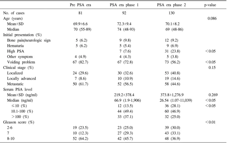
Table 1. Demographics of patients with prostate cancer in the pre and post PSA eras
Pre PSA era PSA era phase 1 PSA era phase 2 p-value
No. of cases 81 92 130
Age (years) 0.086
Mean±SD 69.9±6.6 72.3±9.4 70.1±8.2
Median 70 (55-89) 74 (48-93) 69 (48-86)
Initial presentation (%)
Bone pain/neurologic sign 5 (6.2) 9 (9.8) 12 (9.2)
Hematuria 5 (6.2) 5 (5.4) 9 (6.9)
High PSA 7 (7.6) 31 (23.8) <0.05
Other symptom 4 (4.9) 4 (4.3) 5 (3.8)
Voiding problem 67 (82.7) 67 (72.8) 73 (56.2) <0.05
Clinical stage (%) 0.15
Localized 24 (29.6) 30 (32.6) 53 (40.8)
Locally advanced 7 (8.6) 10 (10.9) 19 (14.6)
Metastatic 50 (61.7) 52 (56.5) 58 (44.6)
Serum PSA level
Mean±SD (ng/ml) 219.2±378.4 373.8±1,276.9 0.269
Median (ng/ml) 66.9 (1.9-1,906) 26.54 (1.07-11,039) <0.05
≤10 (%) 12 (13.5) 36 (28.1) <0.05
10.1-100 (%) 44 (49.4) 60 (46.9)
>100 (%) 33 (37.1) 32 (25.0)
Gleason score (%) <0.01
2-6 19 (23.5) 23 (25.0) 39 (30.0)
7 10 (12.3) 27 (29.3) 43 (33.1)
8-10 52 (64.2) 42 (45.7) 48 (36.9)
PSA: prostate-specific antigen, SD: standard deviation
본 연구의 목적은 PSA의 도입 전후 기간에 있어서 전립 선암 환자의 임상 특징 및 예후 변화에 대해서 알아보고자 하였다.
대상 및 방법
본 연구는 1982년부터 2005년 사이에 본원에서 전립선암 으로 진단 받은 환자 303명 (평균나이 71세 [48-93])을 대상 으로 후향적 조사를 시행하였다. 전립선암은 전립선침생검 또는 경요도전립선절제술을 통한 조직검사로 확진하였다.
본원에서 PSA 검사는 1996년 1월부터 가능하였기 때문에, 그 이전에 내원한 환자들에 대해서는 경직장초음파 (trans- rectal ultrasound; TRUS) 또는 직장수지검사 (digital rectal examination; DRE)에서 이상 소견을 보이는 경우에 전립선 조직검사를 시행하였으며, PSA 검사가 가능해진 이후로는 DRE 또는 TRUS에서 이상소견이 보이거나, PSA가 4.0 ng/
ml보다 높게 측정된 경우에 전립선 조직검사를 시행하였 다. 총 대상 환자 303명을, PSA가 도입되기 전인 1996년 1월 이전에 진단받은 환자 81명 (26.7%) (pre-PSA era군)과, PSA 가 도입된 이후인 1996년 1월 이후에 진단받은 환자 222명
(73.3%) (PSA-era군)으로 분류하였다. 또한 PSA-era군을 각 각 동일한 기간으로 분류하여 1996년 1월과 2000년 12월 사 이에 진단된 환자 92명 (30.4%) (PSA era phase 1군)과, 2001 년 1월과 2005년 12월 사이에 진단된 환자 130명 (42.9%) (PSA era phase 2군)으로 세분하였다.
각 환자들의 진료기록을 통해 나이, 임상적 병기, Gleason score, 암 진단 당시 PSA치, 치료 방법, 예후를 조사하여 임 상적으로 T1/T2인 환자들을 국소전립선암으로 분류하였다.
국소진행성전립선암 환자는 DRE에서 임상적으로 T3/ T4에 해당되거나, MRI에서 립선피막외로 진행된 소견을 보이는 경우로 분류하였다. CT에서 골반림프절의 직경이 1 cm 이 상인 림프절병증이 동반된 환자, 또는 골반외전이를 동반 한 환자, 특히 골주사 검사에서 골전이 소견이 있는 환자는 전이성전립선암으로 분류하였다. 조직검사에 의한 암의 등 급은 WHO 분류 시스템에 따라 분류했다.
통계분석은 SPSS software package version 14.0 (Statistical Package for Social SciencesTM, Chicago, USA)을 이용하였다.
Pre-PSA era군과 PSA era군 간의 주소, 나이, 임상병기, Gleason score, 암 진단 당시 PSA치, 치료 방법, 예후 등에 따른 환자들의 특징들을 chi-square 검정과 분산분석
Table 2. Comparison of treatment options in the pre and post PSA eras
Pre-PSA era PSA era phase 1 PSA era phase 2
T L M T L M T L M
No. of cases 81 31 50 92 40 52 130 72 58
Surveillance 7 (8.6) 7 (22.6) 0 (0.0) 5 (5.4) 5 (12.5) 0 (0.0) 6 (4.6) 5 (6.9) 1 (1.7) Radical prostatectomya 0 (0.0) 0 (0.0) 0 (0.0) 6 (6.5) 5 (12.5) 1 (1.9) 26 (20.0) 24 (33.3) 2 (3.4) Surgical castrationb 32 (39.5) 8 (25.8) 24 (48.3) 35 (38.0) 13 (32.5) 22 (42.3) 18 (13.8) 8 (11.1) 10 (17.2) LHRH and/or
antiandrogenb 37 (45.7) 14 (45.2) 23 (46.5) 45 (48.9) 16 (40.0) 29 (55.8) 69 (53.1) 31 (43.1) 38 (65.5) Radiation therapy 5 (6.2) 2 (6.5) 3 (6.2) 1 (1.1) 1 (2.5) 0 (0.0) 11 (8.5) 4 (5.6) 7 (12.1) PSA: prostate-specific antigen, LHRH: luteinizing hormone-releasing hormone, T: total patient (n=202), L: localized or locally advanced prostate cancer patients (n=143), M: metastatic prostatic cancer patients (n=160), Statistical significance: a: p<0.01, b: p<0.05
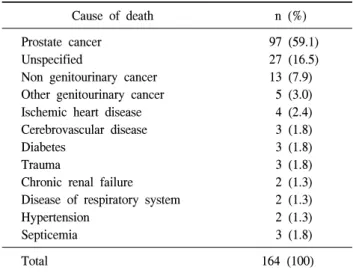
Table 3. Cause of death (n=164)
Cause of death n (%)
Prostate cancer Unspecified
Non genitourinary cancer Other genitourinary cancer Ischemic heart disease Cerebrovascular disease Diabetes
Trauma
Chronic renal failure Disease of respiratory system Hypertension
Septicemia
97 (59.1) 27 (16.5) 13 (7.9) 5 (3.0) 4 (2.4) 3 (1.8) 3 (1.8) 3 (1.8) 2 (1.3) 2 (1.3) 2 (1.3) 3 (1.8)
Total 164 (100)
(ANOVA)을 이용하여 비교하였다. 전체 생존율과 암특이 생존율 및 병기 분류는 Kaplan-Meier 방법과 log-rank test를 이용해 통계적으로 분석하였으며, p값이 0.05 미만인 경우 통계적으로 유의한 것으로 판정하였다.
결 과
Pre-PSA era군 및 PSA era군의 전립선암 환자들에 대한 인구학적 특성을 조사하였다 (Table 1). 전립선암의 진단 당 시 호소한 주 증상은 pre-PSA era군과 PSA era군 모두 배뇨 문제였으며, PSA era군에서 배뇨문제의 비율이 더 감소했 다. PSA era군 내에서 증상 없이 건강검진에서 PSA 상승으 로 전립선암을 발견하는 비율은 PSA era phase 1군은 92명 중 7명 (7.6%)에서, PSA era phase 2군에서는 130명 중 31명 (23.8%)으로 증가하였다. 국소 전립선암이 PSA era phase 1 군 (32.6%)에 비해 PSA era phase 2군 (40.8%)에서 증가하는 양상을 보였지만, 나이와 임상병기에서 세 군 간의 통계적 으로 유의한 차이는 없었다. 나이를 구간별로 나누어 시행 한 비교에서도 차이는 나타나지 않았다. 그러나 pre-PSA era 군에 비해 PSA era군에서 유의하게 낮은 조직학적 등급을 보였다. 높은 조직학적 등급을 보인 경우 (Gleason score 8- 10) 역시 pre-PSA era군 (64.2%), PSA era phase 1군 (45.7%), PSA era phase 2군 (36.9%) 사이에 유의한 차이가 있었다 (p<0.01). PSA era phase 1군에 비해 PSA era phase 2군에서 낮은 PSA를 보였으며 (p<0.05), 특히 PSA가 10 이하인 환 자는 PSA era phase 1군 (13.5%)보다 PSA era phase 2군 (28.1%)에서 증가하였다.
치료 방법에 따라 각 군을 비교하였을 때 (Table 2), 근치 적전립선절제술을 시행한 비율은 pre-PSA era군 (0%)에 비 해 PSA era군 (PSA era phase 1군 6.5%, PSA era phase 2군 20%)에서 증가하였다 (p<0.01). 고환절제술을 받은 비율은
pre-PSA era군 (39.5%)에 비해 PSA era군 (PSA era phase 1군 38%, PSA era phase 2군 13.8%)에서 감소하였다.
이를 국소전립선암 또는 국소진행성전립선암, 전이성전 립선암으로 나누어 초기 치료 방법을 비교하였을 때 (Table 2), 국소전립선암, 또는 국소진행성전립선암에서 근치적전 립선절제술을 시행 받은 비율은 pre-PSA era군 (0%)에 비해 PSA era군 (PSA era phase 1군 12.5%, PSA era phase 2군 33.3%)에서 증가하였으며 (p<0.01), 전이성전립선암에서 고환절제술을 시행한 비율은 pre-PSA era군 (48.3%)에 비해 PSA era군 (PSA era phase 1군 42.3%, PSA era phase 2군 17.2%)에서 감소하였다 (p<0.01).
전체 대상 환자들 중 164명 (54.1%)이 사망했으며, 97명 (59.1%)은 전립선암으로 사망하였다 (Table 3).
Pre PSA era군과 PSA era군 (phase 1, 2)의 비교에서 생존 율의 유의한 차이는 나타나지 않았다. PSA era군을 phase 1, 2로 나누어 비교하였을 때, 전체 생존율은 PSA era phase
Table 4. Cancer-specific survival rate according to clinical stage (%) Pre PSA era PSA era phase 1 PSA era phase 2
All patients 69.7 65.8 82.2
Local or locally
advanced 71.0 77.5 94.4
Metastatic 44.0 30.8 51.1
PSA: prostate-specific antigen
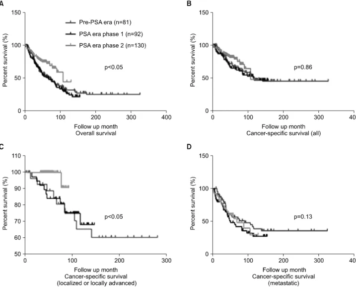
Fig. 1. Kaplan-Meier survival curves of prostate cancer in the pre and post prostate-specific antigen (PSA) eras. (A) Overall survival curves of all cases. (B) Cancer-specific survival curves of all cases. (C) Cancer-specific survival curves of localized or locally advanced prostate cancer. (D) Cancer-specific survival curves of metastatic prostate cancer.
2군에서 pre-PSA era군 및 PSA era phase 1군에 비해 유의하 게 높았으나 (p<0.05), 전체 암특이생존율의 차이는 없었다 (Fig. 1). 또한 전이성전립선암인 경우 pre-PSA era군과 PSA era군 간의 암특이생존율에는 차이가 없었으나, 국소전립선 암 또는 국소진행성전립선암에서 암특이생존율은 pre-PSA era군과 PSA era phase 1군보다 PSA era phase 2군에서 증가 하였다 (p<0.05) (Fig. 1). Pre-PSA era군, PSA era phase 1군, PSA era phase 2군의 전체 5년 암특이생존율은 각각 69.7%, 65.8%, 82.2%였으며, 국소전립선암 및 국소진행성전립선암 은 각각 71.0%, 77.5%, 94.4%, 전이성전립선암은 각각 44.0%, 30.8%, 51.1%였다 (Table 4).
고 찰
전립선암은 1984년 이래 미국에서 발생하는 가장 흔한 악성종양으로, 전체 암의 1/3을 차지하고 있다.8 미국에서 전립선암의 발생률은 PSA가 선별 검사로 도입된 지 5년째
인 1992년에 최고치를 보인 이후, 1995년까지 가파르게 감 소하다 이후 PSA 도입 전과 비슷한 속도로 서서히 증가했 으며, 국소전립선암 및 국소진행성전립선암의 발생률이 증 가한 반면 전이성전립선암의 발생률은 감소했다.6,9,10 또한 DRE에서 종괴가 촉지되지 않아 전적으로 PSA에 의존하여 진단된 경우가 1987년부터 2001년 사이에 새롭게 전립선암 으로 진단된 경우의 75%를 차지했다.11 이에 따라 임상적으 로 국소전립선암으로 진단되어 근치적전립선절제술을 시 행 받은 환자들의 비율이 점점 증가하고 있다.10 국소전립선 암의 증가는 5년, 10년 생존율의 증가와 연관이 있어, 각각 100%, 92%로 보고되고 있다.12 결과적으로 미국에서 PSA 검사로 전립선암의 조기발견이 증가하여 방사선치료 및 근 치적전립선절제술의 빈도를 증가시켜 암특이생존율이 향 상되었다.10,12
본 연구에서는 PSA 도입으로 인한 전립선암의 병기나 전 립선암으로 진단된 환자의 평균 연령의 차이는 없었다.
PSA 검사가 도입된 이후, 국소 및 국소진행성전립선암이 38.2% (pre-PSA era)에서 55.4% (PSA era phase 2)로 증가하 는 경향을 보였지만, 전이성전립선암 환자의 비율이 상대 적으로 줄었으나 높았다. 연구 대상의 대부분이 배뇨증상 으로 인해 전립선암으로 진단되었으며, 건강검진에서 PSA 가 상승하여 전립선암을 진단 받은 경우는 적었다. 이것은 연구 기간이 아직 PSA 선별검사가 정착되지 않아 대상 환 자들이 전립선 검진을 목적이 아닌 배뇨증상으로 내원한 경우가 많았기 때문으로 생각한다. 실제로 국내에서 PSA 선별검사를 받은 50세 이상 남성은 약 15%로, 미국의 75%
에 비해 무척 낮다.4,13
Okihara 등14이 일본에서 시행한 연구 결과에 따르면 1,125 명의 전립선암 환자들을 PSA 도입 이전 (1975년에서 1988 년 사이)에 치료받은 군 (n=182), PSA 도입 초기 (1988년에 서 1997년 사이)에 치료받은 군 (n=301; PSA era)과 PSA 도 입 후기 (1998년에서 2002년 사이)에 치료받은 군 (n=642;
PSA era)으로 나누었을 때, 미국에 비해 전립선암의 발생률 이 낮았음에도 불구하고 국소 전립선암의 비율이 PSA 도입 전 (21%)에 비해 PSA 도입 후 (PSA 도입 초기 35%, PSA 도입 후기 48%) 유의하게 증가했다. 일본에서 PSA 선별검 사를 받은 남자들의 수가 44.8%임을 고려할 때, 한국의 전 립선암 발견율은 최근 급격히 증가한 것으로 생각할 수 있 으며, PSA 검사가 서구나 일본 수준으로 좀 더 활성화 된다 면 앞으로 임상병기의 하향과 진단 시점의 연령을 낮출 수 있을 것이다.
본 연구에서 전체 생존율은 PSA era phase 2군에서 유의 하게 높았음에도 불구하고, 암특이생존율은 전체 전립선암 과 전이성전립선암에서는 PSA 도입 전후 기간 사이에 유의
한 차이가 없었다. 그러나 국소전립선암 또는 국소진행성 전립선암에서 암특이생존율은 PSA era phase 2군에서 다른 군에 비해 유의하게 높았다. Jung 등5의 연구에 따르면 한국 에서 생존율 향상이 가장 높았던 암은 전립선암이며, 5년 전체 생존율은 1993년과 2002년 사이에 59.1%에서 70.6%로 증가했다. 저자들의 연구에서도 전립선암의 전체 생존율은 PSA era phase 2군에서 유의하게 높았으나, 암특이생존율은 차이가 없었다. 이러한 결과는 영양상태의 향상과 더불어 의료기술의 발달로 전립선암 환자들이 전립선암과 관련된 원인으로 인한 사망보다 전립선암 외적인 원인에 의해 사 망할 위험성이 높아 나타난 것으로 생각하나, PSA 선별검 사로 진단된 전립선암이 적어 이에 대한 추가적인 연구가 필요할 것이다.15 본 연구에서는 PSA 검사 도입에 따른 임 상병기와 전립선암으로 진단받은 환자의 나이의 변화는 없 었으며, 전체 환자와 전이성전립선암에서 암특이생존율의 의미 있는 차이는 없었다. 또한 전이성전립선암군의 치료 방법에 있어서 PSA era phase 2군에서 고환절제술이 감소하 고 호르몬치료의 비중이 증가한 것 외에는 의미 있는 변화 는 없었으며, 이는 전이성전립선암의 비율에 큰 변화가 없 었기 때문으로 생각한다.
본 연구 결과 국소 및 국소진행성전립선암 특이생존율은 PSA era phase 2군에서 다른 군에 비해 유의하게 높게 나타 났으며, PSA era phase 2군에서 근치적전립선절제술을 받은 비율이 의미 있게 상승하였다. 이는 서구와 마찬가지로 근 치적전립선절제술과 같은 치료방법들이 조기암에 있어서 효과적이라고 생각한다.16
본 연구는 다기관, 전향적 연구가 아니며, 시기적으로 서 로 다른 기간에 시행하여, 결과에 대한 직접비교가 어려울 수 있다는 한계점이 있다. 또한, 암특이생존율이 국소전립 선암 또는 국소적진행성전립선암에서 유의하게 높게 나타 나 PSA 도입으로 인한 생존율 증가가 관찰되지만, 이러한 결과는 length bias와 lead-time bias에 의한 것일 가능성도 있 다. 또한, 본 연구에서는 PSA 선별검사를 통해 전립선암으 로 진단받은 환자는 단지 23.8% (130명 중 31명)로 나타나, PSA 검사 도입으로 인한 생존율 향상에 대한 평가를 위해 서는 대규모 집단에서 PSA 선별검사를 시행한 결과와 비교 해 볼 필요가 있다. 마지막으로, 본 연구에서는 PSA 절단치 4.0 ng/ml를 기준으로 하였으나 최근 보고들에 의하면 더 낮은 PSA 절단치를 기준으로 한 연구도 필요하다.17-19 한국인에서 전립선암의 발생률과 사망률은 급속히 증가 하고 있다. 미국에서는 PSA를 이용한 조기진단으로 전립선 암 생존율을 증가시키려는 노력을 하고 있다.10,12 따라서 국 내에서도 PSA를 이용한 전립선암 선별검사를 확대할 필요 가 있다.
결 론
본 연구에서 PSA 도입으로 인해 전립선암의 조직학적 분 화도의 감소는 관찰되었지만, 진단 당시 연령 및 임상병기 의 변화는 관찰되지 않았으며, 그 결과 전체적인 암특이생 존율의 차이도 없었다. 그러나 아직까지 한국에서는 PSA 선별검사가 널리 도입되지 않았고, 비록 단일기관의 연구 이기는 하지만 저자들의 연구 결과 PSA 선별검사가 조기 암의 암특이생존율을 증가시킨 것으로 나타나, 향후 PSA 선별검사의 활성화와 전립선암 생존율의 변화에 대한 보다 대규모의 추가적인 연구가 필요할 것으로 생각한다.
REFERENCES
1. Shin HR, Won YJ, Jung KW, Kong HJ, Yim SH, Lee JK, et al. Nationwide cancer incidence in Korea, 1999-2001: first result using the national cancer incidence database. Cancer Res Treat 2005;37:325-31
2. Korean National Statistical Office. The cause of death statistics, 1983-2003. Seoul: Korean National Statistical Office; 2005 3. Sim HG, Cheng CW. Changing demography of prostate cancer
in Asia. Eur J Cancer 2005;41:834-45
4. Park SK, Sakoda LC, Kang D, Chokkalingam AP, Lee E, Shin HR, et al. Rising prostate cancer rates in South Korea. Prostate 2006;66:1285-91
5. Jung KW, Yim SH, Kong HJ, Hwang SY, Won YJ, Lee JK, et al. Cancer survival in Korea 1993-2002: a population-based study. J Korean Med Sci 2007;22(Suppl):S5-10
6. Newcomer LM, Stanford JL, Blumenstein BA, Brawer MK.
Temporal trends in rates of prostate cancer: declining inci- dence of advanced stage disease, 1974 to 1994. J Urol 1997;
158:1427-30
7. Okihara K, Kitamura K, Okada K, Mikami K, Ukimura O, Miki T. Ten year trend in prostate cancer screening with high prostate-specific antigen exposure rate in Japan. Int J Urol 2008;15:156-60
8. Jemal A, Tiwari RC, Murray T, Ghafoor A, Samuels A, Ward E, et al. Cancer statistics, 2004. CA Cancer J Clin 2004;54:8- 29
9. Stephenson RA, Smart CR, Mineau GP, James BC, Janerich DT, Dibble RL. The fall in incidence of prostate carcinoma.
On the down side of a prostate specific antigen induced peak in incidence--data from the Utah Cancer Registry. Cancer 1996;77:1342-8
10. Hankey BF, Feuer EJ, Clegg LX, Hayes RB, Legler JM, Prorok PC, et al. Cancer surveillance series: interpreting trends in prostate cancer--part I: evidence of the effects of screening in recent prostate cancer incidence, mortality, and survival rates. J Natl Cancer Inst 1999;91:1017-24
11. Derweesh IH, Kupelian PA, Zippe C, Levin HS, Brainard J, Magi-Galluzzi C, et al. Continuing trends in pathological stage migration in radical prostatectomy specimens. Urol Oncol 2004;22:300-6
12. American Cancer Society. Cancer facts and figures 2005. Atlanta:
American Cancer Society; 2005
13. Sirovich BE, Schwartz LM, Woloshin S. Screening men for prostate and colorectal cancer in the United States: Does prac- tice reflect the evidence? JAMA 2003;289:1414-20
14. Okihara K, Nakanishi H, Nakamura T, Mizutani Y, Kawauchi A, Miki T. Clinical characteristics of prostate cancer in Japanese men in the eras before and after serum prostate-specific antigen testing. Int J Urol 2005;12:662-7
15. Simone NL, Singh AK, Cowan JE, Soule BP, Carroll PR, Litwin MS. Pretreatment predictors of death from other causes in men with prostate cancer. J Urol 2008;180:2447-51 16. Desireddi NV, Roehl KA, Loeb S, Yu X, Griffin CR, Kundu
SK, et al. Improved stage and grade-specific progression-free survival rates after radical prostatectomy in the PSA era. Uro- logy 2007;70:950-5
17. Park HK, Hong SK, Byun SS, Lee SE. Comparison of the rate of detecting prostate cancer and the pathologic characteristics of the patients with a serum PSA level in the range of 3.0 to 4.0 ng/ml and the patients with a serum PSA level in the range 4.1 to 10.0 ng/ml. Korean J Urol 2006;47:358-61 18. Sohn DW, Byun SS, Lee SE. Predictive factors and characte-
ristics of the prostate cancer in patients with serum PSA levels equal or less than 4.0 ng/ml. Korean J Urol 2005;46:565-8 19. Lee SW, Byun SS, Lee SE. The diagnostic significance of
abnormal findings on transrectal ultrasonography in patients with serum prostate-specific antigen levels equal or less than 4.0 ng/ml. Korean J Urol 2006;47:752-6