R E S E A R C H A R T I C L E
Open Access
G-protein coupled receptor 64 (GPR64) acts
as a tumor suppressor in endometrial
cancer
Jong Il Ahn
1,2†, Jung-Yoon Yoo
3†, Tae Hoon Kim
4, Young Im Kim
1,2, Russell R. Broaddus
5, Ji Yeon Ahn
1,2,
Jeong Mook Lim
1,2*and Jae-Wook Jeong
4*Abstract
Background: Endometrial cancer is the most common gynecological cancer. G-protein coupled receptor 64 (GPR64) belongs to a family of adhesion GPCRs and plays an important role in male fertility. However, the function of GPR64 has not been studied in endometrial cancer. Our objective is to investigate the role of GPR64 in endometrial cancer. Methods: We examined the levels of GPR64 in human endometrioid endometrial carcinoma by immunohistochemistry analysis. To determine a tumor suppressor role of GPR64 in endometrial cancer, we used a siRNA loss of function approach in human endometrial adenocarcinoma cell lines.
Results: GPR64 levels were remarkably lower in 10 of 21 (47.62%) of endometrial carcinoma samples compared to control. Depletion ofGPR64 by siRNA transfection revealed an increase of colony formation ability, cell proliferation, cell migration, and invasion activity in Ishikawa and HEC1A cells. The expression ofConnexin 43 (Cx43), a member of the large family of gap junction proteins, was reduced through activation of AMP-activated protein kinase (AMPK) in Ishikawa cells withGPR64-deficicy.
Conclusions: These results suggest that GPR64 plays an important tumor suppressor role in endometrial cancer. Keywords: Endometrial cancer, Tumor suppressor, GPR64, Connexin 43
Background
Endometrial cancer is the most common gynecologic malignancy, with an estimated 63,230 new cases in 2018
[1]. The most common type of endometrial cancer is
endometrioid adenocarcinoma, which originates from
endometrial epithelial cells [2]. The development of
endometrial hyperplasia, a proliferative process in the epithelium, is the abnormal thickening of the lining of the uterus due to an increase in the number of endomet-rial glands. It is a critical risk factor for endometrioid endometrial carcinoma [3]. Despite most cases being di-agnosed in the early stages of endometrial cancer, a
subset of these patients have poor outcomes and a high
rate of recurrence and metastasis [4]. Recently, many
studies have focused on targeted molecular therapies for controlling endometrial malignancies [5], however they are still insufficient. Therefore, it is important to identify molecular mechanisms involved in the development and progression of endometrial cancer.
G-protein coupled receptor 64 (GPR64) is a member of GPCR superfamily, which is crucial for male fertility [6–8]. GPR64 was expressed in the proximal epididymis and efferent ductule regions with are responsible for spermatozoa maturation and rete testis fluid reabsorp-tion [6–8]. In addition, expression of GPR64 was found in fibroblast-like synovial cells in osteoarthritis [9]. The
level ofGPR64 was higher in ewing sarcoma than other
mesenchymal neoplasms, and GPR64 induces placental growth factor (PGF) and metalloproteinase (MMP1) ex-pression [10]. Loss ofGPR64 in ewing sarcoma cell line leads to decreased PGF and MMP1 expression and reduced © The Author(s). 2019 Open Access This article is distributed under the terms of the Creative Commons Attribution 4.0
International License (http://creativecommons.org/licenses/by/4.0/), which permits unrestricted use, distribution, and reproduction in any medium, provided you give appropriate credit to the original author(s) and the source, provide a link to the Creative Commons license, and indicate if changes were made. The Creative Commons Public Domain Dedication waiver (http://creativecommons.org/publicdomain/zero/1.0/) applies to the data made available in this article, unless otherwise stated. * Correspondence:[email protected];[email protected]
†Jong Il Ahn and Jung-Yoon Yoo contributed equally to this work. 1Department of Agricultural Biotechnology, Seoul National University, Seoul 08826, Republic of Korea
4Department of Obstetrics and Gynecology & Reproductive Biology, College of Human Medicine, Michigan State University, 400 Monroe Avenue NW, Grand Rapids, MI 49503, USA
cellular growth with induced TRAIL dependent apoptosis [10]. Also,GPR64 knock-down in an ewing sarcoma tumor model in immune deficient mice, reduced metastasis and invasiveness to the liver and lung [10]. GPR64 can activate G-proteins GS/Gq when over-expressed in xenopus
mela-nophores [11]. Furthermore, GPR64 was identified as a
target gene of β-catenin/T-cell factor (TCF) in ovarian
endometrioid adenocarcinoma [12]. However, the role of GPR64 in endometrial cancer is unknown.
Connexin 43 (Cx43) is a member of the large family of gap junction proteins [13]. Gap junctions are intercellu-lar plasma membrane proteins that provide for the ex-change of ions and small molecules between adjacent
cells [14]. Some studies have indicated that the Cx43
channel was localized at the plasma membrane, but not
involved in Gap junction formation [15]. The Cx43
channel may regulate cell growth by transportation of calcium ions or other ions between intracellular cyto-plasm and the extracellular environment [15,16]. Other studies suggest that Cx43 can regulate cell growth and death by direct interaction with regulated cell cycle proteins including cyclin A, cyclin D1, p21, and p27 [17, 18]. Dysregulation of gap junction intercellular communication was to linked several human diseases such as cancer, cardiac ischemia, Charcot-Marie-Tooth (CMT), and Visceroatrial Heterotaxia Syndrom (VAH) [19, 20]. Cx43 is ubiquitously expressed in human tissues and con-trols cell growth and differentiation via multiple mecha-nisms. Attenuation of Cx43 is frequently observed in cancers, resulting in loss of gap junctional intercellular
communication [21, 22]. Activation of Cx43 in cancer
cells derived from various tissue types has been shown to result in restoration of normal cell growth and
differenti-ation [23]. Small interfering RNA (siRNA)-mediated
knockdown of Cx43 results in a more aggressive growth
of breast cancer cells [24]. Moreover, knock-out of Cx43 in mice results in increased susceptibility to chemically in-duced lung adenomas [25]. There is an inverse correlation between Cx43 expression and tumor grade in endometrial cancer [26]. These observations suggest that Cx43 has a tumor suppression function and is a potential target in cancer therapy.
In this study, we examined the levels of GPR64 in human endometrioid endometrial carcinoma. To
investi-gate the function of GPR64 in endometrial cancer, we
used GPR64 siRNA in human endometrial cancer cell
lines. Our results showed a new tumor suppressor role
forGPR64 in endometrial cancer.
Methods
Human endometrium samples
The human endometrioid endometrial carcinoma sam-ples were obtained from The University of Texas MD Anderson Cancer Center. The control endometrial
samples were obtained from hysterectomies (e.g., due to leiomyoma or a uterus prolapse). All patients with endo-metrial carcinoma underwent surgery. Twenty four con-trols and 21 endometrial cancer samples (not paired) were fixed in 10% buffered formalin prior to embedding in paraffin wax.
Immunohistochemistry analysis
Immunohistochemistry analysis was performed as previ-ously described [27]. Uterine cross sections from
paraf-fin-embedded tissue were cut into 6μm sections,
mounted on saline-coated slides, deparaffinized and rehydrated in a graded alcohol series. For antigen re-trieval, heat-induced epitope retrieval was performed using a pressure cooker with antigen unmasking solution (H− 3300; Vector Laboratories, Burlingame, CA) and then sections were pre-incubated with 10% normal rabbit serum in phosphate-buffered saline (PBS; pH 7.5) then incubated with anti-GPR64 (Sc-69,492; Santa Cruz Bio-technology, Dallas, TX) antibody in PBS supplemented with 10% normal rabbit serum overnight at 4 °C. The next day, sections were washed with PBS and incubated with secondary antibody conjugated to horseradish peroxidase (Vector Laboratories, Burlingame, CA) for 1 h at room temperature. Immunoreactivity was detected using diaminobenzidine (SK-4100; Vector Laboratories) then counterstained with hematoxylin and coverslipped with permount. Imunnostaining was analyzed using microscopy software from NIS Elements, Inc. (Nikon Instruments Inc., Melville, NY). A semi-quantitative grading system (H-score) was used to compare the im-munohistochemical staining intensities.
Cell culture and siRNA transfection
Human endometrial adenocarcinoma Ishikawa and
HEC1A cells were maintained in phenol red–free DMEM/F12 medium (Gibco, Grand Island, NY) contain-ing 0.1 mM sodium pyruvate (Gibco), 10% fetal bovine serum (FBS; Gibco), and 1% penicillin streptomycin (P/S; Gibco). Cells were cultured in monolayer at 37 °C in 5%
CO2.GPR64 siRNA was obtained from Dharmacon
(Lafa-yette, CO), RNAi Technologies. Human GPR64 siRNA
was transfected using Lipofectamine 2000 reagent
(Invitrogen Crop., Carlsbad, CA) prior to in vitro culture. Colony forming assay
After siRNA treatment, Ishikawa and HEC1A cells were seeded into 6-well plates at a density of 2 × 10²cells per 2 ml cell culture medium and media was changed every 72 h for 14 days. Upon completion of culture, the plate wells were washed with PBS, fixed with 4% paraformal-dehyde and permeabilized with 100% methanol. Colonies were stained with 1% crystal violet and images were
captured via microscopy (Nikon Instruments Inc.) using software from NIS Elements, Inc.
Cell proliferation assay
Cell proliferation was measured with a cell count kit-8 (Dojindo molecular technologies, Kumamoto, Japan) assay according to the manufacturer’s instructions. After siRNA treatment, 1 × 10⁴cells were seeded into 24-well plate and cell proliferation was documented every 24 h for 10 days.
Wound healing assay
Cell migration was measured by a wound healing assay.
1 × 10⁵Ishikawa and HEC1A cells were seeded into
12-well plate and humanGPR64 siRNA was transfected into
cells. After siRNA treatment, cells were incubated for 24 h. After 24 h, a pipette tip was used to create a scratch through the cell monolayer and cells were maintained in
growth medium at 37 °C in 5% CO2. Cell migration was
measured after 48 h, via inverted microscopy (Nikon In-struments Inc.) and distance was determined by Image J software (National Institute of Health, USA). Cell migra-tion rate was converted to a percentage that measured the area compared to directly after the scratch.
Invasion assay
For the transwell invasion assay, Ishikawa and HEC1A cells treated with siRNA were plated in the top chamber of a matrigel-coated membrane (24-well insert; pore size, 8μm; BD Biosciences) at a density of 2.5 × 10⁵per 200μl serum-free culture medium and culture medium with 10% serum was used as a chemoattractant in the lower
chamber. The cells were incubated at 37 °C in 5% CO2
for 48 h and cells that did not invade through the pore were removed by a cotton swab. Cells on the lower surface of the membrane were fixed with 4% paraformal-dehyde and permeabilized with 100% methanol. Cells were stained with 1% crystal violet, and images were captured via fluorescent microscopy (Nikon Instruments Inc.) using software from NIS Elements, Inc.
Annexin V/PI assay
Apoptotosis was measured with a FITC Annexin V Apoptosis Detection Kit I (BD Pharmigen, San Diego, CA) assay according to the manufacturer’s instructions.
After siRNA treatment, 1 × 10⁶ cells were washed twice
with cold PBS and resuspend cells in 1X binding buffer. 100 ul of the solution with 1 × 10⁵cells was transfered to a 5 ml culture tube and added 5 ul of FITC Annexin V and 5 ul PI. The cells were gently vortexed and incu-bated at room temperature in the dark. After 15 min, 400 ul of 1X binding buffer was added to culture tube and analyzed by Flow cytometry (FACScalibur; Becton
Dickinson, San Jose, CA). The data was analyzed using BD cell/Quest Pro software (Becton Dickinson).
RNA isolation and quantitative RT-qPCR
Total RNA was isolated using the RNeasy total RNA iso-lation kit (Qiagen, Valencia, CA) according to the manu-facturer’s instructions. As a template for quantitative RT-qPCR, cDNAs were synthesized using quantitative PCR random hexamers and MMLV Reverse
Transcript-ase (Invitrogen Crop.). The expression of Cx43 was
quantified by real-time PCR using a CFX96 Real-time Detection System (Bio-Rad Laboratories, Hercules, CA) and iQ™ SYBR Green Supermix (Bio-Rad Laboratories). RPL7 expression was included in each treatment group for normalization. The sequences of the primers used for GPR64 were 5′-CTGCAGGATCCCATTGTCTG-3′ and 5′-TGAAAGGGGTTGAATCTCCC-3′, for Cx43 were 5′-ATGAGCAGTCTGCCTTTCGT-3′ and 5′-TCTGCT TCAAGTGCATGTCC-3′, and for RPL7 were 5′-AAGA AGCGAATTGCTTTGAC-3′ and 5′-CAAATCCTCCAT GCAGATGA-3′.
Western blot analysis
Western blot analyses were performed as described previously [27]. Briefly, twenty five micrograms of protein lysates were electrophoresed via SDS-PAGE and trans-ferred onto polyvinylidene difluoride membrane (Milli-pore Corp., Bedford, MA). The membrane was blocked with Casein (0.5% v/v) prior to exposure to anti-GPR64 (sc-69,492 Santa Cruz Biotechnology), anti-phospho-AMPK (Thr 172, #5235; Cell Signaling, Danvers, MA), anti-AMPK (#2532, Cell Signaling), anti-Cx43 (#3512, Cell Signaling), and anti-β-actin (SC-47778; Santa Cruz Biotechnology) antibodies. Immunoreactivity was visual-ized by incubation with a horseradish peroxidase-linked secondary antibody followed by exposure to electro-chemiluminescence reagents (ECL) according to the manufacturer’s instructions (GE Healthcare Biosci-ences, Piscataway, NJ).
Immunofluorescence
Immunofluorescence was performed as described previ-ously [27]. Ishikawa cells were grown on glass coverslips
and transfected withGPR64 siRNA. Upon completion of
growth, coverslips were washed with PBS, fixed with 4% paraformaldehyde and permeabilized with 0.1% of Triton X-100 (Sigma-Aldrich, St. Louis, MO). After further washing, Ishikawa cells were exposed to anti-GPR64 (sc-69,492; Santa Cruz Biotechnology) and anti-Cx43 (#3512; Cell Signaling) antibodes overnight at 4 °C and secondary antibodes for 2 h at room temperature. Washed coverslips were then mounted onto microscope slides with a DAPI-impregnated mounting media (Vec-tor Labora(Vec-tories) to enable nuclear visualization, and
images were captured via a Zeiss LSM700 confocal micro-scope (Carl Zeiss microImaging GmBH, Jena, Germany) using ZEN 2009 software (Carl Zeiss microImaging). Statistical analysis
Cell experiments were measured in triplicate and aver-aged to achieve a single value for each combination of treatment, time point, and cell line. Statistical analyses
were performed using one-way ANOVA analysis,
Tukey’s post hoc multiple range test or Student’s t-tests using the Instat package from GraphPad (San Diego, CA).p < 0.05 was considered statistically significant.
Results
The levels of GPR64 are altered in a subset of endometrial cancer
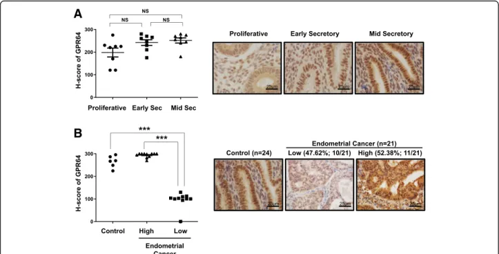
To determine the levels of GPR64 in endometrial cancer, we performed immunohistochemical analysis using tis-sue from 24 controls and 21 endometrioid endometrial carcinoma samples. In control endometrium, GPR64 proteins were strongly detected in the nucleus, cytosol and apical membranes of stromal and epithelial cells of endometrium from the proliferative phase and secretory phases in women and the levels of GPR64 proteins were not significantly changed during the menstrual cycle (Fig. 1a). Interestingly, the levels of GPR64 were signifi-cantly lower in 47.62% (10/21) of endometrial cancer
tissue compared with controls (p < 0.001). However, levels of GRP64 were unchanged in 52.38% (11/21) of
endometrial cancer samples (Fig. 1b; p = 0.0841).
Epi-didymal tissue was included for the
immunohistochem-istry of GPR64 as a positive control [28, 29]. The
expression of GPR64 was detected in apical membranes as well as in some nuclei of epididymal duct epithelial cells (Additional file 1: Figure S1). These results suggest that GPR64 may play a tumor suppressor role in certain cases of endometrial cancer.
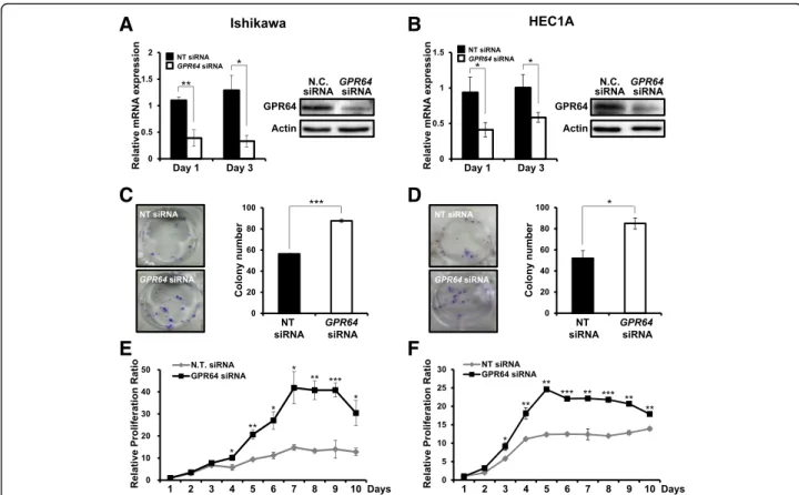
Attenuation ofGPR64 increases colony forming ability and cell proliferation
The continual unregulated proliferation of cells is essen-tial to cancer development [30, 31]. To characterize the proliferative role of GPR64 in endometrial cancer, we performed a colony forming assay and a cell prolifera-tion assay in Ishikawa and HEC1A cells transfected with GPR64 siRNA. First, we confirmed the decrease of GPR64 mRNA and protein levels by GPR64 siRNA transfection compared to non-targeting pool siRNA by RT-qPCR and western blot analysis, respectively (Fig.2a and b). The colony formation assay enables us to deter-mine the survival and proliferation of cells [32]. Colony formation ability was significantly increased in Ishikawa
and HEC1A cells transfected with GPR64 siRNA
compared to control (Fig. 2 c and d). Furthermore, the
A
B
Fig. 1 Levels of GPR64 in human endometrial cancer. a Representative immunohistochemistry andsemiquantitative analysis for GPR64 expression in control endometrium from women in proliferative, early, and mid-secretory phases. b Representative immunohistochemistry and
semiquantitative analysis for GPR64 expression in endometrial cancer tissues. The semiquantitative analysis of immunohistochemistry analysis was calculated by H-score among 24 control endometrium and 21 endometrial cancer tissues. High level of GPR64 was observed in 100% control endometrium (6/6) and 52.38% endometrial cancer (11/21), and low level was observed in 47.62% endometrial cancer (10/21). ***,p < 0.001
proliferation levels of Ishikawa cells were significantly
increased by GPR64 siRNA transfection after 4 days
(Fig. 2e). The proliferation of HEC1A cells was also
significantly increased by GPR64 siRNA transfection
after 3 days (Fig. 2f). Next, we examined whether
GPR64 regulates cell apoptosis using an Annexin V/ PI assay. The ratio of apoptotic cells was not different
between GPR64 siRNA-transfected cells and
non-tar-geting pool siRNA transfected cells of Ishikawa and
HEC1A cells (Additional file 1: Figure S2). These
re-sults suggest that GPR64 suppresses epithelial prolif-eration of endometrial cancer cells.
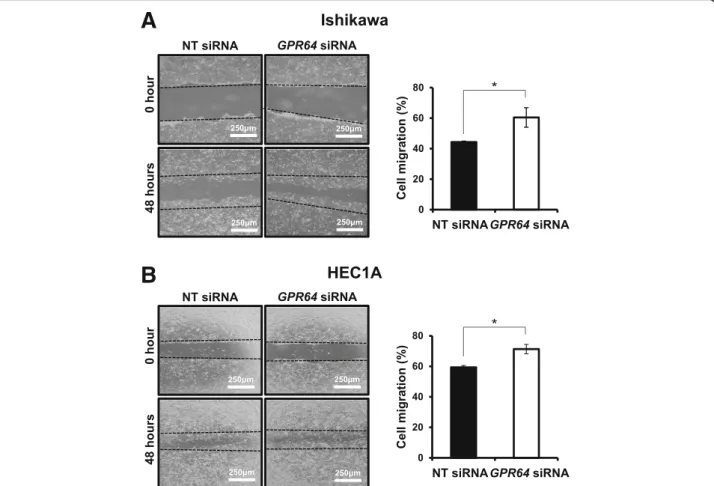
Attenuation ofGPR64 increases cell migration and invasion
We examined the effect of GPR64 attenuation in cell mi-gration and invasion. Our wound healing assay revealed
that reduction of GPR64 by RNA interference
signifi-cantly increased the migration ability of Ishikawa and HEC1A cells (Fig.3). TheGPR64-deficient Ishikawa cells recovered the wound over 61% after 48 h, while control cells healed less than 44% of wound distance (Fig. 3a).
The HEC1A cells withGPR64 knockdown recovered the
wound over 71% but the control cells healed only less than 59% of wound distance (Fig.3b).
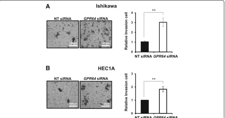
Attenuation ofGPR64 exhibited a significantly higher
infiltration rate in the transwell invasion assay of Ishikawa and HEC1A cells compared to siRNA con-trols. Invasion ability of GPR64-deficient Ishikawa cells was increased by more than three-times compared to
control cells (Fig. 4a), and invasion ability of GPR64
knockdown HEC1A cells was increased by more than two-times (Fig.4b).
Attenuation of GPR64 reduces the expression of Connexin 43 trough activation of AMPK
Activation of AMP-activated protein kinase (AMPK) plays a critical role in induction of EMT in multiple
cancer cell types [37]. To determine whether AMPK
ac-tivity is regulated by depletion of GPR64, we examined
phosphorylation level of AMPK (Thr 172) in Ishikawa
cells transfected with GPR64 siRNA. The levels of
pho-phorylated AMPK were increased by GPR64
knock-down compared to control, however total AMPK levels
A
B
C
D
E
F
Fig. 2 Effect ofGPR64 loss on cell growth in human endometrial cancer cells. a and b The expression of GPR64 mRNA and protein in Ishikawa (a) and HEC1A (b) cells transfected with non-targeting pool (NT) siRNA orGPR64 siRNA was examined by RT-qPCR and Western blot analysis, respectively. c and d Colony formation assay of Ishikawa (c) and HEC1A (d) cells transfected with NT siRNA orGPR64 siRNA. Samples from each treatment were transferred to flat-bottomed 24-well plates and incubated. Cells were fixed and stained with crystal violet. The average colony formation number was quantified with crystal violet stained cells. e and f Cell proliferation assay of Ishikawa (e) and HEC1A (f) cells transfected with NT siRNA orGPR64 siRNA. The results represent the mean ± SEM. *, p < 0.05; **, p < 0.01; and ***, p < 0.001
were not different between control and GPR64-deficient cells (Fig.5a).
Connexin proteins are frequently dysregulated in tu-mors, resulting in loss of gap junctional intercellular
communication [21, 22]. Many tumors with decreased
Cx43 expression exhibited dysfunctional gap junctional intercellular communication [19,38,39]. The expression and function of Cx43 have been correlated with carcino-genesis in endometrial cancer [26, 40]. Therefore, we
examined the levels ofCx43 proteins and mRNA in
Ishi-kawa cells with GPR64 knockdown using RT-qPCR and
Western Blot analysis. Depletion of GPR64 significantly reduced the levels of Cx43 proteins on day 1 and day 6 compared with non-targeting pool siRNA by Western
blot analysis (Fig. 5 a and b). The expression of Cx43
mRNA was significantly reduced in the Ishikawa cells
treated withGPR64 siRNA on day 3 and day 6 compared
with non-targeting pool siRNA (Fig.5c).
Next, we performed double immunofluorescence for
GPR64 and Cx43 after GPR64 knock-down (Fig. 6).
GPR64 is colocalized with Cx43 in Ishikawa cells.
However, the expression of Cx43 protein was
remark-ably reduced in GPR64-deficient cells by GPR64 siRNA
compared to non-targeting pool siRNA.
Discussion
G protein-coupled receptors (GPCRs) are transmem-brane receptors and play important roles in multiple biological processes. Aberrant expression of these recep-tors has been linked to cancer development and
progres-sion [43–45]. However, the molecular function of
GPCRs is not known in endometrial cancer. The present study shows that the expression of GPR64 was distinctly lower in a subset of endometrioid endometrial carcin-oma. These results suggest a tumor suppressor role of GPR64 in endometrial cancer.
Recently, Richter et al. identified that GPR64 is specif-ically overexpressed in Ewing sarcoma (ES) but is also up-regulated in prostate, kidney, and lung carcinoma [10]. This study identified that suppression ofGPR64 in ES by siRNA, led to impaired colony formation, cell growth, and metastasis in Rag2(−/−)γC(−/−)mice. Moreover,
A
B
Fig. 3 Ability of cell migration associated withGPR64 expression in endometrial cancer. The wound healing assay was performed in Ishikawa (a) and HEC1A (b) cells transfected with NT siRNA orGPR64 siRNA. After scratch, cells were incubated for 48 h to determine the cell migration ability. Representative result of wound healing assay and quantification of cell migration by wound healing assay during in vitro culture. The results represent the mean ± SEM. *,p < 0.05
suppression of GPR64 induced TRAIL mediated apoptosis
and reduced PGF and MMP1 expression [10]. GPR64 is
also involved in cell adhesion and migration through acti-vation of serum response element (SRE) and nuclear factor kappa-light-chain-enhancer of activated B cells (NFκB) and its knockdown by siRNA in the highly motile breast cancer cell lines results in a reduction in cell adhesion and migra-tion [46]. However, our results showed that depletion of GPR64 increases the cell proliferation, migration, and inva-sion of endometrial cancer cells. These results suggest the
role of GPR64 as a tumor suppressor in endometrial can-cer. It remains possible thatGPR64 plays dual functions in different cancer conditions depending on the spatial and
temporal distribution and abundance of different GPR64
downstream targets and factors that regulate GPR64. Our
results suggest a new role of GPR64 as a tumor suppressor in endometrial cancer, but further studies are needed to provide a conclusive answer.
Unfortunately, we could not characterize the subtype of endometrial cancers where GPR64 could be a tumor
A
B
Fig. 4 An increase of cell invasion byGPR64 loss in endometrial cancer cells. Representative result of transwell invasion assays of Ishikawa (a) and HEC1A (b) cells transfected with NT siRNA orGPR64 siRNA. Quantification of invasion through matrigel and transwell membrane in Ishikawa and HEC1A cells with or withoutGPR64 siRNA treatment. The results represent the mean ± SEM. **, p < 0.01
A
B
C
Fig. 5 The reduction of Cx43 levels byGPR64 loss through AMPK activation in Ishikawa cells. a The Western blot analysis of phospho-AMPKThr172, Total AMPK, and Cx43 proteins in Ishikawa cells transfected with NT siRNA orGPR64 siRNA on day 1 and day 6. Actin was used as sample-loading control. b Quantification of western blot analysis of Cx43. c The expression ofCx43 gene during in vitro culture after GPR64 siRNA transfection. Cx43 was significantly decreased on day 3 and day 6 in cells transfected with GPR64 siRNA. The results represent the mean ± SEM. *, p < 0.05; and **,p < 0.01
suppressor. The levels of GPR64 were not correlated with grade or stage of endometrial endometrioid adeno-carcinoma which is the most frequently occurring endo-metrial cancer cell type [47]. Cancer classification in the clinic is primarily based on histological analysis in the proper clinical context. Although highly informative, histopathology can be hampered by limitation in its ability to distinguish subtypes of cancers and molecular signatures. Although we could not characterize the sub-type of endometrial tumors based on GPR64 levels in this study, composite molecular profiling of tumor speci-mens is increasingly becoming recognized as an adjunct
to traditional histopathology. Therefore, the molecular characterization of tumor types using GPR64 expression may help to identify additional objective tools to en-hance the classification of endometrial cancer. However, there is a need to investigate a tumor suppressor role of GPR64 in specific subtypes of endometrial cancer.
Several adhesion GPCRs have been shown to be involved in cell adhesion and migration, hereby influen-cing tumor progression [46]. GPR26 is a potent regulator of energy homeostasis through controlling hypothalamic
AMP-activated protein kinase (AMPK) activation [48].
AMPK inhibits Cx43 expression in bladder smooth
muscle cells [36]. While total AMPK levels were not
different between control and GPR64-deficient cells, the
levels of phophorylated AMPK were increased byGPR64
knock-down compared to control. AMPK is associated with cancer development as an important mediator in maintaining cellular energy homeostasis [33, 34], and its activity is increased by extracellular changes such as de-pletion of ATP, low glucose, and changes of NADPH levels [35]. Additionally, AMPK inhibits Cx43 expression in bladder smooth muscle cells [36]. Cx43 expression has been associated with a wide variety of cancers, including liver tumor, colon cancer, breast cancer, ovarian carcin-oma, and endometrial cancer [41]. Its role in controlling cell motility and polarity contributes to cancer develop-ment and metastasis. Especially, Cx43 decreased with poor differentiation of endometrial cancer [26], and Cx43 is considered to be weakened in progression of carcinogen-esis [40, 42]. Thus, our results suggest that GPR64 is a tumor suppressor in endometrial cancer by regulating Cx43 expression through regulation of AMPK activity.
Conclusions
We found an attenuation of GPR64 expression in a subset of endometrial cancer. We demonstrated that
depletion of GPR64 induced tumorigenic potentials by
promoting cell proliferation, migration, and invasion in endometrial cancer cells. GPR64 regulates the expression
ofCx43 and AMPK activity in endometrial cancer cells.
These results suggest that GPR64 acts as a tumor sup-pressor in endometrial cancer.
Additional file
Additional file 1:Figure S1. Expression of GPR64 in mouse epididymis. The immunohistochemistry for GPR64 was performed in mouse epididymis as a positive control. Immunohistochemical staining of mouse epididymis shows membranous and nuclear positivity in epididymal duct epithelial cells. Figure S2. Effect of GPR64 on cell apoptosis in human endometrial cancer cells. Annexin V/PI assay were performed in Ishikawa (A) and HEC1A (B) cells transfected with with non-targeting pool (NT) siRNA or GPR64 siRNA to determine the effect of GPR64 on cell apoptosis. The apoptotic cells were analyzed by Flow cytometry. No difference was found between there were no significant difference between NT siRNA and GPR64 siRNA treatments. (PPTX 330 kb) Fig. 6 The colocalization of GPR64 with Cx43 in Ishikawa cells. The
colocalization of GPR64 (red) and Cx43 (green) were analyzed in Ishikawa cells transfected with NT siRNA orGPR64 siRNA by fluorescence microscopy. GPR64 overlaps with Cx43, but its colocalization was affected by reduction ofGPR64. Nuclei were counterstained with DAPI staining
Abbreviations
CMT:Charcot-Marie-Tooth; Cx43: Connexin 43; DAB: Diaminobenzidine; ES: Ewing sarcoma; ESR1: Estrogen receptorα; GPCR: G protein-coupled receptors; GPR64: G-protein coupled receptor 64; MMP: Metalloproteinase; PBS: Phosphate-buffered saline; PGF: Placental growth factor;
PGR: Progesterone receptor; siRNA: Small interfering RNA; TCF: T-cell factor; VAH: Visceroatiral Heterotaxia Syndrom
Authors’ contributions
JIA and JYY conceived and designed the experimental approach, performed experiments and prepared the manuscript. THK, YIK, and JYA performed the experiments and analyzed the results. RRB provided the human endometrial specimens and performed analysis and interpretation of data. JML and JWJ conceived and designed the experimental approach, performed data analysis and prepared the manuscript. All authors have read and approved the final version of manuscript.
Funding
Grant numbers and sources of support:
The design, data collection, data analysis, and data interpretation of this study were supported by Bio-industry Technology Development Program (IPET312060–5), Ministry for Food, Agriculture, Forestry and Fisheries, Republic of Korea (to J.M.L.), and NIH R01 HD084478 (to J.W.J.). The analysis and inter-pretation of data and writing support of this manuscript were supported by Basic Science Research Program through the National Research Foundation of Korea (NRF-2016R1D1A1B03934346), Ministry of Education, Science and Technology, Republic of Korea (to J.Y.Y.) and Grant Number P50CA098258 from the National Cancer Institute (to R.R.B. and T.H.K.).
Availability of data and materials
The raw data available upon reasonable request from the corresponding authors.
Ethics approval and consent to participate
The study has been approved by Institutional Review Committee of University of Texas MD Anderson Cancer Center (Houston, TX), and written informed consent was obtained from all participants.
Consent for publication Not applicable.
Competing interests
The authors declare that they have no competing interests.
Author details
1Department of Agricultural Biotechnology, Seoul National University, Seoul 08826, Republic of Korea.2Research Institute of Agriculture and Life Sciences, Seoul National University, Seoul 08826, Republic of Korea.3Department of Biochemistry and Molecular Biology, Brain Korea 21 PLUS Project for Medical Sciences, Yonsei University College of Medicine, Seoul 03722, Republic of Korea.4Department of Obstetrics and Gynecology & Reproductive Biology, College of Human Medicine, Michigan State University, 400 Monroe Avenue NW, Grand Rapids, MI 49503, USA.5Pathology, University of Texas M.D. Anderson Cancer Center, Houston, TX 77030, USA.
Received: 9 January 2019 Accepted: 30 July 2019
References
1. Siegel RL, Miller KD, Jemal A. Cancer statistics, 2018. CA Cancer J Clin. 2018; 68(1):7–30.
2. Di Cristofano A, Ellenson LH. Endometrial Carcinoma. Annu Rev Pathol. 2007;2:57–85.
3. Kurman RJ, Kaminski PF, Norris HJ. The behavior of endometrial hyperplasia. A long-term study of“untreated” hyperplasia in 170 patients. Cancer. 1985; 56(2):403–12.
4. Dedes KJ, Wetterskog D, Ashworth A, Kaye SB, Reis-Filho JS. Emerging therapeutic targets in endometrial cancer. Nat Rev Clin Oncol. 2011;8(5): 261–71.
5. Thanapprapasr D, Thanapprapasr K. Molecular therapy as a future strategy in endometrial cancer. Asian Pac J Cancer Prev. 2013;14(6):3419–23.
6. Gottwald U, Davies B, Fritsch M, Habenicht UF. New approaches for male fertility control: HE6 as an example of a putative target. Mol Cell Endocrinol. 2006;250(1–2):49–57.
7. Kirchhoff C, Obermann H, Behnen M, Davies B. Role of epididymal receptor HE6 in the regulation of sperm microenvironment. Mol Cell Endocrinol. 2006;250(1–2):43–8.
8. Davies B, Baumann C, Kirchhoff C, Ivell R, Nubbemeyer R, Habenicht UF, Theuring F, Gottwald U. Targeted deletion of the epididymal receptor HE6 results in fluid dysregulation and male infertility. Mol Cell Biol. 2004;24(19): 8642–8.
9. Galligan CL, Baig E, Bykerk V, Keystone EC, Fish EN. Distinctive gene expression signatures in rheumatoid arthritis synovial tissue fibroblast cells: correlates with disease activity. Genes Immun. 2007;8(6):480–91.
10. Richter GH, Fasan A, Hauer K, Grunewald TG, Berns C, Rossler S, Naumann I, Staege MS, Fulda S, Esposito I, et al. G-protein coupled receptor 64 promotes invasiveness and metastasis in Ewing sarcomas through PGF and MMP1. J Pathol. 2013;230(1):70–81.
11. Foord SM, Jupe S, Holbrook J. Bioinformatics and type II G-protein-coupled receptors. Biochem Soc Trans. 2002;30(4):473–9.
12. Schwartz DR, Wu R, Kardia SL, Levin AM, Huang CC, Shedden KA, Kuick R, Misek DE, Hanash SM, Taylor JM, et al. Novel candidate targets of beta-catenin/T-cell factor signaling identified by gene expression profiling of ovarian endometrioid adenocarcinomas. Cancer Res. 2003; 63(11):2913–22.
13. Sohl G, Willecke K. An update on connexin genes and their nomenclature in mouse and man. Cell Commun Adhes. 2003;10(4–6):173–80. 14. Saez JC, Berthoud VM, Branes MC, Martinez AD, Beyer EC. Plasma
membrane channels formed by connexins: their regulation and functions. Physiol Rev. 2003;83(4):1359–400.
15. Decrock E, Vinken M, De Vuyst E, Krysko DV, D'Herde K, Vanhaecke T, Vandenabeele P, Rogiers V, Leybaert L. Connexin-related signaling in cell death: to live or let die? Cell Death Differ. 2009;16(4):524–36.
16. De Vuyst E, Wang N, Decrock E, De Bock M, Vinken M, Van Moorhem M, Lai C, Culot M, Rogiers V, Cecchelli R, et al. Ca(2+) regulation of connexin 43 hemichannels in C6 glioma and glial cells. Cell Calcium. 2009;46(3):176–87. 17. Chen SC, Pelletier DB, Ao P, Boynton AL. Connexin43 reverses the
phenotype of transformed cells and alters their expression of cyclin/cyclin-dependent kinases. Cell Growth Differ. 1995;6(6):681–90.
18. Sanchez-Alvarez R, Paino T, Herrero-Gonzalez S, Medina JM, Tabernero A. Tolbutamide reduces glioma cell proliferation by increasing connexin43, which promotes the up-regulation of p21 and p27 and subsequent changes in retinoblastoma phosphorylation. Glia. 2006;54(2):125–34. 19. Naus CC, Laird DW. Implications and challenges of connexin connections to
cancer. Nat Rev Cancer. 2010;10(6):435–41.
20. Kumar NM, Gilula NB. The gap junction communication channel. Cell. 1996; 84(3):381–8.
21. Leithe E, Sirnes S, Omori Y, Rivedal E. Downregulation of gap junctions in cancer cells. Crit Rev Oncog. 2006;12(3–4):225–56.
22. Mesnil M, Crespin S, Avanzo JL, Zaidan-Dagli ML. Defective gap junctional intercellular communication in the carcinogenic process. Biochim Biophys Acta. 2005;1719(1–2):125–45.
23. Kardami E, Dang X, Iacobas DA, Nickel BE, Jeyaraman M, Srisakuldee W, Makazan J, Tanguy S, Spray DC. The role of connexins in controlling cell growth and gene expression. Prog Biophys Mol Biol. 2007;94(1–2): 245–64.
24. Shao Q, Wang H, McLachlan E, Veitch GI, Laird DW. Down-regulation of Cx43 by retroviral delivery of small interfering RNA promotes an aggressive breast cancer cell phenotype. Cancer Res. 2005;65(7):2705–11.
25. Avanzo JL, Mesnil M, Hernandez-Blazquez FJ, Mackowiak II, Mori CM, da Silva TC, Oloris SC, Garate AP, Massironi SM, Yamasaki H, et al. Increased susceptibility to urethane-induced lung tumors in mice with decreased expression of connexin43. Carcinogenesis. 2004;25(10):1973–82.
26. Schlemmer SR, Novotny DB, Kaufman DG. Changes in connexin 43 protein expression in human endometrial carcinoma. Exp Mol Pathol. 1999;67(3): 150–63.
27. Kim TH, Yoo JY, Kim HI, Gilbert J, Ku BJ, Li J, Mills GB, Broaddus RR, Lydon JP, Lim JM, et al. Mig-6 suppresses endometrial cancer associated with Pten deficiency and ERK activation. Cancer Res. 2014;74(24):7371–82.
28. Obermann H, Samalecos A, Osterhoff C, Schroder B, Heller R, Kirchhoff C. HE6, a two-subunit heptahelical receptor associated with apical membranes of efferent and epididymal duct epithelia. Mol Reprod Dev. 2003;64(1):13–26.
29. Kirchhoff C, Osterhoff C, Samalecos A. HE6/GPR64 adhesion receptor co-localizes with apical and subapical F-actin scaffold in male excurrent duct epithelia. Reproduction. 2008;136(2):235–45.
30. Thompson HJ, Strange R, Schedin PJ. Apoptosis in the genesis and prevention of cancer. Cancer Epidemiol Biomarkers Prev. 1992;1(7):597–602. 31. Campisi J. Cellular senescence as a tumor-suppressor mechanism. Trends
Cell Biol. 2001;11(11):S27–31.
32. Jeong JW, Lee KY, Kwak I, White LD, Hilsenbeck SG, Lydon JP, DeMayo FJ. Identification of murine uterine genes regulated in a ligand-dependent manner by the progesterone receptor. Endocrinology. 2005;146(8):3490–505. 33. Zadra G, Batista JL, Loda M. Dissecting the dual role of AMPK in Cancer:
from experimental to human studies. Mol Cancer Res. 2015;13(7):1059–72. 34. Li W, Saud SM, Young MR, Chen G, Hua B. Targeting AMPK for cancer
prevention and treatment. Oncotarget. 2015;6(10):7365–78. 35. Jeon SM, Chandel NS, Hay N. AMPK regulates NADPH homeostasis to
promote tumour cell survival during energy stress. Nature. 2012;485(7400): 661–5.
36. Zhang X, Yao J, Gao K, Chi Y, Mitsui T, Ihara T, Sawada N, Kamiyama M, Fan J, Takeda M. AMPK suppresses Connexin43 expression in the bladder and ameliorates voiding dysfunction in cyclophosphamide-induced mouse cystitis. Sci Rep. 2016;6:19708.
37. Saxena M, Balaji SA, Deshpande N, Ranganathan S, Pillai DM, Hindupur SK, Rangarajan A. AMP-activated protein kinase promotes epithelial-mesenchymal transition in cancer cells through Twist1 upregulation. J Cell Sci. 2018;131(14).
38. Mehta PP, Bertram JS, Loewenstein WR. Growth inhibition of transformed cells correlates with their junctional communication with normal cells. Cell. 1986;44(1):187–96.
39. Nicolson GL. Cancer metastasis: tumor cell and host organ properties important in metastasis to specific secondary sites. Biochim Biophys Acta. 1988;948(2):175–224.
40. Saito T, Nishimura M, Kudo R, Yamasaki H. Suppressed gap junctional intercellular communication in carcinogenesis of endometrium. Int J Cancer. 2001;93(3):317–23.
41. Chevallier D, Carette D, Segretain D, Gilleron J, Pointis G. Connexin 43 a check-point component of cell proliferation implicated in a wide range of human testis diseases. Cell Mol Life Sci. 2013;70(7):1207–20.
42. Wincewicz A, Koda M, Sulkowska M, Kanczuga-Koda L, Sulkowski S. Comparison of STAT3 with HIF-1alpha, Ob and ObR expressions in human endometrioid adenocarcinomas. Tissue Cell. 2008;40(6):405–10.
43. Li S, Huang S, Peng SB. Overexpression of G protein-coupled receptors in cancer cells: involvement in tumor progression. Int J Oncol. 2005;27(5): 1329–39.
44. Whitehead IP, Zohn IE, Der CJ. Rho GTPase-dependent transformation by G protein-coupled receptors. Oncogene. 2001;20(13):1547–55.
45. Balenga N, Azimzadeh P, Hogue JA, Staats PN, Shi Y, Koh J, Dressman H, Olson JA Jr. Orphan adhesion GPCR GPR64/ADGRG2 is overexpressed in parathyroid tumors and attenuates calcium-sensing receptor-mediated signaling. J Bone Miner Res. 2016;32(3):654-66.
46. Peeters MC, Fokkelman M, Boogaard B, Egerod KL, van de Water B, AP IJ, Schwartz TW. The adhesion G protein-coupled receptor G2 (ADGRG2/ GPR64) constitutively activates SRE and NFkappaB and is involved in cell adhesion and migration. Cell Signal. 2015;27(12):2579–88.
47. Morice P, Leary A, Creutzberg C, Abu-Rustum N, Darai E. Endometrial cancer. Lancet. 2015;366(9484):491–505.
48. Chen D, Liu X, Zhang W, Shi Y. Targeted inactivation of GPR26 leads to hyperphagia and adiposity by activating AMPK in the hypothalamus. PLoS One. 2012;7(7):e40764.
Publisher’s Note
Springer Nature remains neutral with regard to jurisdictional claims in published maps and institutional affiliations.