pISSN: 0378-6471 eISSN: 2092-9374 DOI : 10.3341/jkos.2011.52.3.293
= 증례보고 =
절반망막정맥폐쇄 황반부종에서의 유리체강내 베바시주맙의 효과와 전방수내 사이토카인 농도
류수정⋅임지원
한림대학교 의과대학 춘천성심병원 안과학교실
목적: 절반망막정맥폐쇄에 의한 황반부종에서 유리체강내 Bevacizumab 주입술의 효과와 전방수내 사이토카인 농도에 대해 알아보고자 하였다.
대상과 방법: 절반망막정맥폐쇄로 인한 황반부종으로 유리체강내 Bevacizumab 주입술을 시행 받은 15명, 15안을 대상으로 12개월 이상 경과관찰을 시행하였다. 유리체강내 주입술 시 전방수를 채취해 multiplex bead assay를 시행하여 대조군과 전방수내 사이토카인 농도를 비교하였다.
결과: 12개월의 경과관찰 기간 동안 평균 4.5회의 유리체강내 주입술이 시행되었으며 12개월후 시력과 중심황반두께 모두 유의하게 호전되었다(p=0.028, p=0.000). 전방수에서 대조군에 비하여 Interleukin-6, vascular endothelial growth factor, monocyte chemoattractant protein-1이 유의하게 높게 측정되었다(p=0.010, p=0.045, p<0.001).
결론: 절반망막정맥폐쇄에서 전방수내 증가된 사이토카인 농도를 확인할 수 있었으며 유리체강내 Bevacizumab 주입술은 황반부종의 감소 및 유의한 시력호전을 이룰 수 있었다.
<대한안과학회지 2011;52(3):293-298>
■ 접 수 일: 2010년 9월 13일 ■ 심사통과일: 2010년 10월 28일
■ 게재허가일: 2011년 1월 17일
■ 책 임 저 자: 임 지 원
강원도 춘천시 교동 153 한림대학교 춘천성심병원 안과
Tel: 033-240-5176, Fax: 033-240-5210 E-mail: [email protected]
망막정맥은 시신경 유두에서 합쳐져서 망막중심정맥을 이루는데 약 20%에서는 시신경유두내 사상판에서 합쳐지 지 않고 각기 존재하게 되며 이 경우 어느 한쪽 정맥이 폐 쇄되는 경우를 절반망막정맥폐쇄라고 하며 임상양상과 합 병증은 중심망막정맥폐쇄와 유사하다.1흔히 다른 망막정맥 폐쇄증과 마찬가지로 허혈로 인한 신경조직 손상과 혈액망 막장벽의 파괴로 인한 황반부종으로 시력저하를 초래하게 되며 신생혈관이 발생할 수 있다.2
망막정맥폐쇄증에서 황반부종에 대한 치료로는 격자광 응고술과 유리체강내 약물주입술이 있으며 최근에는 항 혈 관내피세포증식인자(Vascular endothelial growth factor;
VEGF) 약물인 bevacizumab (Avastin®, Genentech Inc., San Francisco, CA, USA)의 유리체강내 주입이 시력호전과 황반 부종에효과적임이 여러 연구에서 발표되어 왔다.3-9
본 연구에서는 절반망막정맥폐쇄에서 황반부종에 대한 유리체강내 Bevacizumab의 치료효과와 전방수내 사이토카 인 농도에 대하여 알아보고자 하였다.
대상과 방법
2008년 7월부터 2009년 6월까지 본원에서 절반망막정 맥폐쇄로 인한 황반부종이 있어 유리체강내 Bevacizumab주 입술을 시행 받은 15명 15안을 대상으로 12개월 이상 경과 관찰을 시행하였다. 모든 환자에게 시술 전 스넬렌 시력표 를 이용한 최대교정시력, 안압검사, 세극등현미경검사 및 안저검사, 형광안저혈관조영술, 빛간섭단층촬영(Optical coherent tomography3; Carl Zeiss Ophthalmic Systems Inc, Dublin, Ireland)을 시행하였다. 최대교정시력은 통계 적 분석을 위하여 logMAR (logarithm of the minimal angle of resolution) scale로 전환하였고, 빛간섭단층 촬영은 fast macular scan mode로 황반부 단면상을 얻은 후 retinal thickness analysis로 0.5 mm radius 값을 분석 하여 황반부 두께를 측정하였다. 이미 다른 망막질환으로 유리체절제술을 포함한 안구내 수술을 시행 받았거나 시력 저하를 유발할 수 있는 다른 질병을 가지고 있는 경우는 제 외하였고 전방수내 사이토카인 농도에 영향을 줄 수 있는 인공수정체안도 제외하였다. 진단 당시 망막이나 홍채, 전 방각에 신생혈관이 있는 경우도 제외하였다.
유리체강내 Bevacizumab 주입술은 빛간섭단층촬영에서 중심황반두께가 250 μm 초과된 황반부종을 대상으로 하였 으며 환자들에게 시술 전 충분한 설명을 통해 동의를 얻은
A B
C D
E F
Figure 1. Right eye of a 74-year-old man treated with intravitreal bevacizumab (A) Fundus photograph presenting hemi-
retinal venous occlusion. (B) Fluorescein angiography showing no neovasculization or extensive non-perfusion areas.Aqueous VEGF level was 65.98 pg/mL and MCP-1 was 1058.33 pg/mL. (C) Optical coherence tomography at baseline.
His visual acuity was 20/100. (D) 2 months after initial intravitreal bevacizumab, optical coherence tomography showed persistant macular edema. The second intravitreal bevacizumab was performed. (E) At month 4, the third intravitreal bev- acizumab was performed. (F) At month 6, his visual acuity improved to 20/40 and the macula was stable up to 12 months.
후 시행하였다. 시술 방법은 먼저점안마취후 수술방의 무 균환경에서 30 gauge로 전방수를 0.05 mL 제거한 후 각막 윤부에서 3.5 mm 떨어진 부위의 섬모체평면부를 통해 1.25 mg/0.05 mL의 Bevacizumab을 유리체강내로 주입하 였다. 전방수내 사이토카인 농도변화를 비교하기 위하여 안 과적 기저질환이 없는 10명, 10안의 백내장 환자에서 백내 장 수술 시행 전에 전방천자를 통하여 0.05 mL의 전방수를 얻었다.
유리체강내 주입술후 6-8주 간격으로 최대교정시력과 빛간섭단층촬영을 시행하였으며 중심황반두께가 250 μm 이상이며 형태학적으로 망막내 부종이 존재하는 경우 추가 주입술을 시행하였으며 추가 주사 간격은 6-8주를 유지하 였다. 황반부종의 소실 후 경과관찰 기간 동안 중심황반두 께가 250 μm 이상으로 황반부종이 재발하는 경우에도 추가 유리체강내 주입술을 시행하였다.
전방수 검체는 milliplex human cytokine panel I-6 plex kit (xMAP; Luminex Corp., Austin, TX, USA)를 이용하여 multiplex bead assay를 시행하였다. 분석 방법은 제조회 사의 검사방침을 준수하여 시행하였으며 interleukin (IL)-6, IL-8, IL-10, interferon gamma (IFN-λ), monocyte chemoattractant protein-1 (MCP-1), tumor necrosis factor-alpha (TNF-α), platelet-derived growth factor (PDGF)-AA, PDGF-AB/BB, VEGF를 포함한 9개의 사이 토카인을 분석하였다. 전방수 표본은 희석하지 않고 모든 과정은 빛이 차단되어 실온 상태에서 시행했으며, 표본은 suspension array system에 의해 입력되었다. 각 사이토카 인의 표준곡선은 제조회사의 reference cytokine concentrations 에의해 만들어졌다. 사이토카인 농도의 정확한 측정을 위 하여 대조군을 포함한 모든 표본은 같은 실행 과정을 두 번 거쳐서 이중으로 측정하였다. 측정가능범위는 1.0-10,000
Table 1. Aqueous humor levels of cytokines in eyes with hemicentral retinal venous occlusion and controls
Cytokines Patients (n = 15) Controls (n = 10) p-value*
IL-6 (Mean ± SD) 8.3 ± 5.5 2.0 ± 1.3 0.010
IL-8 (Mean ± SD) 5.8 ± 6.0 2.8 ± 1.5 0.051
VEGF (Mean ± SD) 64.1 ± 39.4 36.5 ± 16.5 0.045
MCP-1 (Mean ± SD) 1,214.3 ± 476.5 412.2 ± 98.4 <0.001
PDGF-AA (Mean ± SD) 36.1 ± 19.9 32.8 ± 6.8 0.127
Data presented as the mean ± standard deviation (pg/mL). IL-10, IFN-γ, PDGF-AB/BB and TNF-α were not presented because the levels were near to or below the limits of detection.
IL = interleukin; VEGF = vascular endothelial growth factor; PDGF = platelet-derived growth factor; MCP = monocyte chemoattractant protein.
*Comparison between retinal venous occlusion and controls (Wilcoxon two-sample test).
pg/mL이었다.
통계 분석은 SPSS 12.0 software (SPSS Inc. Chicago, IL, USA)을 사용하였다. 단변량분석은 Two paired t-test, Wilcoxon signed-rank test, Chi-square test로 검정하였 고, 인자들간의 상관관계는 Pearson correlation analysis를 이용하였다. p<0.05인 경우 통계적으로 유의하다고 평가 하였다.
결 과
대상환자 15명의 평균연령은 62.7 ± 14.6세(44-75세) 였으며 남자가 7명, 여자가 8명이었다. 내원 시 시력저하의 평균 발현기간은 10.8 ± 5.2주(4-20주)였다. 환자들이 가 지고 있는 전신질환은 고혈압이 8명(53%), 당뇨가 2명 (13%)이었다. 초진 시 시력(logMAR)은 0.65 ± 0.36였고 중심황반두께(um)는 470.6 ± 108.9였다. 형광안저촬영에 서 심한 모세혈관비관류나 황반허혈을 보인 경우는 없었다.
대조군 10명의 평균연령은 65.4 ± 10.4세였고 남자가 5명, 여자가 5명이었다. 환자군과 대조군의 연령과 성비의 유의 한 차이는 없었다(p=0.458, p=0.249). 대조군 중 당뇨환 자는 없었으며 고혈압은 4명이 있었다.
12개월의 경과관찰기간 동안 4.5 ± 2.4회(3-7회)의 유 리체강내 주입술을 시행하였다. 황반부종이 재발되어 다시 유리체강내 주입술을 시행 받은 환자는 4명(26%)으로 이전 주입술 후 평균 10주(8-16주)에 재발하였다. 망막박리나 안내염 등의 유리체강내 주입술과 관련된 합병증이 발생한 예는 없었다. 시술 후 12개월에 시력은 0.32 ± 0.32, 중심 황반두께는 285.1 ± 85.6으로 초진과 비교하여 유의하게 호전되었다(p=0.028, p=0.000). 경과관찰기간 동안 2안 (13%)에서는 형광안저촬영상 광범위한 비관류가 발견되 어 망막광응고술을 시행하였으며 신생혈관이 발생한 경우 는 없었다. 3안(20%)에서는 12개월 후에도 황반부종이 완 전히 소실되지 않아 1안은 유리체강내 스테로이드 주입술 을, 2안은 유리체강내 스테로이드주입술과 격자레이저광응
고술 병합술을 시행하였다. 대표적인 환자의 경과를 Fig. 1 에 기술하였다.
전방수내 사이토카인 분석에서 IL-6, VEGF, MCP-1이 환자군에서 대조군에 비해 유의하게 높게 측정되었다 (p=0.010, p=0.045, p<0.001) (Table 1). IL-8의 경우 는 유의한 차이가 없었다. IL-10, IFN-γ, PDGF-AB/BB, TNF-α는 환자군과 대조군 모두 측정 하한치 경계역 혹은 아래로 측정되었다. 초진 시력과 사이토카인들의 유의한 상 관관계는 발견되지 않았다. 그러나 중심황반두께는 사이토 카인 중 VEGF 와만 유의한 상관관계를 보였다(Fig. 2). 사 이토카인 사이의 상관관계 분석에서는 MCP-1과 VEGF가 유의한 양의 상관관계를 보였으며 다른 사이토카인들간의 유의한 상관관계는 없었다(Fig. 3). 경과관찰기간중 망막광 응고술을 시행한 2안과 12개월 후에도 황반부종이 관해되 지 않아 다른 치료를 시행한 3안의 전방수내 사이토카인은 환자군과 비교하여 유의한 차이를 보이지 않았다.
고 찰
정맥폐쇄와 동반된 혈관손상은 안구내의 혈관증식 및 염 증과 관련된 사이토카인의 변화를 야기하게 되며 전방수내 사이토카인 농도는 안구 및 유리체강내 사이토카인 농도를 반영하게 된다.10-12따라서 전방수내 사이토카인의 변화를 분석함으로서 치료의 주된 대상을 정하여 효율적인 치료방 법을 정할 수 있다. 본 연구에서는 전방수내의 어떤 인자들 이 절반망막폐쇄 후 증가하는지를 분석하였으며 VEGF, IL-6, MCP-1이 대조군보다 유의하게 증가되어 있음을 알 수 있었다. VEGF는 혈관증식과 혈관누출과 관련된 대표적 인 사이토카인으로 혈관손상후 발현이 증가하여 황반부종 을 일으키는데 관여한다.13,14 또한 IL-6는 급성 염증기에 증가하여 염증반응과 혈관생성을 촉진시키는 인자로서 VEGF의 발현 증가와도 관련이 있다고 알려져 있다.15 기존 연구에서 망막정맥페쇄증에서의 VEGF와 IL-6의 증가는 보고된 바 있으나 연구 결과가 다양하고 대상안이 많지 않
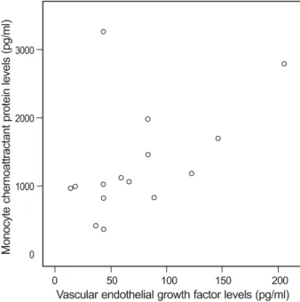
Figure 3. Correlation analysis of the monocyte chemoattractant
protein-1 and vascular endothelial growth factor in aqueous hum or in patient w ith hem icentral retinal vein occlusion (Pearson correlation, r = 0.509; p = 0.031).Figure 2. Correlation analysis of the vascular endothelial growth
factor in aqueous humor and the central macular thickness in patient with hemicentral retinal vein occlusion (Pearson corre- lation, r = 0.413; p = 0.047).으며 절반망막폐쇄를 단독으로 분석한 경우는 없었다.16-18 본 연구에서의 전방수내 VEGF와 IL-6 농도는 기존 중심 망막정맥폐쇄에서의 보고보다 약간 낮은 수준이었다. 이는 중심망막정맥폐쇄보다 절반망막폐쇄가 망막병변의 크기가 작아 상대적으로 사이토카인의 발현이 적은 것과 가장 관 련이 있을 것이다. 또한 본 연구에서는 심한 모세혈관 비관 류나 황반허혈을 보이지 않은 비허혈성 절반망막폐쇄를 분 석하였는데 허혈성 중심망막정맥폐쇄가 비허혈성보다유의 하게 높은 VEGF와 IL-6 농도를 보였음을 고려할 때 허혈 이 심하지 않은 군이 대상으로 된 것도 낮은 전방수내 사이 토카인 농도에 관련이 있을 것이다.18
그 외에 MCP-1은 화학주성 사이토카인의 일종으로 혈 관손상후 혈관벽의 단핵구 보충에 주된 역할을 담당한다.19 또한 MCP-1은 망막정맥폐쇄에서 혈관손상 후 혈관재형성 과정을 담당하는 호산구를 증가시키는 기능이 있다.20 호산 구는 VEGF의 주된 생성 부위 중 하나이기에 MCP-1과 VEGF 농도 사이에 상관관계가 있었던 것으로 생각된다.
본 연구에서 시력과 사이토카인은 상관관계가 없었으나 중 심황반두께는 VEGF 농도와 유의한 상관관계가 있었다. 따 라서 VEGF가 황반부종의 정도와 관련이 깊은 것을 알 수 있었으며 Bevacizumab을 주입함으로써 황반부종에 가장 중요한 원인인자인 VEGF를 집중적으로 억제시켜 손상된 혈액망막장벽의 안정화와 황반부의 누출을 막아 효율적으 로 황반부종을 치료 할 수 있는 이론적 배경이 되겠다.
절반망막정맥폐쇄증에서 유리체강내 Bevacizumab의 정
해진 치료방침은 아직까지 확립되지 않았다. 인체내에서 유 리체강내 Bevacizumab의 지속기간은 6-8주로 보고 되었 다.21 망막정맥폐쇄증에서 단일주사로도 재발 없이 황반부 종이 치료되는 경우가 있으며 유리체강내 반복주입을 최소 화 하기 위하여 본 연구에서는 빛간섭단층촬영을 이용하여 추가주사치료를 결정하였다. Hoeh et al6의 망막중심정맥 폐쇄증 보고와는 다르게 단일 주사로 치료된 경우는 없었 으며 최소 3회의 주사가 황반부종의 관해에 요구되었다. 망 막정맥폐쇄증중 절반망막폐쇄만을 따로 분석하여 보고된 발표는 아직까지 없어 본 연구 결과의 비교는 어려우나 최 근의 Choi et al22과 Wu et al23의 망막중심정맥폐쇄증 결과 와 비교하면 시력호전 정도와 황반부종 소실 정도는 유사 하였다. 따라서 절반망막폐쇄가 병변의 크기는 망막중심정 맥페쇄보다 제한되어 있으며 전방수내 사이토카인 농도는 약간 낮은 수준이었음에도 황반부종의 치료 후 시력개선과 치료과정은 결코 더 양호하지 않으며 25%에서 주사 후 수 개월 내에 재발하여 추가 주사를 요하였을 뿐 아니라 20%
에서 치료에 대한 불응이 있었던 점을 고려할 때 절반망막 폐쇄의 황반부종 치료는 단일치료나 단기간 경과관찰로는 이루어지기 어려움을 주지해야 하겠다.
망막정맥폐쇄증의 황반부종 치료 결과는 혈관손상부위 정도나 황반허혈과 황반부종의 정도, 경과관찰기간에 따라 서 다양하게 보고되고 있으며 아직 무작위의 제한된 비교 대조연구는 없다. 본 연구의 대상안은 비허혈성 환자만을 대상으로 하였고 황반허혈이 심한 환자는 없었음을 고려하
여 치료 결과를 해석하여야 한다. 추후 유리체강내 주사에 대한 치료방침은 보다 많은 환자수를 대상으로 한 대규모 연구가 필요할 것으로 생각된다.
본 연구의 제한점으로는 환자수가 한정되어 있으며 전방 수내 사이토카인 농도를 치료 후에는 측정하지 않은 점이 다. 전방수내 사이토카인 농도가 유리체강내 사이토카인 농 도를 반영하지만 유리체강내 사이토카인 분석이 더 정확하 게 안구내 사이토카인 양상을 반영할 것이다. 또한 고정된 치료계획을 가지고 시행한 것이 아니라 경과관찰을 시행하 면서 추가 유리체강내 주사를 시행한 점을 알아야 하겠다.
요약하면 절반망막정맥폐쇄증으로 인한 황반부종에서 VEGF를 포함한 사이토카인의 농도가 전방수에서 증가되 어 있었으며 VEGF을 억제하는 Bevacizumab의 유리체강내 반복 주입술은 유의한 시력호전과 해부학적 호전을 이룰 수 있었다.
참고문헌
1) Hayreh SS, Hayreh MS. Hemi-central retinal vein occulsion.
Pathogenesis, clinical features, and natural history. Arch Ophthalmol 1980;98:1600-9.
2) Chopdar A. Hemi-central retinal vein occlusion. Pathogenesis, clinical features, natural history and incidence of dual trunk central retinal vein. Trans Ophthalmol Soc U K 1982;102:241-8.
3) Rosenfeld PJ, Fung AE, Puliafito CA. Optical coherence tomog- raphy findings after an intravitreal injection of bevacizumab (avastin) for macular edema from central retinal vein occlusion.
Ophthalmic Surg Lasers Imaging 2005;36:336-9.
4) Ferrara DC, Koizumi H, Spaide RF. Early bevacizumab treatment of central retinal vein occlusion. Am J Ophthalmol 2007;144:864-71.
5) Lee HK, Kim HW, Yun IH. Intravitreal bevacizumab injection for macular edema secondary to branch retinal vein occlusion. J Korean Ophthalmol Soc 2009;50:1800-8.
6) Hoeh AE, Ach T, Schaal KB, et al. Long-term follow-up of OCT-guided bevacizumab treatment of macular edema due to reti- nal vein occlusion. Graefes Arch Clin Exp Ophthalmol 2009;
247:1635-41.
7) Nghiem-Buffet S, Cohen SY. Retinal vein occlusion: anti-VEGF treatments. J Fr Ophtalmol 2009;32:679-86.
8) Fish GE. Intravitreous bevacizumab in the treatment of macular edema from branch retinal vein occlusion and hemisphere retinal vein occlusion (an AOS thesis). Trans Am Ophthalmol Soc 2008;
106:276-300.
9) Hayashi A, Ueta Y, Yunoki T, et al. Short-term effects of intra-
vitreal injection of bevacizumab on macular edema due to central retinal vein occlusion. Nippon Ganka Gakkai Zasshi 2010;114:
347-55.
10) Noma H, Funatsu H, Yamasaki M, et al. Aqueous humour levels of cytokines are correlated to vitreous levels and severity of macular oedema in branch retinal vein occlusion. Eye 2008;22: 42-8.
11) Funatsu H, Yamashita H, Noma H, et al. Aqueous humor levels of cytokines are related to vitreous levels and progression of diabetic retinopathy in diabetic patients. Graefes Arch Clin Exp Ophthalmol 2005;243:3-8.
12) Bakri SJ, Snyder MR, Reid JM, et al. Pharmacokinetics of intra- vitreal bevacizumab (Avastin). Ophthalmology 2007;114:855-9.
13) Aiello LP, Avery RL, Arrigg PG, et al. Vascular endothelial growth factor in ocular fluid of patients with diabetic retinopathy and other retinal disorders. N Engl J Med 1994;331:1480-7.
14) Boyd SR, Zachary I, Chakravarthy U, et al. Correlation of in- creased vascular endothelial growth factor with neovascularization and permeability in ischemic central vein occlusion. Arch Ophthalmol 2002;120:1644-50.
15) Cohen T, Nahari D, Cerem LW, et al. Interleukin 6 induces the ex- pression of vascular endothelial growth factor. J Biol Chem 1996;
271:736-41.
16) Funk M, Kriechbaum K, Prager F, et al. Intraocular concentrations of growth factors and cytokines in retinal vein occlusion and the ef- fect of therapy with bevacizumab. Invest Ophthalmol Vis Sci 2009;50:1025-32.
17) Noma H, Funatsu H, Yamasaki M, et al. Pathogenesis of macular edema with branch retinal vein occlusion and intraocular levels of vascular endothelial growth factor and interleukin-6. Am J Ophthalmol 2005;140:256-61.
18) Noma H, Funatsu H, Mimura T, et al. Vitreous levels of inter- leukin-6 and vascular endothelial growth factor in macular edema with central retinal vein occlusion. Ophthalmology 2009;116:87-93.
19) Schober A, Zernecke A. Chemokines in vascular remodeling.
Thromb Haemost 2007;97:730-7.
20) Hoshino M, Takahashi M, Aoike N. Expression of vascular endo- thelial growth factor, basic fibroblast growth factor, and angioge- nin immunoreactivity in asthmatic airways and its relationship to angiogenesis. J Allergy Clin Immunol 2001;107:295-301.
21) Krohne TU, Eter N, Holz FG, Meyer CH. Intraocular pharmacoki- netics of bevacizumab after a single intravitreal injection in humans.
Am J Ophthalmol 2008;146:508-12.
22) Choi SW, Kim HW, Yun IH. Intravitreal bevacizumab treatment of macular edema in central retinal vein occlusion. J Korean Ophthalmol Soc 2010;51:707-15.
23) Wu L, Arevalo JF, Berrocal MH, et al. Comparison of two doses of intravitreal bevacizumab as primary treatment for macular edema secondary to central retinal vein occlusion: results of the pan American collaborative retina study group at 24 months. Retina 2010;30:1002-11.
=ABSTRACT=
Intravitreal Bevacizumab for Treatment of Macular Edema Secondary to Hemicentral Retinal Venous Occlusion and Concentrations of
Cytokine in Aqueous Humor
Su Jeong Ryu, MD, Ji Won Lim, MD, PhD
Department of Ophthalmology, Hallym University Chuncheon Sacred Heart Hospital, Hallym University College of Medicine, Chuncheon, Korea
Purpose: To evaluate the effects of intravitreal bevacizumab and to investigate the concentrations of cytokine in the aque- ous humors of patients with macular edema secondary to hemicentral retinal venous occlusion.
Methods: Fifteen eyes of 15 patients with macular edema secondary to hemicentral retinal venous occlusion received in- travitreal bevacizumab injections and completed 12 months of follow-up. Cytokine levels were measured in the aqueous humors of these patients using a multiplex bead assay and the levels were compared with those of the controls.
Results: During 12 months of follow-up, a mean of 4.5 intravitreal bevacizumab injections were performed. The visual acui- ty and the central macular thickness improved significantly (p = 0.028, p = 0.000) after treatment. The levels of inter- leukin-6, vascular endothelial growth factor, and monocyte chemoattractant protein-1 were increased in the aqueous hu- mor compared with the levels in the control group (p = 0.010, p = 0.045, p < 0.001).
Conclusions: Elevated cytokine levels were identified in the aqueous humor after hemicentral retinal venous occlusion.
Intravitreal bevacizumab injections were effective for improvements in visual acuity and macular edema due to hemicentral retinal venous occlusion.
J Korean Ophthalmol Soc 2011;52(3):293-298
Key Words: Cytokine, Hemicentral retinal vein occlusion, Intravitreal bevacizumab, Macular edema
Address reprint requests to Ji Won Lim, MD, PhD
Department of Ophthalmology, Hallym University Chuncheon Sacred Heart Hospital
#153 Gyo-dong, Chuncheon 200-060, Korea
Tel: 82-33-240-5176, Fax: 82-33-240-5210, E-mail: [email protected]