Pseudostenosis at the Origin of the Vertebral Artery on Contrast-enhanced MRA: Correlation with Aortic Motion on Dynamic 3D Time-Resolved
Contrast-Enhanced MRA
Seonmun Kim, Sungwon Lee, Hyun Seok Choi, So-Lyung Jung, Kook-Jin Ahn, Bum-soo Kim Department of Radiology, Seoul St. Mary’s Hospital, College of Medicine, The Catholic University of Korea
Purpose : The origin of the vertebral artery (VA) is a frequent site of pseudostenosis on contrast-enhanced MRA (CE- MRA). The purpose of this study is to evaluate the relationship between the motion of the aortic arch and pseudostenosis at the origin of the VA.
Materials and Methods: Our study had approval of our institutional review board. 47 patients underwent CT angiogra- phy (CTA), CE-MRA, and 3D time-resolved contrast-enhanced MRA (TR-CEMRA) within 6.87±9.89 days (mean±SD). Percent stenosis using the NASCET criteria was measured on CTA and CE-MRA. CTA was used as a refer- ence standard to classify the CE-MRA into pseudostenosis and control group. Pseudostenosis was determined as 50%- 99% stenosis observed on CE-MRA but normal to less than 50% stenosis on CTA. Aortic motion (distance between the highest position and lowest position of aortic arch) was measured on TR-CEMRA. Age, route of intravenous contrast media, motion of aortic arch, and normal distal diameter of VA were compared between the two groups.
Results: There were 17 patients and 23 vertebral arteries of pseudostenosis. Patients with pseudostenosis showed more aortic motion (3.61±1.88 vs. 2.05±1.97 mm) and older age (71.29±8.88 vs. 62.32±13.19 year-old). Route of intra- venous contrast media and normal distal diameter of VA were not associated with pseudostenosis.
Conclusion: VA origin is a frequent site of pseudostenosis. Older age and more motion of aortic arch are associated with pseudostenosis on CE-MRA.
Index words : Pseudostenosis∙Neck MR angiography∙Vertebral artery∙Time-resolved contrast enhanced MRA Aortic motion
Although digital subtraction angiography (DSA) is still the standard method in evaluating the supraaortic arteries, contrast-enhanced MR angiography (CE-
MRA) is also widely used and has improved in image quality and accuracy (1-5). Stenosis of the proximal vertebral artery is a frequent cause of symptomatic stroke with a high prevalence (6-8). However, there are some limitations in evaluating the vertebral artery with CE-MRA. CE-MRA has a tendency to overesti- mate the degree of stenosis at the origin of the vertebral artery (9-12). The cause of this phenomenon is thought to be due to weak spatial resolution, intravoxel dephasing, and motion artifact caused by cardiac pulsation and respiration (11-14). 3D time- resolved contrast-enhanced MRA (TR-CEMRA) offers both anatomic and hemodynamic information of the INTRODUCTION
�Received; August 13, 2012�Revised; September 28, 2012
�Accepted; September 29, 2012
Corresponding author : Hyun Seok Choi, M.D.
Department of Radiology, Seoul St. Mary’s Hospital, 222, Banpo-daero, Seocho-gu, Seoul 137-701, Korea.
Tel. 82-10-2517-0806, Fax. 82-2-599-6771 E-mail : [email protected]
Original Article
supraaortic arteries using low dose gadolinium based contrast media (15, 16). In our institution, we have been performing low dose TR-CEMRA after CE-MRA for the evaluation of craniocervical vessels. By doing so, we have noticed a tendency of aortic arch pulsation in the TR-CEMRA in patients with pseudostenosis at the origin of the vertebral artery on CE-MRA, especially in elderly patients. The purpose of this study is to evaluate the relationship between the motion of the aortic arch and pseudostenosis at the origin of the vertebral artery. We compared CE-MRA with CT angiography (CTA) to enroll more patients. This is predicated upon high accuracy of CTA in determining the pathology of vertebral arteries (17-19).
Patient selection
This retrospective study was approved by our Institutional Review Board, and the requirement for informed consent was waived. We used our computer- ized hospital information system to identify all patients who underwent CE-MRA, TR-CEMRA, and CT angiography within 40 days, during the last 3 years (between March 2009 and February 2012). Among 3719 patients, only 78 patients underwent all three, and 18 patients were excluded for an interval of more than 40 days. 13 patients who had limited scan range, severe calcification in the aortic arch, and various artifacts causing limitation for measurement of arterial diameter on MRA or CTA were also excluded. The final number of patients included were 47 (mean age 66.74 years old, 26 man and 21 woman) and the interval between MRA and CTA was 6.87 ± 9.89 days (mean intervals ± standard deviation). The reasons for MRA and CTA were brain infarction in 41 patients; TIA in four; cerebral hemorrhage in one; and for headache in one.
Imaging technique
CTAs were performed using a 64 channel multi- detector row CT (Sensation 64, Siemens, Erlangen, Germany) with contrast injection of 70 mL at a rate of 4 mL/sec with a power injector (Stellant; Medrad, Pittsburg, PA, USA), followed by 50 mL of saline chaser injected at a rate of 4 mL/sec. Automatic triggering at enhancement at the bifurcation level of
the common carotid artery over 70 Hounsfield unit was used to start CTA. Pre-contrast scan was obtained for subtraction of bones, and both pre-contrast and post-contrast scan were obtained ranging from the level below the aortic arch to above the circle of Willis. The scan was performed caudal to cranial direction with the following parameters: field of view, 256 × 256 mm;
section thickness, 0.6 mm; imaging time, 10 seconds.
CE-MRAs were performed with 3T MRI (Verio, Siemens’, Erlangen, Germany). 16 channel head and neck coil was used. CE-MRA was performed with the following parameters: repetition time, 3.24 ms; echo time, 1.23 ms; flip angle, 25�; field of view, 352 × 218 mm; matrix, 448 × 358; voxel size after zero interpolation, 1.0 × 0.8 × 0.6 mm; number of acquisition, 1; acquisition time, 60 seconds. MR fluoroscopic triggering was used at the level of common carotid artery for proper timing of initiating CE-MRA. The centric phaseordering scheme was used for k-space filling. 0.1 mL/kg of contrast media (Gadovist [1.0 mmol/mL], Bayer, Berlin, Germany) was infused at a rate of 1.5 mL/s with power injector (Spectris Solaris; Medrad, Pittsburg, PA, USA).
TR-CEMRAs were performed with the following parameters: repetition time, 2.57 ms; echo time, 0.97 ms; flip angle, 19�; field of view, 420 × 341 mm;
matrix, 448 × 318; voxel size after zero interpolation, 1.3 × 0.9 × 1.6 mm; number of acquisition, 2;
acquisition time, 202 seconds; temporal resolution, 2 seconds/phase. 2 mL of contrast media (Gadovist [1.0 mmol/mL], Bayer, Berlin, Germany) was infused at a rate of 1.5 mL/s with power injector (Spectris Solaris;
Medrad, Pittsburg, PA, USA). Each bolus was immedi- ately followed by a 25mL saline flush. The patients breathed freely during scanning.
CTA images were processed by subtraction of post- contrast CTA from pre-contrast CT and then reconstructed by maximum-intensity-projection (MIP).
Lateral rotational projection images of every 10�from -90�to +90�were reconstructed and displayed on picture archiving and communicating system (PACS).
MRA images were reconstructed by using the MIP algorithm and lateral rotational projections of every 10�from -90�to +90�were reconstructed and displayed on PACS.
Image analysis: Percent stenosis in CTA
Image analysis was performed using picture archiv- MATERIALS AND METHODS
ing and communicating system (PACS). CTA was considered as the gold standard for evaluating stenosis at the origin of the vertebral artery. The MIP and lateral projection images of the CTA were used to evaluate the origin of the vertebral artery, and when the images were unclear in CTA, we interpreted with the source images and multi-planar reconstruction with proper window level and width. Percent stenosis at the origin of the vertebral artery and the diameter of normal distal vertebral artery was measured by a radiologist (S.M.K.; 4-years of experience) using NASCET criteria (20). The degree of stenosis was classified; Normal to less than 50% stenosis, 50-99%
stenosis, and occlusion.
Image analysis: Percent stenosis in CE-MRA Percent stenosis at the origin of the vertebral artery and the diameter of normal distal vertebral artery on contrast-enhanced MRA was also measured by a radiologist (S.M.K.; 4-years of experience) using NASCET criteria after 2 weeks.
The degree of stenosis was classified; Normal to less than 50% stenosis, 50-99% stenosis, and occlusion.
The percent stenosis CTA and CE-MRA were compared and classified into two groups:
Pseudostenosis group [50-99% stenosis was observed on CE-MRA but normal to less than 50% stenosis on CTA] and control group [All other patients without pseudostenosis].
Image analysis: Motion of aortic arch in TR- CEMRA
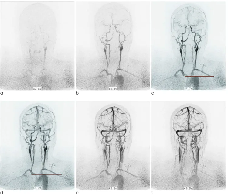
Motion of aortic arch was measured by another radiologist (H.S.C., 6-year experienced neuroradiolo- gist). By reviewing the multi-phase coronal images of TR-CEMRA, the upper border of aortic arch was detected and highest position of upper border of aortic arch and lowest position of aortic arch were marked by horizontal lines. The distance between the horizontal lines (highest and lowest position of aortic arch) was considered as the degree of aortic arch motion and measured on PACS (Fig. 1).
Statistical analysis
The accuracy of CE-MRA was obtained in each percent stenosis group, with CTA as the gold standard.
Independent two sample t-test was performed with age, motion of aortic arch, and diameter of normal distal vertebral artery between the pseudostenosis group and control group. Chi-Square test was performed with sex and route of intravenous contrast media between the pseudostenosis group and control group. P-value for statistical significance was set at P <
0.05.
A total of 94 vertebral arteries (47 right vertebral arteries and 47 left vertebral arteries from 47 patients)
RESULTS
Table 1. Comparison of Results between Contrast-enhanced MR Angiography (MRA) and CT Angiography (CTA)
Results of CTA Results of MRA
Normal to stenosis less than 50% 50-99% stenosis Occlusion Total
Normal to stenosis less than 50% 62 0 0 62
50-99% stenosis 23 6 0 29
Occlusion 00 0 3 03
Total 85 6 3 94
Table 2. Comparison of Results between Pseudostenosis Group and Control Group and True Stenosis Group
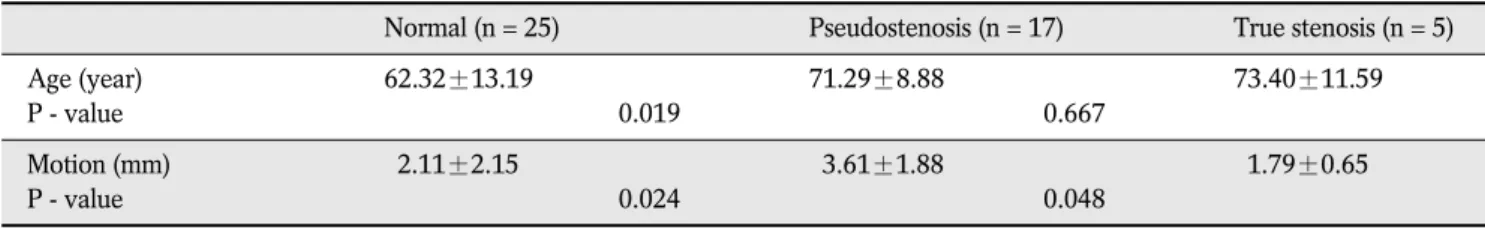
Normal (n = 25) Pseudostenosis (n = 17) True stenosis (n = 5)
Age (year) 62.32±13.19 71.29±8.88 73.40±11.59
P - value 0.019 0.667
Motion (mm) 2.11±2.15 3.61±1.88 1.79±0.65
P - value 0.024 0.048
were all interpretable on CTA and CE-MRA. Percent stenosis on CTA, resulted in 85 cases of normal to stenosis less than 50%, six cases of 50-99% stenosis, and three cases of occlusion by NASCET criteria.
Precent stenosis on CE-MRA, resulted in 62 cases of normal to stenosis less than 50%, 29 cases of 50-99%
stenosis, and three cases of occlusion by NASCET criteria (Table 1). The accuracy of CE-MRA was 74.73% for 50-99% stenosis, 100% for occlusion (overall 75.55%).
There were 17 patients in the pseudostenosis group (2 in right; 8 in left; and 7 in both vertebral arteries).
25 patients were classified to the control group who have no true stenosis and no pseudostenosis.
Remaining 5 patients were classified to true stenosis group who show true stenosis. 23 vertebral arteries shows pseudostenosis (Fig. 2, Table 1). The age of pseudostenosis group was higher than that of control group, but not significantly different than true stenosis group. The motion of aortic arch of pseudostenosis group was higher than that of control group and that of true stenosis group (Table 2). There were no statisti- cal difference in diameter of normal distal vertebral artery, sex, and intravenous route of contrast media
a b c
d e f
Fig. 1. a-f. serial images of dynamic 3D time-resolved contrast-enhanced MRA.
c. Lowest position of upper border of aortic arch are marked by red horizontal line.
d. Highest position of upper border of aortic arch. Distance between these two horizontal lines was considered as motion of aortic arch
between pseudostenosis group and control group.
CTA has been known to be highly accurate in determining the pathology of vertebral arteries (17- 19). CTA also has better accessibility and short scanning time than MRA. However, MRA has the advantage of radiation avoidance and good soft tissue contrast, but has a tendency in overestimation of arterial stenosis (10, 13, 18). When consideration motion artifacts, CTA is less affected by the motion of cardiac pulsation and respiration because of innate excellent temporal resolution. Recent multi-detector row CT have shorter gantry rotation time (less than 500 msec) and advanced reconstruction algorithm, being able to overcome these motion induced artifacts (21, 22). On the other hand, CE-MRA has frequently shown pseudostenosis of vertebral artery origin because of weak spatial resolution, intravoxel dephas- ing, and motion artifact caused by cardiac pulsation and respiration.
Among suggested reasons of pseudostenosis, spatial resolution and intravoxel dephasing are MR system-
dependent factors. Large voxel size relative to the diameter of vertebral artery resulted intravoxel dephasing. Partial volume effect can overestimate severe stenosis. Future advanced MR angiographic sequence is needed to minimize these MR system- dependent factors. However, motion artifact is a patient-dependent factor and inevitable. In our study, older age and more motion of aortic arch were associ- ated with pseudostenosis. Other factors such as diameter of normal distal vertebral artery, sex, and intravenous route of contrast media were not associ- ated with pseudostenosis. The age of pseudostenosis group was higher than that of control group (71.29 ± 8.88 versus 62.32 ± 13.19 year-old; mean±standard deviation; p = 0.019). Older patients are known to may have tortuous aortic arch and great vessels (23).
In this situation, delayed passage of contrast media along with the tortuous vascular anatomy may induce high concentration of gadolinium and cause signal loss from T2* decay. However, the age of pseudostenosis group was not significantly different than that of true stenosis group. We think that this is probably due to higher incidence of vertebral artery stenosis in older age. Motion from cardiac pulsation and respiration can affect motion of aortic arch, thus the image quality of DISCUSSION
a b
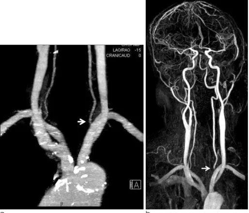
Fig. 2. a. CTA shows no stenosis of the origin of the left vertebral artery.
b. MRA of same patient after 5 days shows pseudostenosis of the origin of the left vertebral artery (arrow).
origin of vertebral artery. Although cardiac pulsation is unavoidable, breath holding can be achieved during short scan time. CE-MRA with breath holding technique demonstrated better image quality of the aortic arch and the origin of the great vessels (24).
However, we did not use breath holding technique because we focused on higher resolution with longer scan time to increase spatial resolution.
Our study protocol of TR-CEMRA was set with a low dose of gadolinium based contrast media (2 mL of Gadovist) and showed acceptable image quality (16).
There was no patient with glomerular filtration rate (GFR) of 30 mL/min or under in this study. Recent results of 3T MRA for supraaortic arteries showed acceptable imaging quality with small amounts of contrast media as low as 0.047 mmol/kg (14).
Therefore, optimization of MRA protocol with adequate usage of gadolinium based contrast media can minimize nephrotoxicity and nephrogenic systemic fibrosis. Recent advanced in non-contrast MRA for supraaortic arteries can also prevent nephro- genic systemic fibrosis (25, 26).
Our study has a few limitations. There can be various factors associated with aortic motions: cardiac disease, hypertension, compliance, stiffness of aorta, and etc.
We did not analyze them but evaluated only motion of aortic arch as the sample size was too small to evaluate other factors causing aortic motion. Secondly, we measured the aortic motion by means of distance of z- axis between the highest and lowest position of the aortic arch on coronal MR images, but the aortic arch actually moves three dimensionally not only along z- axis but also x-, and y- axis and this was not reflected.
Thirdly, this study could not evaluate MR systemdepen- dent causes of pseudostenosis at the origin of vertebral artery. Because the study design was retrospective, we could not handle acquisition parameters such as repeti- tion time, echo time, voxel size, methods of contrast injection, and number of signal average. Further researchs involving newly developed MR angiographic sequences is needed. Finally, CTA has some limitations as a gold standard, as calcification, beam hardening artifact, and contrast in venous structure can affect interpretation of the images. To overcome this we used the source image and multiplanar reconstruction with proper window level and width when the origin of vertebral artery was not clear on MIP.
The origin of the vertebral artery is a frequent site of pseudostenosis. Older age and the motion of aortic arch are associated with pseudostenosis on CE-MRA.
References
1. Khan S, Rich P, Clifton A, Markus HS. Noninvasive detection of vertebral artery stenosis: a comparison of contrast-enhanced MR angiography, CT angiography, and ultrasound. Stroke 2009;40:3499-3503
2. Anzidei M, Napoli A, Marincola BC, et al. Gadofosveset- enhanced mr angiography of carotid arteries: does steady-state imaging improve accuracy of firstpass imaging? comparison with selective digital subtraction angiography. Radiology 2009;251:457-466
3. Wardlaw JM, Chappell FM, Best JJ, Wartolowska K, Berry E.
Non-invasive imaging compared with intra-arterial angiography in the diagnosis of symptomatic carotid stenosis: a meta- analysis. Lancet 2006;367:1503-1512
4. Yang CW, Carr JC, Futterer SF, et al. Contrast-enhanced MR angiography of the carotid and vertebrobasilar circulations.
AJNR Am J Neuroradiol 2005;26:2095-2101
5. Remonda L, Senn P, Barth A, Arnold M, Lovblad KO, Schroth G. Contrastenhanced 3d MR angiography of the carotid artery:
comparison with conventional digital subtraction angiography.
AJNR Am J Neuroradiol 2002;23:213-219
6. Kim SH, Lee JS, Kwon OK, Han MK, Kim JH. Prevalence study of proximal vertebral artery stenosis using high-resolution contrast-enhanced magnetic resonance angiography. Acta Radiologica 2005;46:314-321
7. Marquardt L, Kuker W, Chandratheva A, Geraghty O, Rothwell PM. Incidence and prognosis of > or = 50% symptomatic vertebral or basilar artery stenosis: prospective population- based study. Brain : a journal of neurology 2009;132:982-988 8. Compter A, van der Worp HB, Algra A, Kappelle LJ.
Prevalence and prognosis of asymptomatic vertebral artery origin stenosis in patients with clinically manifest arterial disease. Stroke 2011;42:2795-2800
9. Khan S, Cloud GC, Kerry S, Markus HS. Imaging of vertebral artery stenosis: a systematic review. J Neurol Neurosurg Psychiatry 2007;78:1218-1225
10. Choi HS, Kim DI, Kim DJ, Kim J, Kim ES, Lee SK. Accuracy of 3 t MR angioraphy in vertebral artery stenosis and coincidence with other cerebrovascular stenoses. Neuroradiology 2010;52:
893-898
11. Leclerc X, Martinat P, Godefroy O, et al. Contrast-enhanced three-dimensional fast imaging with steady-state precession (fisp) MR angiography of supraaortic vessels: preliminary results. AJNR Am J Neuroradiol 1998;19:1405-1413
12. Randoux B, Marro B, Koskas F, Chiras J, Dormont D, Marsault C. Proximal great vessels of aortic arch: comparison of three- dimensional gadolinium-enhanced MR angiography and digital subtraction angiography. Radiology 2003;229:697-702
13. Cosottini M, Calabrese R, Puglioli M, et al. Contrast-enhanced three-dimensional MR angiography of neck vessels: does dephasing effect alter diagnostic accuracy? Eur Radiol 2003;13:
CONCLUSION
571-581
14. Tomasian A, Salamon N, Lohan DG, Jalili M, Villablanca JP, Finn JP. Supraaortic arteries: contrast material dose reduction at 3.0-t high-spatial-resolution MR angiography--feasibility study.
Radiology 2008;249:980-990
15. Lohan DG, Tomasian A, Saleh RS, Singhal A, Krishnam MS, Finn JP. Ultra-low-dose, time-resolved contrast-enhanced magnetic resonance angiography of the carotid arteries at 3.0 tesla. Invest Radiol 2009;44:207-217
16. Lee YJ, Laub G, Jung SL, et al. Low-dose 3d time-resolved magnetic resonance angiography (MRA) of the supraaortic arteries: correlation with high spatial resolution 3d contrast- enhanced MRA. J Magn Reson Imaging 2011;33:71-76
17. Randoux B, Marro B, Koskas F, et al. Carotid artery stenosis:
prospective comparison of CT, three-dimensional gadolinium- enhanced MR, and conventional angiography. Radiology 2001;220:179-185
18. Vertinsky AT, Schwartz NE, Fischbein NJ, Rosenberg J, Albers GW, Zaharchuk G. Comparison of multidetector CT angiogra- phy and MR imaging of cervical artery dissection. AJNR Am J Neuroradiol 2008;29:1753-1760
19. Forsting M. Cta of the ica bifurcation and intracranial vessels.
Eur Radiol 2005;15 Suppl 4:D25-27
20. Beneficial effect of carotid endarterectomy in symptomatic
patients with highgrade carotid stenosis. North american symptomatic carotid endarterectomy trial collaborators. The New England Journal of Medicine 1991;325:445-453
21. Roos JE, Willmann JK, Weishaupt D, Lachat M, Marincek B, Hilfiker PR. Thoracic aorta: motion artifact reduction with retrospective and prospective electrocardiography-assisted multi-detector row CT. Radiology 2002;222:271-277
22. Morgan-Hughes GJ, Owens PE, Marshall AJ, Roobottom CA.
Thoracic aorta at multi-detector row CT: motion artifact with various reconstruction windows. Radiology 2003;228:583-588 23. Lin SC, Trocciola SM, Rhee J, et al. Analysis of anatomic factors
and age in patients undergoing carotid angioplasty and stenting.
Annals of Vascular Surgery 2005;19:798-804
24. Carr JC, Ma J, Desphande V, Pereles S, Laub G, Finn JP. High- resolution breath-hold contrast-enhanced MR angiography of the entire carotid circulation. AJR Am J Roentgenol 2002;178:
543-549
25. Takei N, Miyoshi M, Kabasawa H. Noncontrast mr angiography for supraaortic arteries using inflow enhanced inversion recovery fast spin echo imaging. J Magn Reson Imaging 2012;
35:957-962
26. Lummel N, Boeckh-Behrens T, Lutz J, Burke M, Linn J.
Evaluation of the supraaortic arteries using non-contrast-enhanced velocity MR angiography “inhance”. Neuroradiology 2012
통신저자 : 최현석, (137-701) 서울시 서초구 반포대로 222, 가톨릭대학교 의과대학 서울성모병원 영상의학과 Tel. 010-2517-0806 Fax. (02) 599-6771 E-mail: [email protected]
조영증강 자기공명 혈관 조영술에서 척추 동맥 기시부의 거짓 협착:
역동적 3D Time-resolved 조영증강 자기공명 혈관 조영술에서 대동맥궁의 움직임과의 연관성
가톨릭대학교 의과대학 서울성모병원 영상의학과 김선문∙이승원∙최현석∙정소령∙안국진∙김범수
목적: 척추동맥 기시부는 조영증강 자기공명 혈관조영술(MRA)에서 거짓 협착이 잘 일어나는 부위이다. 이 연구의 목표는 척추 동맥 기시부의 거짓 협착과 대동맥궁의 움직임과의 연관성에 대해 평가하는 것이다.
대상과 방법: 47명의 환자가 CT혈관 조영술(CTA)과 MRA, 그리고 3D time-resolved MR 혈관 조영술(TR- MRA)을 6.87±9.89 일 (mean±SD) 이내에 시행하였다. 협착의 정도는 NASCET 분류에 따라 CTA와 MRA에서 평가 하였다. CTA를 비교 기준으로 하여 MRA에서의 척추 동맥 기시부의 협착 정도에 따라 거짓 협착군과 대조군으 로 분류하였다. 거짓 협착군은 MRA에서 50-99%의 협착이 있으나 CTA에서 50% 미만의 협착이 있는 환자로 정의 하였다. 대동맥궁의 움직임은 TR-MRA에서 동맥궁이 가장 높을 때와 가장 낮을 때의 거리로 측정하였다. 연령, 조영 제의 주입 경로, 대동맥궁의 움직임 그리고 척추 동맹의 원위부 정상 직경을 비교 분석하였다.
결과: 17명의 환자에서, 그리고 23개의 척추 동맥에서 거짓 협착이 있었다. 거짓협착을 보이는 환자에서, 더 큰 대동 맥궁의 움직임(3.61 ± 1.88 vs. 2.05 ± 1.97 mm)을 보였고 더 고령이었다(71.29 ± 8.88 vs. 62.32 ± 13.19 year-old). 조영제의 주입 경로나 척추 동맥의 원위부 정상 직경은 거짓 협착과는 연관성이 없었다.
결론: 척추동맥의 기시부는 거짓 협착이 자주 일어나는 곳이다. 연령이 높을수록 그리고 대동맥궁의 움직임의 클수록 MRA에서 거짓 협착이 일어날 가능성이 높다.
대한자기공명의과학회지 16:236-242(2012)