Received on September 19, 2011. Revised on September 29, 2011. Accepted on October 4, 2011.
CC This is an open access article distributed under the terms of the Creative Commons Attribution Non-Commercial License (http://creativecommons.org/licenses/by-nc/3.0) which permits unrestricted non-commercial use, distribu- tion, and reproduction in any medium, provided the original work is properly cited.
*Corresponding Author. Tel: 82-43-261-2826; Fax: 82-43-268-2732; E-mail: [email protected] Keywords: Nanoliposome, Poly(aspartic acid), L-Lysine, Dendritic cell, Immunomodulation.
Nanoliposomes of L-lysine-conjugated poly(aspartic acid) Increase the Generation and Function of Bone Marrow- derived Dendritic Cells
Sun-A Im1, Ki-Hyang Kim1, Hong-Geun Ji2, Hyoung-Gyoung Yu2, Sun-Ki Park3 and Chong-Kil Lee1*
1College of Pharmacy, Chungbuk National University, Cheongju 361-763, 2H&A PharmaChem, R&D Center, Bucheon 421-808,
3Cosmeca, R&D Center, Eumseung 869-821, Korea
Background: Biodegradable polymers have increasingly been recognized for various biological applications in recent years.
Here we examined the immunostimulatory activities of the novel poly(aspartic acid) conjugated with L-lysine (PLA).
Methods: PLA was synthesized by conjugating L-lysine to as- partic acid polymer. PLA-nanoliposomes (PLA-NLs) were prepared from PLA using a microfluidizer. The immunostim- ulatory activities of PLA-NLs were examined in mouse bone marrow-derived dendritic cells (BM-DCs). Results: PLA-NLs increased the number of BM-DCs when added to cultures of GM-CSF-induced DC generation on day 4 after the initiation of cultures. Examination of the phenotypic properties show- ed that BM-DCs generated in the presence of PLA-NLs are more mature in terms of the expression of MHC class II mol- ecules and major co-stimulatory molecules than BM-DCs generated in the absence of PLA-NLs. In addition, the BM-DCs exhibited enhanced capability to produce cyto- kines, such as IL-6, IL-12, TNF-α and IL-1β. Allogeneic mixed lymphocyte reactions also confirmed that the BM- DCs were more stimulatory on allogeneic T cells. PLA- NL al- so induced further growth of immature BM-DCs that were harvested on day 8. Conclusion: These results show that PLA-NLs induce the generation and functional activities of BM-DCs, and suggest that PLA-NLs could be immunostim- ulating agents that target DCs.
[Immune Network 2011; 11(5):281-287]
INTRODUCTION
The importance of polymers with biodegradability and bio- compatibility has increasingly been recognized for various bi- ological applications. Biodegradable polymers based on both natural and synthetic polymers are widely used as viscosity agents, emulsifying agents, and carriers for delivery of drugs or active ingredients in medical, cosmetic, and other industrial fields (1,2).
Poly-glutamic acid (PGA), for instance, is an anionic poly- mer that is composed of D- and L-glutamic acid units (3,4).
PGA is produced by Bacillus subtilis during fermentation of soybeans. Recent reports have demonstrated immune modu- latory activities of PGA or its derivatives (5-7). Nanoparticles composed of PGA and L-phenylalanine ethylester served as an adjuvant that was able to activate dendritic cells and en- hance adaptive immune responses against antigens carried by the nanoparticles (8,9).
Poly(aspartic acid), which is one of the poly(amino acid)s and biodegradable synthetic polymers, is a water-soluble pol- yamide containing carboxylic pendants that can be obtained from the hydrolysis of polysuccinimide (10,11), the thermal polycondensation product of L-aspartic acid (12,13). Due to their amphiphilic uniqueness, amino acid polymers contain- ing both hydrophilic and hydrophobic components can be used to stabilize dispersions, emulsions, and polymer blends (14,15).
Table I. Recipes used to prepare PLA-NLs
PLA-NL-1 PLA-NL-2
A) Lecithin (%) 3.00 -
Phosphatidyl
serine (%) - 6.00
Lysolecithin (%) 1.00 -
Caprylic/Capric
Triglyceride (%) - 10.00
Sucrose palmitate (%) 3.00 3.00
Ethanol (%) 5.00 5.00
H2O Make to 100 ml Make to 100 ml
B) EDTA-2Na (%) 0.01 0.01
Sodium ascorbyl
phosphate (%) 0.30 0.30
B-1) Ethanol (%) 10.00 10.00
PLA (%) 10.00 10.00
(16-18). L-lysine is also important to the collagen structure in connective tissue in skin, ligaments, cartilage, vertebral discs, joint linings, capillary walls, bones and teeth (16). L-ly- sine components conjugated to the polymer backbone have specific biological functions in vivo and a unique pH-sensitive property (19,20). Poly (L-lysine) has been widely used as a gene delivery carrier due to its strong ability to protect DNA from nuclease attack (21).
Liposomes, spherical nanoparticles with an aqueous com- partment, possess the unique ability to effectively deliver both water-soluble compounds (in the aqueous phase) and lip- id-soluble compounds (in the lipid bilayer) (22,23). More- over, liposomes rapidly penetrate the dermal barrier, making the delivery of water-soluble compounds as effective as that of lipid-soluble compounds (24). In contrast with other drug carriers such as DMSO (25), liposomes are entirely non-toxic to human skin since they are composed of naturally occurring biodegradable compartments (26).
In this work, novel liposomes composed of biodegradable polymers based on poly(aspartic acid) conjugated with L-ly- sine (PLA-NLs) were prepared from PLA using a micro- fluidizer. The immunomodulatory activities of PLA-NLs on bone marrow derived-dendritic cells (BM-DCs) were then investigated. The present study showed that PLA-NLs induce the generation and functional activities of BM-DCs.
MATERIALS AND METHODS
Preparation of PLA-nanoliposomes (PLA-NLs) Polymers of L-aspartic acid and L-lysine-conjugated poly (aspartic acid), PLA, were prepared as described previou- sly (2,27). The powdery product was filtered out and dried under vacuum at 40oC for three days to obtain PLA as a yellowish white powder (yield 81%).
PLA-NLs were prepared from PLA as described in Table I.
The phases described in Table I, A, B and B-1, were dis- solved at 70oC, respectively. The dissolved A phase was add- ed to the mixed phase of B and B-1 and then this mixed solution was passed through a microfluidizer (M110F, Micro- fluidic, USA) three times at 1000 bar to prepare PLA-NLs.
The average size of PLA-NL-1 and PLA-NL-2 was 70 nm and 200 nm, respectively, when measured using a particle size analyzer (ELS-Z, Photal, Japan).
Cell culture
Cells were cultured in Dulbecco's modified Eagle's medium (Hyclone Laboratories, Inc., UT, USA) supplemented with 10% heat-inactivated fetal bovine serum (Hyclone), 100 U/ml penicillin and 100μg/ml streptomycin (Hyclone), and 50μM 2-mercaptoethanol (SIGMA-ALDRICH, Inc., St. Louis, MO, USA) at 37oC in a 5% CO2 atmosphere.
Generation of bone marrow derived dendritic cells (BM-DCs)
BM-DCs were generated from total bone marrow cells as de- scribed previously (28). Briefly, bone marrow cells obtained from femurs of C57BL/6 mice were cultured in 6-well plates (5×10⁶/well) in culture medium supplemented with 40 ng/ml GM-CSF (CreaGene, Seongnam, Korea). At days 3 and 4 after the initiation of the culture, nonadherent cells were discarded by gentle shaking and replacing the culture medium with fresh medium containing GM-CSF. BM-DCs were harvested by gentle pipetting on day 8. In experiments examining the effects of PLA-NLs, PLA-NLs were added to the culture on day 4 after the initiation of the culture, and the resultant BM-DCs were harvested on day 8 and used for phenotypic analysis and functional assays.
Proliferation assay
BM-DCs harvested on day 8 were cultured in the presence
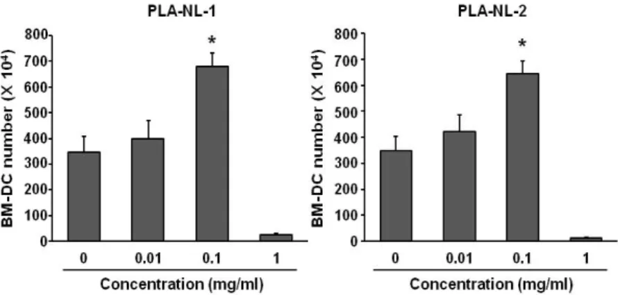
Figure 1. PLA-NLs increase the generation of BM-DCs. Mouse BM-DCs were generated from C57BL/6 mouse BM cells. BM cells were cultured with the indicated concentration of PLA-NLs and GM-CSF (40 ng/ml). PLA-NLs were added to the culture on day 4 after the initiation of the BM cell culture, and the resultant BM-DCs were harvested on day 8 and counted using a hemocytometer. Results are mean±SD. *p<0.05 compared with untreated control.
of different concentrations of PLA-NLs in 96-well microtiter plates (2×10⁴ cells/well). DNA synthesis was measured by [3H]-thymidine (PerkinElmer) incorporation (0.5μCi/well) for the final 16 h of the 2-day culture period. The cells were har- vested onto glass fiber filter paper using a cell harvester. The filters were washed, dried, and then counted in a microbeta liquid scintillation counter (Wallac, USA).
Allogeneic mixed lymphocyte reaction (MLR) BM-DCs were treated with mitomycin C (SIGMA-ALDRICH) for 20 min at 37oC, washed three times with culture medium, and then adjusted to 5×10⁴ cells/ml. Responder T cells were isolated from spleens of BALB/c mice by a nylon wool-en- richment technique and adjusted to 5×10⁵ cells/ml. Each 100 μl of cell suspension was mixed and cultured in 96-well mi- crotiter plates. DNA synthesis was measured by [3H]- thymi- dine (PerkinElmer) incorporation (0.5μCi/well) for the final 16 h of the 3-day culture period. The cells were harvested onto glass fiber filter paper using a cell harvester. The filters were washed, dried, and then counted in a microbeta liquid scintillation counter (Wallac, USA).
Cytokine production
BM-DCs were cultured in the presence of different con- centrations of PLA-NLs with OVA-containing nanoparti- cles (50μg/ml as OVA) in 12-well microtiter plates (1×
106 cells/well/2 ml). Nanoparticles containing OVA were prepared using a homogenization/solvent evaporation meth- od using poly(lactic-co-glycolic acid), as described previously (29). After 24-h incubation, the culture supernatants were col- lected, and the amounts of IL-12, IL-6, IL-1β and TNF-α were measured using commercial immunoassay kits (BD Bio- sciences, San Diego, CA, USA) according to the manufac-
turer’s instructions.
Flow cytometry
Cells were stained with monoclonal antibodies recognizing murine cell surface markers as described previously (30), and flow cytometric analysis was performed on a FACSCanto II (BD Biosciences). The monoclonal antibodies, anti-ICAM-1 (clone 3E2), anti-I-Ab (clone AF6-120.1), anti-B7-1 (clone 16-10A1), anti-B7-2 (clone GL1), and isotype-matched control antibodies were purchased from BD Biosciences. Dead cells were gated out by their low forward angle light scatter in- tensity. In most analyses, 10,000 cells were scored.
Statistical analysis
Data were expressed as mean±SD. The statistical significance of the difference between the control group and treatment group was assessed by a one-way ANOVA followed by a Student's t-test.
RESULTS
PLA-NLs increase the generation of BM-DCs from BM cells
To examine the effects of PLA-NLs on the growth and matura- tion of BM-DCs, PLA-NLs were added to the cultures of BM cells on day 4 after the initiation of the culture, and the re- sultant BM-DCs were harvested on day 8. As shown in Fig.
1A, addition of PLA-NL-1 (0.1 mg/ml) to cultures of BM cells increased the number of BM-DCs that were harvested on day 8 by approximately 76% compared to the GM-CSF only control. PLA-NL-2 (0.1 mg/ml) also increased the number of BM-DCs by approximately 74% compared to the GM-CSF only control (Fig. 1). Addition of 1 mg/ml of PLA-NLs to BM cells
Figure 2. PLA-NLs induce phenotypic maturation of BM-DCs. PLA-NLs (0.1 mg/ml) were added to BM cell culture together with GM- CSF (40 ng/ml) on day 4 after the initiation of cultures, and the resultant BM-DCs were harvested on day 8, washed, and then used for immunophenotypic analysis. Levels of expression (thin line) in BM-DCs generated in the presence of GM-CSF and PLA-NLs are illustrated in comparison with those in BM-DCs generated in the presence of GM-CSF only (shaded line).
Figure 3. DCs generated in the presence of PLA-NLs exert increased capability to produce cytokines.
PLA-NLs (0.1 mg/ml) were added to BM cell cultures together with GM-CSF (40 ng/ml) on day 4 after the initiation of cultures, and the resultant BM-DCs were harvested on day 8 and then cultured with OVA-conta- ining nanoparticles (50μg/ml as OVA). After 24 h, cyto- kines were measured by ELISA. Results are mean±SD.
*p<0.05, **p<0.01 compared with untreated control.
Because increased expression of cell surface molecules could be a marker of dendritic cell activation, we examined the ef- fect of PLA-NLs on the expression levels of MHC class II and co-stimulatory molecules in BM-DCs that were harvested on day 8. As shown in Fig. 2, BM-DCs that were generated in medium containing GM-CSF without PLA-NLs stimulation ex- hibited characteristics of immature DCs based on staining with I-Ab, B7-1, B7-2 and ICAM-1 molecules. In contrast, the BM-DCs generated in the presence of PLA-NLs exhibited more mature phenotypes. The expression levels of I-Ab, B7-1, B7-2 and ICAM-1 molecules were significantly increased in
DCs generated in the presence of PLA-NLs exhibit an increased capability to produce cytokines
Because PLA-NLs appeared to induce differentiation of BM-DCs, the cytokine producing capability of BM-DCs that were generated in the presence of PLA-NLs was compared with that of control BM-DCs. BM-DCs that were stimulated with OVA-containing poly(lactic-co-glycolic acid) nano- particles (50μg/ml as OVA) for 24 h, culture supernatants were collected from the stimulated BM-DCs, and the amounts of IL-6, IL12, IL-1β and TNF-α were measured by ELISAs.
Figure 5. PLA-NLs induce the proliferation of GM-CSF-generated BM-DCs. The DCs generated in the presence of 40 ng/ml of GM-CSF only were harvested on day 8, and then cultured with the indicated concentration of PLA-NLs. After 2 days, the growth of BM-DCs was measured by [3H]-thymidine incorporation. Results are mean±SD. *p
<0.05, **p<0.01 compared with untreated control.
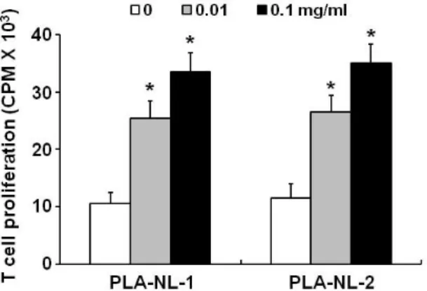
Figure 4. DCs generated in the presence of PLA-NLs exert increased allostimulatory capacity. PLA-NLs (0.1 mg/ml) were added to BM cell cultures together with GM-CSF (40 ng/ml) on day 4 after the initiation of cultures, and the resultant BM-DCs were harvested on day 8. All BM-DCs were treated with mitomycin C, washed, and then cultured with allogeneic mouse lymphocytes. After 3 days, cell growth was measured by [3H]-thymidine incorporation. Results are mean±SD. *p
<0.01 compared with untreated control.
As shown in Fig. 3, the BM-DCs that were generated in the presence of PLA-NLs produced significantly enhanced levels of all the cytokines that were examined.
It has previously been shown that DCs cultured in the pres- ence of OVA-containing poly(lactic-co-glycolic acid) nano- particles produce large amounts of cytokines (31). The pres- ent study shows that BM-DCs generated in the presence of PLA-NLs produced even further increased levels of cytokines when stimulated with OVA-containing poly(lactic-co-glycolic acid) nanoparticles.
DCs generated in the presence of PLA-NLs exhibit increased allostimulatory capacity
The allostimulatory capacity of BM-DCs generated in the pres- ence of PLA-NLs was examined in allogeneic mixed lympho- cyte reactions. As shown in Fig. 4, control BM-DCs that were generated in medium containing GM-CSF only induced low to moderate proliferation of allogeneic T cells. In contrast, BM-DCs that were generated in the presence PLA-NLs ex- hibited significantly enhanced allostimulatory capacity. The proliferation of allogeneic T cells was increased by approx- imately 220% when BM-DCs generated in the presence of PLA-NLs were used as stimulatory cells compared to BM-DCs generated in GM-CSF only. Since all the BM-DCs were treated with mitomycin C and subsequently washed thoroughly to re- move mitomycin C, the T cell proliferation must be a re- flection of increased BM-DC function.
PLA-NLs induce further proliferation of BM-DCs To examine the effects of PLA-NLs on the further growth of immature BM-DCs, immature BM-DCs were harvested on day 8 and then cultured for 2 days in the presence or absence of PLA-NLs. During this 2-day culture period, GM-CSF was not added to the culture medium. As shown in Fig. 5, BM-DCs that were harvested on day 8 did not proliferate ac- tively when cultured in GM-CSF-free medium. In contrast, BM-DCs that were harvested on day 8 proliferated sig- nificantly when cultured in the presence of PLA-NLs (1 mg/ml).
DISCUSSION
The present study shows that PLA-NLs exert significant im- munostimulating activity on DCs. It has been well-established that DCs can be generated from bone marrow stem cells by culturing in the presence of a high dose of GM-CSF (32). To examine the effects of PLA-NLs on the generation of BM-DCs from BM cells, PLA-NLs were added to BM cells cultures to- gether with GM-CSF on day 4 after the initiation of cultures.
BM-DCs were harvested on day 8, and the numbers, pheno- type and functional characteristics of the cells were exam- ined.
The present study shows that PLA-NLs when added to BM cell cultures together with GM-CSF induce phenotypic and functional maturation of the BM-DCs. In addition, PLA-NLs in-
tiation of culture, but also when PLA-NLs were added to BM-DCs that were harvested on day 8. When added to cul- tures of immature DCs, PLA-NLs induced phenotypic matura- tion as evidenced by up-regulation of class II MHC molecules and co-stimulatory molecules such as I-Ab, B7-1, B7-2 and ICAM-1.
The DCs cultured with PLA-NLs (0.1 mg/ml) exhibited more potent allogeneic T cell stimulatory activity and induced the proliferation of GM-CSF-generated BM-DCs. DCs can rec- ognize and process antigens in the periphery and then mi- grate to secondary lymphoid organ where they prime primary T cell responses (33). The capability of DCs to activate naive T cells in a primary response has been explained by their abil- ity to express high levels of class II MHC and co-stimulatory molecules (34). The immature DCs must be matured or further activated in order to fully perform accessory cell functions (35). Maturation signals appear to be very diverse and include inflammatory cytokines such as TNF-α and IL-1β (36). It is noteworthy that 1 mg/ml PLA-NLs appeared to be toxic to the cells when they were added to very early stage of DC generation (day 4 from the initiation of the culture) (Fig. 1).
However, PLA-NLs did not exert cytotoxic activity on im- mature BM-DCs that were harvested on day 8 from the ini- tiation of the culture (Fig. 5). At 1 mg/ml concentration, PLA-NLs even increased the proliferation of day-8 immature DCs. The underlying mechanism for the differential effects is not clear at present.
These results show that PLA-NLs induce the generation and functional activities of BM-DCs. Both types of PLA-NLs, PLA-NL-1 and PLA-NL-2, exerted similar stimulatory activity on BM-DCs.
ACKNOWLEDGEMENTS
This research was financially supported by the Ministry of Knowledge Economy (MKE) and Korea Institute for Advance- ment of Technology (KIAT) through the Research and Devel- opment for Regional Industry.
CONFLICTS OF INTEREST
The author have no financial conflict of interest.
1. Min SK, Kim SH, Kim JH: Preparation and swelling behav- ior of biodegradable poly(aspartic acid)-based hydrogel. J Ind Eng Chem 6;276-279, 2000.
2. Goddard JM, Hotchkiss JH: Polymer surface modification for the attachment of bioactive compounds. Progress in Polymer Science 32;698-725, 2007.
3. Champion JA, Walker A, Mitragotri S: Role of particle size in phagocytosis of polymeric microspheres. Pharm Res 25;
1815-1821, 2008.
4. Elamanchili P, Diwan M, Cao M, Samuel J: Characterization of poly(D,L-lactic-co-glycolic acid) based nanoparticulate system for enhanced delivery of antigens to dendritic cells.
Vaccine 22;2406-2412, 2004.
5. Akagi T, Shima F, Akashi M: Intracellular degradation and distribution of protein-encapsulated amphiphilic poly(ami- no acid) nanoparticles. Biomaterials 32;4959-4967, 2011.
6. Foged C, Sundblad A, Hovgaard L: Targeting vaccines to dendritic cells. Pharm Res 19;229-238, 2002.
7. Panyam J, Labhasetwar V: Biodegradable nanoparticles for drug and gene delivery to cells and tissue. Adv Drug Deliv Rev 55;329-347, 2003.
8. Matsusaki M, Hiwatari K, Higashi M, Kaneko T, Akashi M:
Stably-dispersed and surface-functional bionanoparticles prepared by self-assembling amphipaethic polymers of hy- drophilic poly(g-glutamic acid) bearing hydrophobic amino acids. Chemistry Letters 33;398-399, 2004.
9. Akagi T, Wang X, Uto T, Baba M, Akashi M: Protein direct delivery to dendritic cells using nanoparticles based on am- phiphilic poly(amino acid) derivatives. Biomaterials 28;
3427-3436, 2007.
10. Giammona G, Pitarresi G, Tomarchio V, Dispenza C, Spadaro G: Synthesis and characterization of water-swel- lable α,β-polyasparthydrazide derivatives. II. Hydrogels at low crosslinking degree as potential systems for anti- cancer drug release. Colloid and Polymer Science 273;
559-564, 1995.
11. Nakato T, Yoshitake M, Matsubara K, Tomida M, Kakuchi T: Relationships between structure and properties of poly(aspartic acid)s. Macromolecules 31;2107-2113, 1998.
12. Nakato1 T, Kusuno A, Kakuchi T: Synthesis of poly(succini- mide) by bulk polycondensation of L-aspartic acid with an acid catalyst. Journal of Polymer Science Part A: Polymer Chemistry 38;117-122, 2000.
13. Matsubara K, Nakato T, Tomida M: 1H and 13C NMR char- acterization of poly(succinimide) prepared by thermal poly- condensation of l-aspartic acid. Macromolecules 30;2305- 2312, 1997.
14. Horgan A, Saunders B, Vincent B, Heenan RK: Poly(butyl methacrylate-g-methoxypoly(ethylene glycol)) and poly (methyl methacrylate-g-methoxypoly(ethylene glycol)) graft copolymers: preparation and aqueous solution properties.
J Colloid Interface Sci 262;548-559, 2003.
15. Zhu G: Micellization of polystyrene-graft-poly(ethylene ox- ide) and its mixtures with polystyrene homopolymer in ethanol. European Polymer Journal 41;2671-2677, 2005.
16. Li P, Yin YL, Li D, Kim SW, Wu G: Amino acids and im- mune function. Br J Nutr 98;237-252, 2007.
17. Kidd MT, Kerr BJ, Anthony NB: Dietary interactions be- tween lysine and threonine in broilers. Poult Sci 76;608- 614, 1997.
18. Konashi S, Takahashi K, Akiba Y: Effects of dietary essen- tial amino acid deficiencies on immunological variables in broiler chickens. Br J Nutr 83;449-456, 2000.
19. Yang SR, Lee HJ, Kim JD: Histidine-conjugated poly(amino acid) derivatives for the novel endosomolytic delivery car- rier of doxorubicin. J Control Release 114;60-68, 2006.
20. Xu Q, An L, Yu M, Wang S: Design and synthesis of a new conjugated polyelectrolyte as a reversible ph sensor. Ma- cromolecular Rapid Communications 29;390-395, 2008.
21. Jeong JH, Park TG: Poly(L-lysine)-g-poly(D,L-lactic-co-gly- colic acid) micelles for low cytotoxic biodegradable gene delivery carriers. J Control Release 82;159-166, 2002.
22. Stone WL, Mukherjee S, Smith M, Das SK: Therapeutic uses of antioxidant liposomes. Methods Mol Biol 199;145-161, 2002.
23. Stone WL, Smith M: Therapeutic uses of antioxidant lipo- somes. Mol Biotechnol 27;217-230, 2004.
24. Chen CH, Liu DZ, Fang HW, Liang HJ, Yang TS, Lin SY:
Evaluation of multi-target and single-target liposomal drugs for the treatment of gastric cancer. Biosci Biotechnol Bio- chem 72;1586-1594, 2008.
25. Koike M, Ishino K, Kohno Y, Tachikawa T, Kartasova T, Kuroki T, Huh N: DMSO induces apoptosis in SV40-trans- formed human keratinocytes, but not in normal kera- tinocytes. Cancer Lett 108;185-193, 1996.
26. Paromov V, Kumari S, Brannon M, Kanaparthy NS, Yang H, Smith MG, Stone WL: Protective effect of liposome- en- capsulated glutathione in a human epidermal model ex- posed to a mustard gas analog. J Toxicol 2011;109516, 2011.
27. Kim SI, Min SK, Kim JH: Synthesis and characterization of novel amino acid-conjugated poly(aspartic acid) deriva- tives. Bull Korean Chem Soc 29;1887-1892, 2008
28. Lee JK, Lee MK, Yun YP, Kim Y, Kim JS, Kim YS, Kim K, Han SS, Lee CK: Acemannan purified from Aloe vera induces phenotypic and functional maturation of immature dendritic cells. Int Immunopharmacol 1;1275-1284, 2001.
29. Lee YR, Lee YH, Im SA, Kim K, Lee CK: Formulation and characterization of antigen-loaded plga nanoparticles for ef- ficient cross-priming of the antigen. Immune Netw 11;
163-168, 2011.
30. Im SA, Lee YR, Lee YH, Oh ST, Gerelchuluun T, Kim BH, Kim Y, Yun YP, Song S, Lee CK: Synergistic activation of monocytes by polysaccharides isolated from Salicornia her- bacea and interferon-gamma. J Ethnopharmacol 111;365- 370, 2007.
31. Lee YH, Lee YR, Kim KH, Im SA, Song S, Lee MK, Kim Y, Hong JT, Kim K, Lee CK: Baccatin III, a synthetic pre- cursor of taxol, enhances MHC-restricted antigen pre- sentation in dendritic cells. Int Immunopharmacol 11;
985-991, 2011.
32. Talmor M, Mirza A, Turley S, Mellman I, Hoffman LA, Steinman RM: Generation or large numbers of immature and mature dendritic cells from rat bone marrow cultures.
Eur J Immunol 28;811-817, 1998.
33. Mora JR: Homing imprinting and immunomodulation in the gut: role of dendritic cells and retinoids. Inflamm Bowel Dis 14;275-289, 2008.
34. Cai Z, Brunmark AB, Luxembourg AT, Garcia KC, Degano M, Teyton L, Wilson I, Peterson PA, Sprent J, Jackson MR:
Probing the activation requirements for naive CD8+ T cells with Drosophila cell transfectants as antigen presenting cells. Immunol Rev 165;249-265, 1998.
35. Drutman SB, Trombetta ES: Dendritic cells continue to cap- ture and present antigens after maturation in vivo. J Immunol 185;2140-2146, 2010.
36. Karlsen M, Hovden AO, Vogelsang P, Tysnes BB, Appel S: Bromelain treatment leads to maturation of mono- cyte-derived dendritic cells but cannot replace PGE2 in a cocktail of IL-1β, IL-6, TNF-α and PGE2. Scand J Immu- nol 74;135-143, 2011.