© 2011 The Korean Academy of Medical Sciences.
This is an Open Access article distributed under the terms of the Creative Commons Attribution Non-Commercial License (http://creativecommons.org/licenses/by-nc/3.0) which permits unrestricted non-commercial use, distribution, and reproduction in any medium, provided the original work is properly cited.
pISSN 1011-8934 eISSN 1598-6357
Refractory Hypertension and Isosexual Pseudoprecocious Puberty Associated with Renin-Secreting Ovarian Steroid Cell Tumor in a Girl
Steroid cell tumor, not otherwise specified (NOS), are rare ovarian tumor, in addition, it is more rare in children. The majority of these tumors produce several steroid hormones, particularly testosterone. Estrogen also secreted by steroid cell tumor, NOS, but it is uncommon. Furthermore, hypertension is an infrequent sign in steroid cell tumor, NOS.
An 8.5-yr-old girl with hypertension and frequent vaginal spotting visited at our clinic. On laboratory evaluation, secondary hypertension due to an elevated plasma renin level and isosexual pseudoprecocious puberty was diagnosed. Right solid ovarian mass was detected in radiologic tests. She underwent a right ooporectomy and it revealed renin and
progesterone receptor positive steroid cell tumor, NOS. After operation, her blood pressure returned to normal level and vaginal bleeding disappeared. Even though this case is very rare, when hypertension coincides with virilization or feminization, a renin-secreting ovarian steroid cell tumor, NOS, should be considered.
Key Words: Pseudo-Precocious Puberty; Hypertension; Ovarian Neoplasms; Renin- Secreting Tumor
Sun Hee Lee1, Mi Seon Kang2,
Gyeong Sin Lee3 and Woo Yeong Chung1 Departments of 1Pediatrics and 2Pathology, College of Medicine, Inje University, Busan; 3Department of Pathology, Dong-eui Medical Center, Busan, Korea Received: 16 December 2010
Accepted: 28 February 2011 Address for Correspondence:
Woo Yeong Chung, MD
Department of Pediatrics, College of Medicine, Inje University Paik Hospital, 75 Bokji-ro, Busanjin-gu, Busan 614-735, Korea Tel: +82.51-890-6280, Fax: +82.51-895-7785
E-mail: [email protected]
DOI: 10.3346/jkms.2011.26.6.836 • J Korean Med Sci 2011; 26: 836-838
CASE REPORT
Pediatrics
INTRODUCTION
Renin-secreting tumors are characterized by high circulating active renin and aldosterone levels, hypokalemia and refractory hypertension (1). The majority of renin-secreting tumors origi- nate in the kidney and extra-renal renin-secreting tumors are very rare (2). Ovarian renin-secreting tumors are uncommon, and only two pediatric cases have been reported (3, 4).
Steroid cell tumors are a group of ovarian tumors composed of cells that resemble steroid hormone-secreting cells; these cells are capable of secreting steroid hormones. These tumors account for only 0.1% of all ovarian neoplasms (5). Patients with steroid hormone-secreting tumors generally present with clinical syn- dromes caused by excessive hormone secretion such as Cush- ing’s syndrome, hypertension, virilization or feminization, or even pseudoprecocious puberty (6, 7).
Here we report an 8.5-yr-old girl with refractory hypertension, hypokalemia and isosexual pseudoprecocious puberty due to a renin-secreting ovarian steroid cell tumor, not otherwise speci- fied (NOS) that was confirmed by immunohistochemical stain- ing for renin and progesterone receptor in the ovarian tissue, and related laboratory findings.
CASE DESCRIPTION
An 8.5-yr-old girl was referred to an endocrine clinic because of
vaginal bleeding and uncontrolled hypertension on January 15, 2004. At 7.5-yr-old, the girl presented with severe headaches and nausea and was diagnosed with hypertension. She was pre- scribed antihypertensive drugs (Unipril 10 mg and Norvasc 10 mg). However, the hypertension remained uncontrolled. Con- currently, she showed breast development, and 3 months later, frequent vaginal spotting was observed. At 8-yr-old, pubic hair was observed. The girl had undergone surgery at 15 months of age due to bilateral grade III vesicourethral reflux but was sub- sequently found to be healthy. Her grandfather had been treat- ed with hypertension.
Upon presentation to our clinic, the patient’s height was 149 cm (> 95 percentile) and her weight was 42 kg (> 95 percentile).
Her arterial blood pressure was 140/90 mmHg. On physical ex- amination, the Tanner stages of breast and pubic hair were at stage III. Neurological examination was normal. Neither andro- genic nor glucocorticoid excess was detected.
Laboratory investigations revealed normal renal function and low serum potassium (2.7 mEq/L). Plasma renin (133.5 ng/mL/
hr; normal: < 4.4 ng/mL/hr), angiotensin II (5,230 pg/mL; nor- mal: 9-47 pg/mL) and aldosterone levels (824.1 pg/mL; normal:
< 130 pg/mL) were extremely elevated. The remaining serum electrolyte and complete blood cell count were normal, but pro- teinuria was detected in the urine analysis. Thyroid function was normal, and there was very mild prolactin elevation (33.69 ng/
mL; normal 0-29.5 ng/mL). There was no elevation in Luteiniz-
Lee SH, et al. • Renin-Secreting Ovarian Steroid Cell Tumor
http://jkms.org 837
DOI: 10.3346/jkms.2011.26.6.836
ing hormone (0.29 mIU/mL) or Follicle-stimulating hormone (< 0.1 mIU/mL); however, estradiol was elevated (290.38 pg/mL).
Prepubertal response was observed in a Gonadotropin-releas- ing hormone (GnRH) stimulation test, and beta HCG was in the normal range.
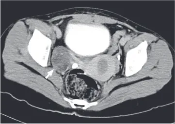
On radiologic tests, the patient’s bone age was advanced (12 yr old by the Greulich-Pyle method). Pelvic computed tomog- raphy confirmed the well-defined right solid ovarian tumor (5.1
× 4.0 cm, Fig. 1). Right oophorectomy was performed and anti- hypertensive treatment was tapered off over several days.
On gross examination, the right ovary measured 5.0 × 4.0 cm and the capsule was intact. The cut surface showed a bright yel- low solid tumor with lobulation. There was no hemorrhage or necrosis.
Histological examination revealed that the ovarian tumor had sheets of clear or eosinophilic cells surrounded with a delicate fibrous stroma. The tumor cells showed small, dark-stained sin- gle nuclei with occasional nucleoli. Cells undergoing mitosis were counted less than 2 per 10 high power fields. Immunohis- tochemistry revealed a diffuse positive reaction for renin, alpha- inhibin in tumor cells. Ovarian stromal cells were positive for progesterone receptor (Fig. 2).
In conjunction with the clinical history, these findings sup- ported a diagnosis of renin-secreting ovarian steroid cell tumor, NOS.
The patient was progressing well 16 months after surgery. Her blood pressure was normal (100/60 mmHg) without medication, and proteinuria had disappeared. Plasma renin, aldosterone and potassium levels had returned to normal, a normal puber- tal response was shown in a GnRH stimulation test, and regular menstruation was present.
Fig. 1. Pelvic computed tomography. A 5.1 × 4 cm-sized, well-defined solid mass with multiple calcifications and fat density is noted in right adnexa (arrow).
A B
C
Fig. 2. Histological appearance. (A) The individual tumor cells are polygonal. The cyto- plasm is clear or eosinophilic, granular and focally show thick eosinophilic cry stalloid materials (H&E staining, 400 × original magnifications). (B) Immunohisto chemical stains. The tumor cells exhibit a diffuse positive reaction for rennin (brown). (C) Immu- nohistochemical stains. Ovarian stromal cells are positive for progesterone receptor (brown to black).
Lee SH, et al. • Renin-Secreting Ovarian Steroid Cell Tumor
838 http://jkms.org DOI: 10.3346/jkms.2011.26.6.836
DISCUSSION
In euvolemic subjects, high levels of renin results in elevated al- dosterone levels, increased blood pressure and hypokalemia.
Therefore, spontaneous hypokalemia in hypertensive patients indicates an abnormality of the renin-angiotensin-aldosterone cascade. Most renin-secreting tumors are renal in origin, al- though rare cases of clear cell carcinoma (8), Wilm’s tumor (8) and mesoblastic nephroma (9) have been reported. Moreover, ovarian renin-secreting tumors are exceptional. The ovarian re- nin-angiotensin system plays a crucial role in reproductive func- tions such as folliculogenesis, oocyte maturation, ovulation, ste- roid synthesis, and the formation of the corpus luteum (10). How- ever, the amount of secreted renin in the ovaries is normally low, so hypertension due to renin derived from the ovaries is noticed only in situations of tumor formation.
In the case presented here, several factors verified the renin- producing ovarian tumor, the elevated plasma renin level be- fore the operation, the presence of a solid ovarian mass as well as the immunohistochemical staining for renin in ovarian tumor tissues, the post-surgical normalization of plasma renin and al- dosterone levels, the correction of hypokalemia and spontane- ous resolution of uncontrolled hypertension after removal of the mass.
Our patient also presented with breast development, preco- cious pubarche, frequent vaginal spotting, advanced bone age, and accelerated height velocity. These clinical features and pre- pubertal response in the GnRH stimulation test suggested go- nadotropin-independent precocious puberty. In girls, this con- dition can be induced by autonomous estrogen secretion by the ovary, adrenal or ovarian tumors, or exogenous estrogen expo- sure (11). Ovarian steroid cell tumors are very rare causes of iso- sexual pseudoprecocious puberty in girls (3, 12, 13). The exis- tence of a solid ovarian mass that was immunoreactive for pro- gesterone receptor in ovarian stromal tissues, elevated estradiol level in spite of prepubertal state and the disappearance of vag- inal bleeding after removal of the mass also confirmed the tu- moral origin of the pseudoprecocious puberty.
Steroid cell tumors have been subdivided into four different subtypes: stromal luteomas, Leyding cell tumors, adrenal corti- cal type tumors, and steroid cell tumors, NOS. The last subtype occupies a large proportion of steroid cell tumors whose cellu- lar origins are uncertain (14). Lin et al. (13) have demonstrated the presence of adrenal-specific steroidogenic P450 enzyme and ACTH receptor mRNAs in ovarian steroid cell tumors and they suggested adrenal rest origin of ovarian steroid tumor. Steroid cell tumors, NOS, may occur at any age but are rarely present in prepubertal girls (4). They are associated with hirsutism or viril- ization in about half of the cases because of elevated testosterone level but may be estrogenic in approximately 10% of cases (12).
Ovarian steroid cell tumor, NOS, is very rare in children and can be difficult to diagnosis. Additionally, when combined with hypertension, it is even less likely that an ovarian tumor would be suspected. However, if secondary hypertension is suspected and it is accompanied by virilization or precocious puberty, a renin-secreting ovarian steroid cell tumor, NOS, should be con- sidered.
REFERENCES
1. Conn JW, Cohen EL, Lucas CP, McDonald WJ, Mayor GH, Blough WM Jr, Eveland WC, Bookstein JJ, Lapides J. Primary reninism, hypertension, hyperreninemia, and secondary aldosteronism due to renin-producing juxtaglomerular cell tumors. Arch Intern Med 1972; 130: 682-96.
2. Anderson PW, Macaulay L, Do YS, Sherrod A, d’Ablaing G, Koss M, Shi- nagawa T, Tran B, Montz FJ, Hsueh WA. Extrarenal renin-secreting tu- mors: insights into hypertension and ovarian renin production. Medicine (Baltimore) 1989; 68: 257-68.
3. Ehrlich EN, Dominquez OV, Samuels LY, Lynch D, Oberhelman H Jr, Warner NE. Aldosteronism and precocious puberty due to an ovarian androblastoma (sertoli cell tumor). J Clin Endocrinol Metab 1963; 23:
358-67.
4. Balu L, Gasc JM, Boccon-Gibod L, De Vries P, Blanc P, Guigonis V, De- schênes G, Bensman A, Ulinski T. Arterial hypertension and ovarian tu- mour in a girl: what is the link? Nephrol Dial Transplant 2005; 20: 231-4.
5. Reedy MB, Richards WE, Ueland F, Uy K, Lee EY, Bryant C, van Nagell JR Jr. Ovarian steroid cell tumors, not otherwise specified: a case report and literature review. Gynecol Oncol 1999; 75: 293-7.
6. Freeman DA. Steroid hormone-producing tumors of the adrenal, ovary, and testes. Endocrinol Metab Clin North Am 1991; 20: 751-66.
7. Kim YT, Kim SW, Yoon BS, Kim SH, Kim JH, Kim JW, Cho NH. An ovar- ian steroid cell tumor causing virilization and massive ascites. Yonsei Med J 2007; 48: 142-6.
8. Tomita T, Poisner A, Inagami T. Immunohistochemical localization of renin in renal tumors. Am J Pathol 1987; 126: 73-80.
9. Bauer JH, Durham J, Miles J, Hakami N, Groshong T. Congenital meso- blastic nephroma presenting with primary reninism. J Pediatr 1979; 95:
268-72.
10. De Nuccio I, Salvati G, Genovesi G, Paolini P, Marcellini L, Schiavello V, Re M. Physiopathology of the renin-angiotensin system in the ovary. Mi- nerva Endocrinol 1999; 24: 77-81.
11. Eugster EA. Peripheral precocious puberty: causes and current manage- ment. Horm Res 2009; 71 Suppl 1: 64-7.
12. Hayes MC, Scully RE. Ovarian steroid cell tumors (not otherwise speci- fied). A clinicopathological analysis of 63 cases. Am J Surg Pathol 1987;
11: 835-45.
13. Lin CJ, Jorge AA, Latronico AC, Marui S, Fragoso MC, Martin RM Carv- alho FM, Arnhold IJ, Mendonca BB. Origin of an ovarian steroid cell tu- mor causing isosexual pseudoprecocious puberty demonstrated by the expression of adrenal steroidogenic enzymes and adrenocorticotropin receptor. J Clin Endocrinol Metab 2000; 85: 1211-4.
14. Harris AC, Wakely PE Jr, Kaplowitz PB, Lovinger RD. Steroid cell tumor of the ovary in a child. Arch Pathol Lab Med 1991; 115: 150-4.