http://dx.doi.org/10.4174/astr.2015.88.5.253 Annals of Surgical Treatment and Research
Oncologic outcomes following metastasectomy in
colorectal cancer patients developing distant metastases after initial treatment
Seung Yeop Oh, Do Yoon Kim, Kwang Wook Suh
Department of Surgery, Ajou University School of Medicine, Suwon, Korea
INTRODUCTION
Colorectal cancer can metastasize to the liver, lung, ovary, peritoneum and other organs systemically. Approximately 20% of patients with colorectal cancer have distant metastasis (DM) at the time of presentation [1]. Additionally, among those patients who undergo curative resection of the primary tumor, nearly a third will develop recurrent disease. DMs in patients with colorectal cancer are the main cause of cancer-related mortality. Although management of metastatic colorectal cancer has been based on systemic chemotherapy, surgical resection
in selected patients with metastatic colorectal cancer offers the only possibility for long-term survival. Traditionally, patients with limited hepatic or pulmonary metastases have been considered the only candidates for surgical resection. Recently, with the advances in chemotherapy, surgical technique and curative surgery for selected patients, outcomes of patients with metastatic colorectal cancer have improved. While most studies on oncologic outcomes in patients with DM have included those with synchronous metastasis [2-5], there are few studies on those who developed metastasis after initial treatment [6,7].
Moreover, the definition of synchronous and metachronous Purpose: We performed a comparative analysis of the clinicopathologic features and oncologic outcomes of colorectal cancer patients with metachronous versus synchronous metastasis, according to the prognostic factors.
Methods: Ninety-three patients who underwent curative resection for distant metastatic colorectal cancer were included in the study between December 2001 and December 2011. We assessed recurrence-free survival and overall survival in patients with distant metastasis who underwent curative surgery.
Results: The most common site of distant metastasis was lung alone (n = 19, 51.4%) in patients with metachronous meta- stasis, while liver alone was most common in those with synchronous metastasis (n = 40, 71.4%). Overall survival rate was significantly different between patients with synchronous metastasis and metachronous metastasis (34.0% vs. 53.7%; P = 0.013). Incomplete resection of the metastatic lesion was significantly related to poor overall survival in both, patients with synchronous metastasis, and metachronous metastasis.
Conclusion: Our study indicates that patients developing distant metastasis after initial treatment show a different metastatic pattern and better oncologic outcomes, as compared to those presenting with distant metastasis. Resection with tumor free margins significantly improves survival in patients with metachronous as well as synchronous metastasis.
[Ann Surg Treat Res 2015;88(5):253-259]
Key Words: Colorectal neoplasms, Metastasis, Metastasectomy
Reviewed January February March April May June July August September October November December
Received September 24, 2014, Revised November 20, 2014, Accepted November 26, 2014
Corresponding Author: Seung Yeop Oh
Department of Surgery, Ajou University School of Medicine, 206 World cup-ro, Yeongtong-gu, Suwon 443-721, Korea
Tel: +82-31-219-4450, Fax: +82-31-219 5755 E-mail: [email protected]
Copyright ⓒ 2015, the Korean Surgical Society
cc Annals of Surgical Treatment and Research is an Open Access Journal. All articles are distributed under the terms of the Creative Commons Attribution Non- Commercial License (http://creativecommons.org/licenses/by-nc/4.0/) which permits unrestricted non-commercial use, distribution, and reproduction in any medium, provided the original work is properly cited.
metastasis was not definite in previous studies [2-7]. Therefore, it is not clear whether patients with metachronous metastasis represent a different category, as compared to those with synchronous metastasis. Very few studies have compared the clinicopathologic features between patients with metachronous metastasis and synchronous metastasis [6,7].
The aim of this study is to compare the clinicopathologic features and oncologic outcomes between patients with metachronous metastasis and synchronous metastasis.
METHODS
Ninety-three patients who underwent surgical resection for distant metastatic colorectal cancer were included for analysis of oncologic outcome between December 2001 and December 2011.
Presenting with DM at initial diagnosis was defined as DM detected by preoperative screening or during resection of the primary tumor. Developing DM was defined as DM detected after primary operation. However, all patients recurred ≥6 months after primary operation. Therefore, patients presenting with DM were considered as synchronous metastasis and those developing DM, as metachronous metastasis. Patients were divided into 2 groups: synchronous metastasis and metachronous metastasis.
Variables included patient demographic factors (e.g., age, gender), tumor factors (e.g., grade, depth of invasion, regional lymph node metastasis, tumor location, tumor size, number of liver or lung metastasis, and site of DM). We assessed the 5-year RFS and OS in patients with DM who underwent surgical resection. The study was approved by our Institutional Review Board.
Lesions were detected using CT, MRI, and PET. Meta- stasectomy was indicated when preoperative staging showed that margin negative resections could be achieved among patients fit to undergo major surgery. A complete examination of the abdomen was performed by intraoperative palpation and inspection to rule out other lesions that were missed by preoperative investigations. Hepatic resections were performed by either anatomic or non-anatomic resections. Intraoperative ultrasound was carried out to confirm the findings of pre- operative imaging and to assist in surgical planning during hepatic resection. Hepatic resection was performed using the Cavi-Pulse ultrasonic surgical aspirator (CUSA, Model 200T, Valley Lab., Boulder, CO, USA). Pulmonary resections were performed by either anatomic or nonanatomic resections using open thoracotomy or video-assisted thoracoscopic surgery.
Following diagnosis of DMs, chemotherapy was given to 90 patients (96.4%); 3 patients (3.6%) refused chmeotherapy. The regimens of first-line chemotherapy for DM were: oxaliplatin combined with infusion of 5-fluorouracil (5-FU)/leucovorin (LV),
n = 62; irinotecan combined with infusion of 5-FU/LV, n = 13;
capecitabine, n = 9; and miscellaneous regimen, n = 6.
The follow-up examinations included physical examinations and serum CEA assay. Chest x-ray, abdominopelvic CT, and colonoscopy were performed 6 months postoperatively and annually thereafter, as well as on suspicion of recurrence.
Chest CT was performed annually after the operation. PET was performed on suspicion of recurrence.
The primary endpoint was recurrence-free survival (RFS) and overall survival (OS). Patients were followed up every 3 months until death or the cutoff date (July, 2014). Median follow-up time was 43.0 months (range, 11–129 months).
Pearson chi-square or Fisher exact test was used to assess differences in the clinicopathologic features. Continuous data were compared by Student t-test. Kaplan-Meier survival analysis with the log-rank test was used to evaluate the prognosis of colorectal cancer with DM. Variables with a P-value < 0.20 in the univariate analysis were included in the multivariate analysis. Multivariate analysis was performed with the Cox regression analysis for OS. Statistical analysis was performed using the SPSS ver. 13.0 (SPSS Inc., Chicago, IL, USA). A P-value of ≤0.05 was considered statistically significant.
RESULTS
Clinicopathologic characteristics
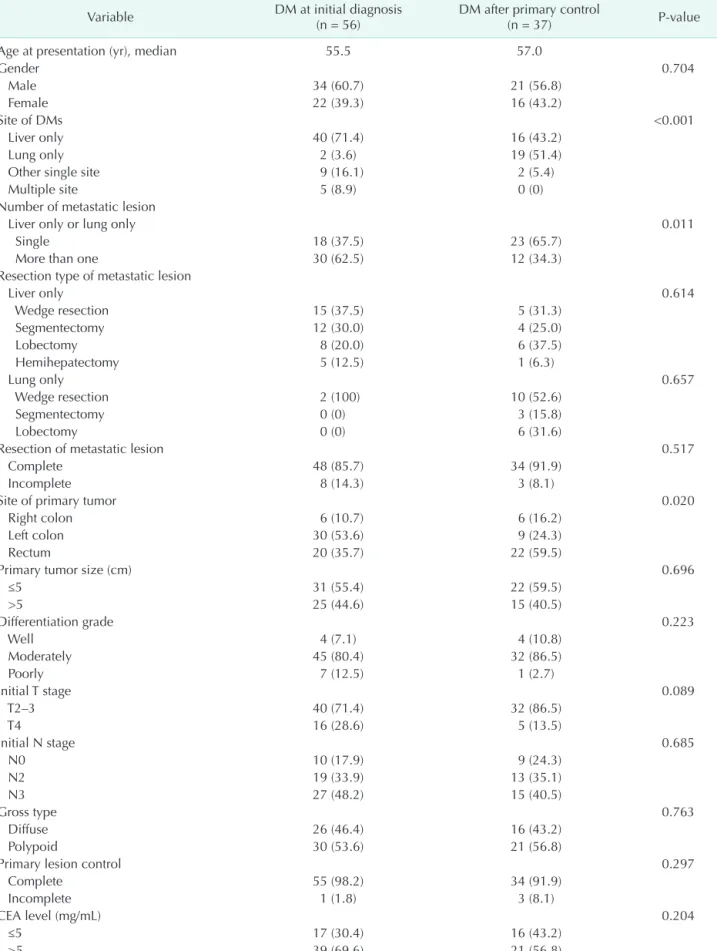
Table 1 summarizes the characteristics of the patients with colorectal cancer DM who underwent metastasectomy from December 2001 to December 2011. Of the 93 patients with colorectal cancers included in the study, 56 patients presented with DM and 37 patients developed DM after resection of primary tumor. The median age of patients with metachronous metastasis was 57.0 years, as compared to 55.5 years for those with synchronous metastasis (P = 0.626).
There were no significant differences between the 2 groups in terms of age, gender, resection margin of metastatic lesion, histological grade, initial T stage, initial N stage, gross type, resection margin of primary lesion, and CEA level. The most common site of DM was lung alone (n = 19, 51.4%) in patients with metachronous metastasis, while liver alone was most common in those with synchronous metastasis (n = 40, 71.4%).
The other single site was ovarian metastasis. Multiple site metastases included the metastases to ≥2 sites among ovary, lateral wall, small bowel, liver, lung, and pelvic wall. Eighty- two patients had complete resection of the metastatic lesion and incomplete resection was done in 11 patients. Primary tumor site was different between the 2 groups. Left colon was most common in patients with synchronous metastasis, while rectum was most common in patients with metachronous metastasis.
Table 1. Clinicopathologic characteristics
Variable DM at initial diagnosis
(n = 56) DM after primary control
(n = 37) P-value
Age at presentation (yr), median 55.5 57.0
Gender 0.704
Male 34 (60.7) 21 (56.8)
Female 22 (39.3) 16 (43.2)
Site of DMs <0.001
Liver only 40 (71.4) 16 (43.2)
Lung only 2 (3.6) 19 (51.4)
Other single site 9 (16.1) 2 (5.4)
Multiple site 5 (8.9) 0 (0)
Number of metastatic lesion
Liver only or lung only 0.011
Single 18 (37.5) 23 (65.7)
More than one 30 (62.5) 12 (34.3)
Resection type of metastatic lesion
Liver only 0.614
Wedge resection 15 (37.5) 5 (31.3)
Segmentectomy 12 (30.0) 4 (25.0)
Lobectomy 8 (20.0) 6 (37.5)
Hemihepatectomy 5 (12.5) 1 (6.3)
Lung only 0.657
Wedge resection 2 (100) 10 (52.6)
Segmentectomy 0 (0) 3 (15.8)
Lobectomy 0 (0) 6 (31.6)
Resection of metastatic lesion 0.517
Complete 48 (85.7) 34 (91.9)
Incomplete 8 (14.3) 3 (8.1)
Site of primary tumor 0.020
Right colon 6 (10.7) 6 (16.2)
Left colon 30 (53.6) 9 (24.3)
Rectum 20 (35.7) 22 (59.5)
Primary tumor size (cm) 0.696
≤5 31 (55.4) 22 (59.5)
>5 25 (44.6) 15 (40.5)
Differentiation grade 0.223
Well 4 (7.1) 4 (10.8)
Moderately 45 (80.4) 32 (86.5)
Poorly 7 (12.5) 1 (2.7)
Initial T stage 0.089
T2–3 40 (71.4) 32 (86.5)
T4 16 (28.6) 5 (13.5)
Initial N stage 0.685
N0 10 (17.9) 9 (24.3)
N2 19 (33.9) 13 (35.1)
N3 27 (48.2) 15 (40.5)
Gross type 0.763
Diffuse 26 (46.4) 16 (43.2)
Polypoid 30 (53.6) 21 (56.8)
Primary lesion control 0.297
Complete 55 (98.2) 34 (91.9)
Incomplete 1 (1.8) 3 (8.1)
CEA level (mg/mL) 0.204
≤5 17 (30.4) 16 (43.2)
>5 39 (69.6) 21 (56.8)
Values are presented as number (%).
DM, distant metastasis.
Survival analysis
Median follow-up time was 43.0 months (range, 11–129 months). There was no postoperative death at 30 days in either group.
Five-year RFS was not significantly different between pati- ents with synchronous metastasis and those metachronous metastasis (28.5% vs. 30.8%, P = 0.291). Five-year OS rate was significantly different between patients with synchronous Table 2. Univariate analysis for recurrence-free and overall survival
Variable
All patients with DM
(n = 93) Synchronous
metastasis (n = 56) Metachronous metastasis (n = 37) 5-yr RFS P-value 5-yr OS P-value 5-yr OS P-value 5-yr OS P-value
Age at presentation (yr) 0.101 0.026 0.095 0.192
≤60 33.4 47.6 38.7 62.7
>60 24.1 26.5 31.8 35.8
Gender 0.903 0.291 0.194 0.947
Male 29.0 46.8 44.3 54.4
Female 29.7 30.9 17.5 53.3
Site of DMs 0.067 0.109 0.286 0.502
Liver only 31.1 50.9 46.4 64.3
Lung only 28.0 40.4 50.0 41.5
Other single site 34.1 24.2 11.1 100
Multiple site 0 0 0
Number of metastatic lesion
Liver only or lung only 0.003 0.017 0.272 0.066
Single 45.2 51.8 40.7 62.4
More than one 10.4 32.9 35.5 31.8
Resection of metastatic lesion <0.001 0.001 0.008 0.083
Complete 32.8 44.7 37.3 55.6
Incomplete 0 10.2 12.5 0
Site of primary tumor 0.490 0.289 0.543 0.025
Right colon 25.0 23.1 25.0 20.8
Left colon 36.0 51.0 43.9 83.3
Rectum 25.3 38.6 26.3 53.4
Differentiation grade 0.924 0.292 0.390 0.638
Well 37.5 65.6 75.0 50.0
Moderately 27.6 38.2 31.0 50.8
Poorly 37.5 37.5 28.6
Initial T stage 0.496 0.111 0.127 0.501
T2–3 30.4 44.3 39.0 52.4
T4 24.6 28.4 21.1 66.7
Initial N stage 0.779 0.826 0.692 0.662
N0 34.8 49.3 56.3 45.7
N1 31.5 28.2 22.7 39.4
N2 25.7 44.4 33.8 64.3
Gross type 0.482 0.768 0.808 0.706
Diffuse 25.1 42.5 39.7 49.9
Polypoid 33.3 39.3 29.2 57.5
Primary lesion control 0.555 0.934 0.239 0.868
Complete 28.7 41.3 34.6 54.5
Incomplete 37.5 33.3 0 50.0
CEA level 0.029 0.052 0.110 0.337
≤5 38.3 55.4 45.2 68.4
>5 24.2 33.9 28.6 46.4
Presentation of DM 0.291 0.013 - -
At initial diagnosis 28.5 34.0 - -
After primary control 30.8 53.7 - -
RFS, recurrence-free survival; OS, overall survival; DM, distant metastasis.
metastasis and those with metachronous metastasis (34.0% vs.
53.7%, P = 0.013).
Univariate analyses were performed to clarify the prognostic factors in all patients with DM, metachronous metastasis and synchronous metastasis (Table 2).
Age, site of DM, incomplete resection of metastatic lesion (hazard ration [HR], 3.452; 95% confidence interval [CI], 1.453–
8.200; P = 0.005), number of metastatic lesion, and CEA level were independent risk factors for OS in patients with DM.
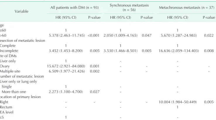
Incomplete resection of metastatic lesion was an independent risk factor for OS in both patients with metachronous metastasis (HR, 16.636; 95% CI, 2.059–134.403; P = 0.008), and synchronous metastasis (HR, 3.530; 95% CI, 1.466–8.501; P = 0.005). Age and site of primary tumor was an independent risk factor for OS in patients with metachronous metastasis, while increased age was an independent risk factor in those with synchronous metastasis (Table 3).
DISCUSSION
We compared clinicopathologic characteristics and oncologic outcomes between patients with metachronous metastasis and those with synchronous metastasis. Although most studies have reported oncologic outcomes in patients who presented with DM, few studies have investigated oncologic outcomes in patients with metachronous metastasis. Firstly, we analyzed a
number of clinicopathologic factors, including the presentation timing of DM by univariate analysis to find prognostic fac- tors in all patients with DM. We found that patients with metachronous metastasis had a better OS rate than those with synchronous metastasis by univariate analysis, but RFS was not different. Using a multivariate analysis, we found that the presentation timing of DM was not an independent predictor of poor OS. This is similar to previous studies, which likewise reported that metachronous metastasis is not a poor prognostic factor for survival [4,6]. However, these previous studies differed from ours in terms of inclusion criteria. Ng et al. [6]
reported that a higher rate of multiple metastatic lesions led to less curative resection. They suggested that the poor prognostic outcome was overcome by perioperative chemotherapy. Van der Pool et al. [4] reported that although patients with synchronous metastases may have poorer biological characteristics, there was no difference in prognosis, as compared to those with metachronous metastases. They furthermore provided the rationale for significantly greater neoadjuvant chemotherapy in patients with synchronous metastasis.
On the contrary, 2 recent studies reported that patients with metachronous tumors showed a significantly better prognosis than those with synchronous lesions [8,9]. Schule et al. [9]
attributed the difference to the lower likelihood of complete resection in synchronous disease.
We investigated the reason for the better OS rate in patients
Table 3. Multivariate analysis for overall survival
Variable All patients with DM (n = 93) Synchronous metastasis
(n = 56) Metachronous metastasis (n = 37)
HR (95% CI) P-value HR (95% CI) P-value HR (95% CI) P-value
Age
≤60 1 1 1
>60 5.378 (2.463–11.745) <0.001 2.050 (1.009–4.165) 0.047 5.670 (1.287–24.983) 0.022 Resection of metastatic lesion
Complete 1 1 1
Incomplete 3.452 (1.453–8.200) 0.005 3.530 (1.466–8.501) 0.005 16.636 (2.059–134.403) 0.008 Site of DMs
Liver only 1 - -
Ovary 15.672 (2.921–84.080) 0.001 - - - -
Multiple site 6.509 (1.977–21.426) 0.002 - - - -
Number of metastatic lesion Liver only or lung only
Single 1 - -
More than one 2.273 (1.100–4.700) 0.027 - -
Location of primary lesion
Right - - - - 10.004 (1.984–50.449) 0.005
Rectum - - 1
CEA level
≤5 1 - -
>5 2.932 (1.268–6.783) 0.012 - - - -
DM, distant metastasis; HR, hazard ratio; CI, confidence interval.
with metachronous metastasis on univariate analysis. We con- cluded that it is due to difference in patients characteristics bet ween the 2 groups. Among the prognostic factors for DM de monstrated by multivariate analysis, site of DM and the number of metastatic lesions were different between patients with metachronous metastasis and synchronous metastasis.
We think that the multiplicity represents more disseminated disease resulting in a poorer prognosis that may contribute to poor OS in patients with DM. These results are compatible with several previous reports that the number of DM is of significant importance to survival, although prognostic factors included are not the same [7,10-13]. Many authors reported that metastatic lesion can be detected at an earlier stage in patients with meta- chronous metastasis [14,15]. This difference may contribute to better OS in patients with metachronous metastasis.
We found that hepatic metastasis was more common in pa tients with synchronous metastasis, while pulmonary me- tastasis was more common in those with metachronous metastasis. Furthermore, DM other than liver and lung were more common in patients with synchronous metastasis. The study demonstrated that the site of metastatic lesion was an independent risk factor of recurrence-free and OS. Our study suggests that this difference in metastatic sites may contribute to the difference in OS in patients with metachronous metastasis.
Recent advances in chemotherapy, surgical technique and cu rative surgery for selected patients have led to improved out- comes in patients with metastatic colorectal cancer. Patients with limited hepatic or pulmonary metastases have been considered the sole candidates for surgical resection. The overall 5-year survival rates are in the range of 37%–58% in the previous studies that reported the results of hepatectomy with curative intent [16-18]. Like hepatic metastases, complete resection of lung metastases is associated with prolonged survival in selected patients [12,19-21]. Ovarian metastases are rare, but easily resected and may benefit from surgical resection.
Although resection of isolated liver or lung or ovarian meta- stases from colorectal cancer appears to be beneficial, the value of aggressive surgical treatment for patients who develop
multiple site DM is not well defined. It has been believed that metastatic involvement of >1 organ represents widespread tumor dissemination and leads to a reluctance in attempting metastasectomy. Our study confirms that metastatic invol- vement of ovary and >1 organ shows worse OS than hepatic involvement alone.
We also investigated the oncological outcome of patients who developed DM after initial surgical resection for DM.
Our study demonstrates that incomplete resection of the metastatic lesion is the most important factor for predicting the prognosis in patients with metachronous metastasis after treatment, as in those with synchronous metastasis. Positive resection margins were previously reported as an unfavorable prognostic factor [9,10,17,22,23].
This is a retrospective analysis of a small sample from our prospective database; we evaluated the clinicopathologic characteristics and oncologic outcomes of advanced colorectal cancer patients with metachronous metastasis, as compared to those with synchronous metastasis. There was thus a likely selection bias on inclusion criteria for surgical re- section. However, our study suggested that the patients with metachronous metastasis can be treated with surgical re- section with acceptable oncologic outcomes.
In conclusion, our study indicates that patients with meta- chronous metastasis show a different metastatic pattern and better oncologic outcome, as compared to those with synchronous metastasis. Resection with negative resection margin significantly improves survival in patients with metachronous metastasis as well as those with synchronous metachronous. We recommend curative surgical resection with negative resection margin in patients with metachronous metastasis. Further clinical trials are needed to identify biologic features in patients with metachronous metastasis.
CONFLICTS OF INTEREST
No potential conflict of interest relevant to this article was reported.
1. Jemal A, Siegel R, Ward E, Hao Y, Xu J, Murray T, et al. Cancer statistics, 2008. CA Cancer J Clin 2008;58:71-96.
2. Manfredi S, Lepage C, Hatem C, Coatmeur O, Faivre J, Bouvier AM. Epidemiology and management of liver metastases from colorectal cancer. Ann Surg 2006;244:254-9.
3. Miller G, Biernacki P, Kemeny NE, Gonen
M, Downey R, Jarnagin WR, et al. Out- comes after resection of synchronous or metachronous hepatic and pulmonary colorectal metastases. J Am Coll Surg 2007;205:231-8.
4. van der Pool AE, Lalmahomed ZS, Ozbay Y, de Wilt JH, Eggermont AM, Jzermans JN, et al. 'Staged' liver resection in synchron-
ous and metachronous colorectal hepatic metastases: differences in clinicopatholo- gical features and outcome. Colorectal Dis 2010;12(10 Online):e229-35.
5. Wang X, Hershman DL, Abrams JA, Feingold D, Grann VR, Jacobson JS, et al.
Predictors of survival after hepatic resec- tion among patients with colorectal liver
REFERENCES
metastasis. Br J Cancer 2007;97:1606-12.
6. Ng WW, Cheung YS, Wong J, Lee KF, Lai PB. A preliminary analysis of combined liver resection with new chemotherapy for synchronous and metachronous colo- rectal liver metastasis. Asian J Surg 2009;
32:189-97.
7. Tsai MS, Su YH, Ho MC, Liang JT, Chen TP, Lai HS, et al. Clinicopathological features and prognosis in resectable synchronous and metachronous colorectal liver meta- stasis. Ann Surg Oncol 2007;14:786-94.
8. Kumar R, Price TJ, Beeke C, Jain K, Patel G, Padbury R, et al. Colorectal cancer sur- vival: An analysis of patients with meta- static disease synchronous and meta- chronous with the primary tumor. Clin Colorectal Cancer 2014;13:87-93.
9. Schule S, Dittmar Y, Knosel T, Krieg P, Albrecht R, Settmacher U, et al. Long-term results and prognostic factors after resec- tion of hepatic and pulmonary metastases of colorectal cancer. Int J Colorectal Dis 2013;28:537-45.
10. Fong Y, Cohen AM, Fortner JG, Enker WE, Turnbull AD, Coit DG, et al. Liver resection for colorectal metastases. J Clin Oncol 1997;15:938-46.
11. Ueno H, Mochizuki H, Hatsuse K, Hase K, Yamamoto T. Indicators for treatment strategies of colorectal liver metastases.
Ann Surg 2000;231:59-66.
12. Saito Y, Omiya H, Kohno K, Kobayashi
T, Itoi K, Teramachi M, et al. Pulmonary metastasectomy for 165 patients with colorectal carcinoma: a prognostic assess- ment. J Thorac Cardiovasc Surg 2002;
124:1007-13.
13. Girard P, Ducreux M, Baldeyrou P, Rou- gier P, Le Chevalier T, Bougaran J, et al.
Surgery for lung metastases from colorec- tal cancer: analysis of prognostic factors. J Clin Oncol 1996;14:2047-53.
14. Jenkins LT, Millikan KW, Bines SD, Sta ren ED, Doolas A. Hepatic resection for meta- static colorectal cancer. Am Surg 1997;
63:605-10.
15. Scheele J, Stang R, Altendorf-Hofmann A, Paul M. Resection of colorectal liver meta- stases. World J Surg 1995;19:59-71.
16. Choti MA, Sitzmann JV, Tiburi MF, Sumet- cho timetha W, Rangsin R, Schulick RD, et al. Trends in long-term survival following liver resection for hepatic colorectal meta- stases. Ann Surg 2002;235:759-66.
17. Fong Y, Fortner J, Sun RL, Brennan MF, Blumgart LH. Clinical score for predicting recurrence after hepatic resection for metastatic colorectal cancer: analysis of 1001 consecutive cases. Ann Surg 1999;
230:309-18.
18. Fernandez FG, Drebin JA, Linehan DC, Dehdashti F, Siegel BA, Strasberg SM. Five- year survival after resection of hepatic metastases from colorectal cancer in
pa tients screened by positron emission tomo graphy with F-18 fluorodeoxyglucose (FDG-PET). Ann Surg 2004;240:438-47.
19. Yedibela S, Klein P, Feuchter K, Hoffmann M, Meyer T, Papadopoulos T, et al. Sur - gical management of pulmonary meta- stases from colorectal cancer in 153 pati- ents. Ann Surg Oncol 2006;13:1538-44.
20. Inoue M, Ohta M, Iuchi K, Matsumura A, Ideguchi K, Yasumitsu T, et al. Benefits of surgery for patients with pulmonary me- tastases from colorectal carcinoma. Ann Thorac Surg 2004;78:238-44.
21. Iwasaki A, Shirakusa T, Yamashita Y, Noritomi T, Maekawa T, Hamada T. Cha- racteristic differences between patients who have undergone surgical treatment for lung metastasis or hepatic metastasis from colorectal cancer. Thorac Cardiovasc Surg 2005;53:358-64.
22. Nuzzo G, Giuliante F, Ardito F, Vellone M, Giovannini I, Federico B, et al. Influence of surgical margin on type of recurrence after liver resection for colorectal meta- stases: a single-center experience. Surgery 2008;143:384-93.
23. Pawlik TM, Scoggins CR, Zorzi D, Abdalla EK, Andres A, Eng C, et al. Effect of surgi- cal margin status on survival and site of recurrence after hepatic resection for colorectal metastases. Ann Surg 2005;
241:715-22.