Annals of Surgical Treatment and Research 105
pISSN 2288-6575 • eISSN 2288-6796 http://dx.doi.org/10.4174/astr.2014.86.2.105 Annals of Surgical Treatment and Research
CASE REPORT
Use of meso-Rex shunt with transposition of the coronary vein for the management of extrahepatic portal vein
obstruction
Yong-Pil Cho, Tae-Yong Ha1, Gi-Young Ko2, Kyung-Mo Kim3, Sung-Gyu Lee1
Division of Vascular Surgery, Department of Surgery, Asan Medical Center, University of Ulsan College of Medicine, Seoul, 1Liver Transplantation Surgery, Department of Surgery, Asan Medical Center, University of Ulsan College of Medicine, Seoul,
Departments of 2Radiology, and 3Pediatrics, Asan Medical Center, University of Ulsan College of Medicine, Seoul, Korea
INTRODUCTION
Patients with extrahepatic portal vein obstruction usually present with symptoms of portal hypertension. Acute bleeding from esophageal and gastric varices is temporarily treated with sclerotherapy or variceal banding. Patients who experience continued bleeding despite medical treatment or who experience clinically significant symptoms of hypersplenism can be referred for surgery. Surgical options have historically included gastric devascularization procedures and various shunt operations [1-8]. The meso-Rex shunt, which was initially indicated for the treatment of extrahepatic portal vein thrombosis following liver transplantation in children, has been successfully used to treat nontransplant patients with
thrombosis due to other etiologies [1-4]. The standard meso- Rex shunt technique uses the patient’s own internal jugular vein conduit between the superior mesenteric vein and the intrahepatic left portal vein to restore hepatopetal flow [1-8].
The bypass can be constructed through the interposition of a conduit using the internal jugular vein, the saphenous vein, or the iliac vein from deceased donors [4-8].
Here, we describe a new method using the coronary vein, which is enlarged in most cases of portal hypertension, as an alternative to the standard meso-Rex shunt technique.
This simplified technique decreases the total operating time, generates a transposed vein of sufficient length, and eliminates the need for simultaneous embolization of collaterals to augment portal flow and the harvest of autologous veins.
The meso-Rex shunt is used to safely and effectively treat patients with portal hypertension due to extrahepatic portal vein obstruction. In the standard meso-Rex shunt technique, the patient’s own internal jugular vein is used as a vascular autograft. Inevitably, such a procedure requires neck exploration and sacrifice of the internal jugular vein. Here, we present a case of a 20-year-old man with idiopathic extrahepatic portal vein obstruction, who was treated with a new technique of transposition of the coronary vein, which is enlarged in most cases of portal hypertension, as an alternative to the standard meso-Rex shunt technique. The transposition of the coronary vein into the Rex recessus is more efficient and less invasive than harvesting an autologous vein graft. Therefore, this technique simplifies the procedure and should be used when possible.
[Ann Surg Treat Res 2014;86(2):105-108]
Key Words: Portal vein, Obstruction, Meso-Rex shunt, Coronary vein, Technique
Received July 11, 2013, Revised August 26, 2013, Accepted August 27, 2013
Corresponding Author: Yong-Pil Cho
Division of Vascular Surgery, Department of Surgery, Asan Medical Center, University of Ulsan College of Medicine, Asanbyeongwon-gil 88 Olympic- ro 43-gil, Songpa-gu, Seoul 138-736, Korea
Tel: +82-2-3010-5039, Fax: +82-2-3010-6701 E-mail: ypcho@amc.seoul.kr
Copyright ⓒ 2014, the Korean Surgical Society
cc Annals of Surgical Treatment and Research is an Open Access Journal. All articles are distributed under the terms of the Creative Commons Attribution Non- Commercial License (http://creativecommons.org/licenses/by-nc/3.0/) which permits unrestricted non-commercial use, distribution, and reproduction in any medium, provided the original work is properly cited.
106
Annals of Surgical Treatment and Research 2014;86(2):105-108
CASE REPORT
A meso-Rex shunt with transposition of the coronary vein was performed in a 20-year-old man. The patient had a previous clinical history of increased abdominal volume and five episodes of gastrointestinal bleeding, and was diagnosed with idiopathic portal vein obstruction at 5 years of age.
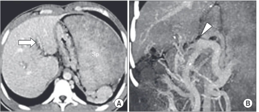
Hypercoagulability studies were performed at the time of the diagnosis. Antithrombin III level was 58% of normal, but other parameters were all unremarkable. One month before the shunt operation, an endoscopy identified aggravated esophageal and paraesophageal varices and hypertensive gastropathy. The size and patency of the umbilical portion of the intrahepatic left portal vein were assessed by preoperative Doppler ultrasonography and computed tomography; there was massive splenomegaly and no detectable intrahepatic left portal vein (Fig.
1). A preoperative percutaneous transhepatic liver needle biopsy showed a noncirrhotic liver without fibrosis, and hepatic venous pressure gradient was 28 mmHg (pressure in the inferior vena cava 7 mmHg, wedged hepatic venous pressure 35 mmHg).
After laparotomy, the round ligament was dissected toward the distal part of the left portal vein at the level of the Rex recessus. The left portal vein was then approached, and its ventral and lateral aspects were dissected over a length of 3-4 cm. The intrahepatic left portal vein was evaluated by surgical exploration, and its diameter was approximately 3 mm.
Splenectomy was performed because of massive splenomegaly.
There was a nest of peripancreatic collaterals with flow toward the gastroesophageal junction through a series of large varices, which included the coronary vein with an adequate diameter and flow in the hepatopetal direction. This vein was fully mobilized, divided, and then transposed across the mesocolon and behind the pylorus to the intrahepatic left portal vein to which it was anastomosed end-to-side using nonabsorbable monofilament interrupted sutures (Fig. 2). Unclamping of the meso-Rex shunt immediately allowed adequate portal flow into the liver, as confirmed by intraoperative portography, and there were no remaining large portosystemic shunts
(Fig. 3). Postoperative recovery was rapid and uneventful, with liver function tests within normal ranges and normal portal flow observed by Doppler ultrasonography and computed tomography (Fig. 4). Follow-up was uneventful, with improvement of the signs of portal hypertension, without any new episodes of gastrointestinal bleeding. Six months after surgery, computed tomography showed preserved portal vein flow via the shunt.
DISCUSSION
Although the incidence and natural history of extrahepatic portal vein obstruction are not completely characterized, morbidity is mainly related to variceal bleeding, hypersplenism, limitations on quality of life, recurrent thrombosis, and symptomatic portal bilopathy [9]. Despite conservative treatment, patients who persistently present with clinically significant symptoms of portal hypertension are considered for surgical options such as gastric devascularization procedures and various shunt operations [1-8]. In the early 1990s, de Ville de Goyet et al. [2] reported the meso-Rex shunt as a therapeutic Fig. 1. (A, B) Preoperative axial and coronal reformatted computed tomo graphy scan images showing an obliterated main (arrowhead) and left (open arrow) intrahepatic portal vein.
Note the massively enlarged spleen (Sp).
Fig. 2. Operative findings showing the transposed coronary vein (white arrows) anastomosed end-to-side to the ventral portion of the extrahepatic left portal vein using nonabsorbable monofilament interrupted sutures.
Annals of Surgical Treatment and Research 107 option to relieve extrahepatic portal hypertension after
partial liver transplantation. Recently, the meso-Rex shunt has been successfully used to treat nontransplant patients with extrahepatic portal vein obstruction due to other etiologies [3,4]. The meso-Rex bypass procedure restores physiologic portal venous blood flow to the liver by connecting the superior mesenteric vein and the intrahepatic left portal vein branch, and can thereby effectively resolve or prevent all of the known complications of extrahepatic portal vein obstruction [9].
For successful application of the meso-Rex shunt, eligible patients must fulfill two preconditions: the hepatic structure must be within normal limits and the umbilical portion of the left portal vein must stay patent. When the intrahepatic left portal vein is permeable without acute liver necrosis or liver fibrosis, despite extrahepatic portal vein thrombosis, a meso- Rex shunt, which physiologically redirects portal flow to the liver, can be indicated. The umbilical portion of the left portal vein is accessible after the division of the liver bridge between segments 3 and 4. This part of the portal vein is generally intact and is not affected by changes to the main trunk of the portal vein following thrombosis [6]. Furthermore, this area is generally not involved in the cavernous transformation that may occur after thrombosis and recanalization [6].
Although extension of thrombosis to the intrahepatic left portal vein sometimes precludes the use of a meso-Rex shunt, this technique is surgically feasible in most patients even when the intrahepatic left portal vein is poorly visible or not seen at all on routine preoperative imaging studies [9]. In such cases, the size and patency of the umbilical portion of the left portal vein must be confirmed by surgical exploration. A distal splenorenal shunt is an alternative to the meso-Rex shunt, but should only be implemented if the use of meso-Rex shunt is clearly not feasible, either owing to anatomical issues or to a lack of a usable portal vein at the time of surgical exploration of the Rex recessus [9]. Although this alternative procedure can be highly efficacious in both preventing recurrence of variceal bleeding and in decompressing the enlarged spleen with resolution of the hypersplenism, it does not restore mesenteric blood flow to the liver [6]. In most published case series in children, the patient’s own internal jugular vein is used as an autograft for the meso-Rex shunt with good results [3-7]. In adult patients, the bypass can be constructed through interposition of a conduit using the patient’s own saphenous vein or the iliac vein from deceased donors [5-7]. However, to harvest the internal jugular or saphenous vein, surgeons must perform neck or inguinal dissection. Iliac veins from deceased Yong-Pil Cho, et al: Meso-Rex shunt for portal vein obstruction
Fig. 4. (A–C) Axial and coronal reformatted computed tomography scan images at the 22-day follow-up showing increased portal flow (open arrows) via the preserved meso-Rex shunt (arrowheads).
Fig. 3. (A, B) Postoperative ve- nogram of the splenic vein showing a brisk flow to the left intrahepatic portal vein (open arrows) via the meso-Rexshunt (arrowheads).
108
Annals of Surgical Treatment and Research 2014;86(2):105-108
1. de Ville de Goyet J, Lo Zupone C, Grimaldi C, D'Ambrosio G, Candusso M, Torre G, et al. Meso-Rex bypass as an alternative technique for portal vein reconstruction at or after liver transplantation in children: review and perspectives. Pediatr Transplant 2013;17:19-26.
2. de Ville de Goyet J, Clapuyt P, Otte JB. Extrahilar mesenterico-left portal shunt to relieve extrahepatic portal hypertension after partial liver transplant.
Transplantation 1992;53:231-2.
3. Salzedas-Netto AA, Duarte AA, Linhares MM, Mattar RH, Medeiros KL, Cury EK, et al. Variation of the Rex shunt for treating concurrent obstruction of the portal and superior mesenteric veins. J Pediatr Surg 2011;46:2018-20.
4. Superina R, Bambini DA, Lokar J, Rigsby C,
Whitington PF. Correction of extrahepatic portal vein thrombosis by the mesenteric to left portal vein bypass. Ann Surg 2006;243:515-21.
5. Luoto T, Pakarinen M, Mattila I, Rintala R. Mesoportal bypass using a constructed saphenous vein graft for extrahepatic portal vein obstruction: technique, feasibility, and outcomes. J Pediatr Surg 2012;47:688-93.
6. Bambini DA, Superina R, Almond PS, Whitington PF, Alonso E. Experience with the Rex shunt (mesenterico-left portal bypass) in children with extrahepatic portal hypertension. J Pediatr Surg 2000;35:13-8.
7. de Ville de Goyet J, Alberti D, Falchetti D, Rigamonti W, Matricardi L, Clapuyt P, et al. Treatment of extrahepatic
portal hypertension in children by mesenteric-to-left portal vein bypass: a new physiological procedure. Eur J Surg 1999;165:777-81.
8. Chiu B, Pillai SB, Sandler AD, Superina RA. Experience with alternate sources of venous inflow in the meso-Rex bypass operation: the coronary and splenic veins.
J Pediatr Surg 2007;42:1199-202.
9. Shneider BL, Bosch J, de Franchis R, Emre SH, Groszmann RJ, Ling SC, et al.
Portal hypertension in children: expert pediatric opinion on the report of the Baveno V Consensus Workshop on Methodology of Diagnosis and Therapy in Portal Hypertension. Pediatr Transplant 2012;16:426-37.
REFERENCES
donors are not readily available and may show an inferior patency rate compared to the use of autologous vein graft because of gradual deterioration and degeneration [1].
In this case report, we propose a new technique for the transposition of the coronary vein for the management of extrahepatic portal vein obstruction without a vascular conduit, which simplified the operative procedures, resulting in reduced total operating time and no need for procurement of autologous veins or for simultaneous embolization of collaterals to
augment portal flow. This alternative technique can be safely used in most cases of portal hypertension with an enlarged coronary vein and was effective in treating portal hypertension in the patient presented here.
CONFLICTS OF INTEREST
The authors declare no potential conflict of interests in relation to this article.