Copyright ⓒ 2019 by Korean Society for Surgery of the Hand, Korean Society for Microsurgery, and Korean Society for Surgery of the Peripheral Nerve. All Rights reserved.
This is an Open Access article distributed under the terms of the Creative Commons Attribution Non-Commercial License (http://creativecommons.org/licenses/by-nc/4.0/)
INTRODUCTION
A malunited forearm fracture is not uncommon after
forearm fracture in children1. Radius malunion can cause distal radioulnar joint (DRUJ) malalignment, which may result in limited forearm rotation and wrist joint pain2-5. Hand and
Microsurgery
원위 요척 관절 불안정성을 동반한 요골 골절 부정 유합의 수술적 치료
심재우ㆍ박민종
성균관대학교 의과대학 삼성서울병원 정형외과학교실
Surgical Treatment for Malunited Radius Fracture with Distal Radioulnar Joint Instability
Jae Woo Shim, Min Jong Park
Department of Orthopedic Surgery, Samsung Medical Center, Sungkyunkwan University School of Medicine, Seoul, Korea
Purpose: The purpose of this study is to report the results of surgical treatment for malunited radius shaft fracture com- bined with distal radioulnar joint (DRUJ) instability.
Methods: A retrospective review of patients with malunited radius shaft fracture with DRUJ instability from 2002 to 2015 was performed. All patients underwent corrective osteotomy for malunited radius. Anatomical distal radioulnar ligament reconstruction was additionally performed in patients who had remaining instability after correction. Preopera- tive and postoperative the anteroposterior stress test, range of motion, pain visual analogue scale, grip strength, modified Mayo wrist score, and disabilities of the arm, shoulder and hand score were analyzed.
Results: A total of 13 patients with an average age of 25 years (range, 15-41 years) were included. The mean follow-up period was 35 months (range, 24-66 months). The DRUJ joint became stable in 4 patients after corrective osteotomy, and thus, no further procedures were required. For 9 patients, additional ligament reconstruction was performed at the same time after osteotomy. DRUJ stability was obtained in 11 patients at the final follow-up. One patient, who failed to achieve DRUJ stability due to insufficient correction, underwent redo-osteotomy. Mild limitation of rotation was observed in 4 patients. Clinical scores were significantly improved after surgery.
Conclusion: Restoration of DRUJ alignment by osteotomy is mandatory for treatment of malunited radius combined with DRUJ instability. Additional anatomical ligament reconstruction may improve DRUJ stability.
Key Words: Wrist, Instability, Malunited, Osteotomy, Reconstruction
Received December 17, 2018, Revised [1] February 4, 2019, [2] February 27, 2019, Accepted March 4, 2019 Corresponding author: Min Jong Park
Department of Orthopedic Surgery, Samsung Medical Center, Sungkyunkwan University School of Medicine, 81 Irwon-ro, Gangnam-gu, Seoul 06351, Korea
TEL: +82-2-3410-3506, FAX: +82-2-3410-0061, E-mail: [email protected]
Original Article
Chronic DRUJ malalignment combined with instabil- ity suggests that the distal radioulnar ligament might be injured6,7. Distal radioulnar ligaments are the most im- portant soft tissue structure providing DRUJ stability8. DRUJ instability associated with chronic distal radioulnar ligament injury can be treated with anatomical ligament reconstruction9-11. In patients with a malunited radius combined with DRUJ instability, correction of bony ma- lalignment by osteotomy may be insufficient to achieve restoration of DRUJ stability12.
Several studies have reported on outcomes after oste- otomies for radius malunion4,12-19. In these studies, indi- cations of corrective osteotomies are limitation of wrist motion, wrist pain, functional deficit, DRUJ instability, and gross deformities. Most studies focused on increasing the range of motion, subjective symptoms, and functional outcomes. There is little information about the outcomes of DRUJ stability. Trousdale and Linscheid4 reported that of 27 consecutive cases of corrective osteotomies, only six were treated for DRUJ instability. Oda et al.12 report- ed that three patients who had volar instability of DRUJ associated with radial shaft malunion were treated with osteotomy.
In the current study, we had performed corrective oste- otomies in patients with a radius malunion. For selected patients, additional ligament reconstruction was per- formed with an attempt to restore normal DRUJ stability.
This study reported 13 consecutive patients who were treated for malunited radius shaft fracture combined with DRUJ instability.
MATERIALS AND METHODS
1. Subjects
Institutional Review Board of Samsung Medical Center approved this retrospective study and waived the require- ment for informed patient consent (IRB No. 2016-09- 143).
This study is a retrospective case series of malunited radius shaft fracture with DRUJ instability. We per- formed a retrospective review of patients who underwent
malunited radius fracture in our center from 2002 to 2015. A total 67 of surgeries for malunited radius were performed during the study period. The inclusion criteria were (1) malunited radius shaft fracture (mid to distal shaft of radius angulation with DRUJ malalignment) and (2) DRUJ instability. DRUJ instability was defined when subluxation of ulna head was observed in stress test. The anteroposterior stress test was performed with the forearm in the neutral, 60° supination, and 60° prona- tion positions, and the result was compared with that of the opposite side. The examiner firmly grasped the radius with one hand, held the ulnar head with the other hand, and applied anteroposterior force20. The test results were classified into grade 0 to 3 as shown in Table 1. Grade 2 and 3 were defined as instability. Twenty one patients of these 67 patients had a diagnosis of malunited radius with DRUJ instability. Of 21 patients, 7 patients were exclud- ed as malunion of radius metaphysis and 1 patient was excluded as pathologic fracture. This group of 13 patients is the focus of the current study.
2. Surgery
With the patient under general anesthesia, stress test was performed prior to surgery and confirmed DRUJ instability in all patients. The approach for osteotomy varied according to the location and severity of the mal- union. An anterior Henry approach was routinely used for radius shaft malunion. The type of osteotomy was in- dividualized to the deformity. The plane of maximal an- gulation was identified and an osteotomy was then made at the malunited site. After the osteotomy, the deformity was corrected according to preoperative radiographic planning using a small laminar spreader or manual reduc-
Table 1. Grading of the distal radioulnar joint stress test
Grade Definition
0 No difference of laxity (symmetric) 1 Laxity with firm end point
2 Translation with unstable end point 3 Subluxation during rotation
tion. Temporary fixation using plate and bone holding clamps or K-wire was performed, and the rotation, gross deformity, and stability of DRUJ were evaluated. An open wedge osteotomy was performed when shortening was expected by performing an approximate correction with preoperative radiographs. In open wedge osteotomy, an autogenous or allogenous corticocancellous iliac bone graft was used. An open wedge osteotomy for length restoration was performed in 7 of 13 patients. In 6 of 13 patients, closed wedge osteotomy without bone graft was performed. The osteotomy and the bone graft were fixed with a plate. Dynamic compression plate was used for radius shaft fixation. In one adolescent patient, K-wire fixation was used.
After correction of the malunion, we performed stress test once again intraoperatively. Anatomical distal radio- ulnar ligament reconstruction was additionally performed at the same time in patients who had remaining instabil- ity. Because of the long duration of DRUJ malalignment, ligament reconstruction was performed rather than repair.
A total of 9 patients who showed DRUJ instability of grade 2 after corrective osteotomy, underwent additional ligament reconstruction described by Adams and Berger9. The radial tunnel was made in parallel with the articular surface of the sigmoid notch. The ulnar tunnel was made through the ulnar fovea toward the ulnar side of the ulnar neck. A palmaris longus tendon was harvested. The ten- don graft was passed through the radial tunnel and both
of its limbs were passed through the ulna tunnel to the ul- nar neck. The tendon graft was fixed with nonabsorbable sutures (Fig. 1).
Postoperatively, a long-arm cast was applied for 4 weeks, followed by range of motion exercise to restore normal joint mobility. Postoperative treatment was same between patients with ligament reconstruction and with- out reconstruction. Normal activity was allowed at 10 to 12 weeks postoperatively, once bony union and normal range of motion was achieved.
3. Evaluation
Preoperative and postoperative data, including pain visual analogue scale (pVAS), range of motion, grip strength assessment using a Jamar dynamometer (Jamar;
Smith and Nephew, Memphis, TN, USA), the modified Mayo wrist score (MMWS), and the disabilities of the arm, shoulder and hand (DASH) score were analyzed.
The anteroposterior stress test was performed to check stability at regular follow-up. Preoperative and postopera- tive radiologic parameters were evaluated. The absolute values of the side-to-side difference in angle were mea- sured. The angle at the longitudinal axis was measured in the radius shaft malunion. The ulnar variance was also measured and compared. Preoperative and postoperative values were analyzed using the Wilcoxon signed rank test.
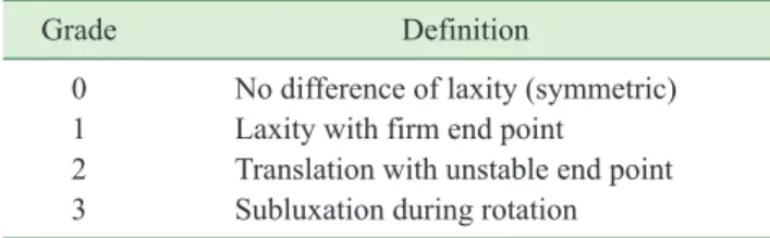
Fig. 1. (A) Preoperative wrist posteroanterior and lateral radiographs of a 24-year-old woman showed distal radioulnar joint subluxation associated with radial malunion. (B) Computed tomography showed volar subluxation of the ulna. This patient was treated with osteotomy and distal radioulnar ligament reconstruction. (C) Radiographs obtained one year after the surgery showed restored alignment. Note that the bone tunnels for ligament reconstruction in the distal radius and ulnar head are visible
RESULTS
Thirteen patients information are described on Table 2. The patient cohort consisted of 10 male and 3 female patients, with an average age of 25 years (range, 15-41 years). The mean follow-up period was 35 months (range, 24-66 months). All patients had pain or discomfort symp-
toms such as the feeling of unstable ulnar head during ro- tation associated with DRUJ instability and three patients had limited range of motion in wrist rotation.
1. Assessment of instability
The anteroposterior stress test at the final follow-up Table 2. Preoperative data of all cases
Case
No. Sex Age
(yr)
Interval between initial fracture and
osteotomy (mo)
Preoperative stress test*
Location of fracture malunion
Preoperative degree of angulation† Lateral view (°),
direction Antero-posterior (°), direction
1 Male 20 96 Grade 3 Distal shaft 11.9, volar 3.9, varus
2 Male 15 15 Grade 3 Distal shaft 15.3, dorsal 4.5, varus
3 Female 16 42 Grade 3 Mid shaft 10.1, dorsal 12.5, valgus
4 Male 24 120 Grade 3 Mid shaft 17.5, dorsal 10.8, valgus
5 Female 39 312 Grade 3 Distal shaft 12.1, dorsal 8.2, valgus
6 Female 15 21 Grade 2 Distal shaft 18.5, dorsal 0.2, valgus
7 Male 22 132 Grade 2 Distal shaft 4.9, dorsal 6.1, valgus
8 Male 41 300 Grade 2 Distal shaft 9.2, dorsal 1.4, valgus
9 Male 32 10 Grade 2 Distal shaft 10.0, dorsal 4.5, valgus
10 Male 20 120 Grade 3 Distal shaft 10.1, dorsal 1.3, varus
11 Male 23 84 Grade 2 Distal shaft 8.5, dorsal 6.6, valgus
12 Male 19 96 Grade 3 Distal shaft 17.0, dorsal 9.8, valgus
13 Male 40 312 Grade 3 Mid shaft 17.9, dorsal 4.6, valgus
Mean 25 128 12.5 5.7
*Grading according to Table 1. †Difference between a malunion site and other, distal fragment displacement direction.
Table 3. Postoperative outcomes of all cases
Case No. Follow-up (mo) DRUJ ligament
reconstruction Postoperative stress test*
MMWS
Preoperative Postoperative
1 50 Yes Grade 0 20 85
2 66 Yes Grade 3† 65 75
3 28 Yes Grade 0 70 85
4 25 Yes Grade 0 50 75
5 25 Yes Grade 1 60 60
6 57 Yes Grade 0 60 60
7 24 No Grade 1 55 85
8 54 No Grade 0 55 95
9 28 Yes Grade 0 50 50
10 26 No Grade 1 85 85
11 28 Yes Grade 0 75 90
12 26 Yes Grade 0 70 90
13 24 No Grade 2 60 70
Mean 35 60 77
DRUJ: distal radioulnar joint, MMWS: modified Mayo wrist score.
*Grading according to Table 1. †Redo osteotomy.
visit indicated grade 0 instability in 8 patients and grade 1 instability in 3 patients. Grade 2 and 3 instability were observed in one patient each. Of the 4 patients who un- derwent corrective osteotomy alone, one patient achieved grade 0 instability. Of the 9 patients who underwent liga- ment reconstruction combined with corrective osteotomy, 7 patients achieved grade 0 instability (Table 3).
The patient who had grade 2 instability had mild symptoms, and thus, did not require further treatment.
The patient who had grade 3 instability after corrective osteotomy and ligament reconstruction failed to restore DRUJ alignment. Corrective osteotomy was performed
again after one year, which improved the alignment and stability. Grade 1 instability was observed at final follow- up (Fig. 2).
2. Clinical outcomes
The pVAS, grip strength, MMWS, and DASH score were significantly improved after surgery (Table 4). Al- though there were statistical improvements after surgery in the whole group, three patients had unsatisfied results in MMWS and DASH. Postoperative MMWS grade was excellent in 3 patients, good in 4 patients, fair in 5 pa-
Table 4. Pre- and postoperative clinical outcomes
Variable pVAS Grip strength (lb) MMWS DASH score
Total
Preoperative 5.4±2.0 57.6±23.1 59.6±15.6 37.9±17.8
Postoperative 2.2±1.6 72.3±20.6 77.3±13.7 21.2±19.8
p-value <0.001 0.021 0.006 <0.001
LR* Preoperative 5.3±1.7 48.8±19.0 57.7±16.6 42.8±18.1
Postoperative 2.6±1.7 65.5±20.2 74.4±14.6 26.1±22.1
p-value 0.011 0.041 0.027 0.011
CO†
Preoperative 5.5±2.9 77.5±20.6 63.7±14.3 26.8±12.5
Postoperative 1.3±0.5 87.5±13.2 83.7±10.3 10.2±5.2
p-value 0.077 0.109 0.109 0.068
Values are presented as mean±standard deviation.
pVAS: pain visual analogue scale, MMWS: modified Mayo wrist score, DASH: disabilities of the arm, shoulder and hand.
Fig. 2. (A) Preoperative wrist posteroanterior and lateral radiographs of a 15-year-old man showed distal radioulnar joint (DRUJ) subluxation associated with radial malunion. (B) After one year after surgery, he still had grade 3 instability after corrective osteotomy, and ligament reconstruction failed to restore DRUJ alignment. Corrective osteotomy was performed again. (C) Radiographs obtained one year after re-do osteotomy show improved alignment and stability.
tients, and poor in 1 patient.
3. Range of motion
The differences between the preoperative and postop- erative ranges of motion were not statistically significant (Table 5). A mild limitation of rotation, which was less than 60°, was observed in four patients. Three patients could not overcome preoperative limitation of rotation.
One patient lost range of supination postoperatively.
Three of these four patients had ligament reconstruction additionally.
4. Radiological measurements
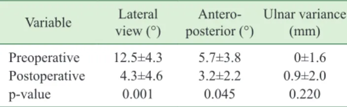
The side-to-side differences in the sagittal and coronal plane significantly reduced, but the ulnar variance was not significantly reduced (Table 6).
5. Complications
Hardware was removed in 4 patients due to irritation symptom. Delayed union was observed in one patient, in whom union was obtained at seven months follow-up.
There were no major complications related to the opera- tion. Three patients reported wrist pain and discomfort over the ulnar head with strenuous use, and they had un- satisfied results in MMWS and DASH score. However, they did not want additional operative treatment and were observed.
DISCUSSION
It is clear that radius malunion changes the DRUJ
alignment and effects the DRUJ stability21-26. The goal of treatment for radius malunion combined with DRUJ in- stability is the restoration of DRUJ stability and painless forearm rotation. Optimal treatment of radius malunion combined with DRUJ instability remains controversial.
Either correction of malunion or restoration of soft tissue stabilizers, or both, have been used. In the current study, we performed corrective osteotomies for DRUJ malalign- ment, with additional ligament reconstruction in cases which DRUJ instability remained after bony correction.
The correction of bony alignment of DRUJ is manda- tory for the restoration of the range of motion and DRUJ stability. In this study, radiological findings improved on both the coronal and sagittal planes, and 4 of the 13 patients regained DRUJ stability after corrective oste- otomy without ligament reconstruction. In these cases, the main cause of instability may not be ligament disrup- tion, but DRUJ malalignment. Chia et al.18 reported six cases of corrective osteotomies for forearm malunion, and the DRUJ stability was partly restored by corrective osteotomy alone. Oda et al.12 reported three cases of volar instability of DRUJ associated radial shaft malunion. In- stability of the DRUJ improved after corrective osteoto- mies without soft tissue procedure, but a slight instability remained in all cases. The results of the current study were similar to previous findings, because corrective os- teotomy without ligament reconstruction enabled 4 of the 13 patients to regain DRUJ stability postoperatively, but at final follow-up, 2 patients had mild DRUJ instability (grade 1) and 1 patient had moderate DRUJ instability (grade 2).
The presence of DRUJ instability after correction of DRUJ alignment indicates that the instability can be at- tributed to distal radioulnar ligament insufficiency. For
Table 5. Pre- and postoperative range of motion Variable Range of motion (°)
Extension Flexion Supination Pronation Preoperative 78.8±7.6 76.5±10.2 82.3±8.5 73.0±20.8 Postoperative 77.6±11.4 74.6±13.4 78.4±8.7 75.7±12.8
p-value 0.703 0.488 0.268 0.383
Values are presented as mean±standard deviation.
Table 6. Pre- and postoperative radiographic data*
Variable Lateral
view (°) Antero-
posterior (°) Ulnar variance (mm) Preoperative 12.5±4.3 5.7±3.8 0±1.6 Postoperative 4.3±4.6 3.2±2.2 0.9±2.0
p-value 0.001 0.045 0.220
Values are presented as mean±standard deviation.
*Difference between a malunion site and other.
these cases, ligament reconstruction is helpful to restore DRUJ stability. Several authors have previously reported the anatomical reconstruction of the distal radioulnar ligament. Teoh and Yam11 reported nine cases of DRUJ ligament reconstruction, of whom seven regained DRUJ stability. Adams and Berger9 reported 14 cases of DRUJ ligament reconstruction, of whom 12 regained DRUJ stability. Two patients underwent concurrent osteotomy for a malunion, resulting in a favorable outcome. In the current study, anatomical ligament reconstruction was performed concurrently with corrective osteotomy in 9 patients, of whom 8 patients regained stability.
In this study, one patient failed to restore DRUJ stabil- ity (Fig. 2). The angular deformity of radius had been properly corrected during initial operation. However, the deformity seemed to recur on follow-up radiographs. In addition, radius shortening was observed after correction and this also could be the cause of instability. As a result, the DRUJ alignment was not restored and DRUJ insta- bility remained despite of the ligament reconstruction.
However, the DRUJ stability improved following re- osteotomy to realign the DRUJ. This suggests that recon- struction of ligament without restoring DRUJ alignment cannot achieve DRUJ stability.
In the current study, three patients had unsatisfied re- sults after surgery. All three patients with poor outcome underwent ligament reconstruction and had DRUJ stabil- ity at final follow-up. One patient had a long duration (26 years) from injury to operation and she was adolescent, which may cause poor results after surgery. Three pa- tients with poor outcomes had mainly ulnar side pain, so the authors thought that triangular fibrous cartilage injury or arthritic change of DRUJ not observed in radiograph caused poor outcomes. There were cases of increased ulnar variance after surgery, but the ulnar variance did not increase in these three patients. It may be helpful to check the DRUJ and triangular fibrous cartilage injury though preoperative magnetic resonance imaging. All pa- tients who underwent corrective osteotomy only showed improvement in functional outcome but there was no sta- tistical significance because the number of patients was 4.
Jupiter and Ring14 reported early reconstruction of mal-
united distal radius led to a better outcome, in comparison to late reconstruction. In their study, the early reconstruc- tion group were treated within an average of two months, while in the current study, treatments were done within an average of 128 months; 12 patients were treated after one year. Thus, the long injury duration in this study could have adversely affected our results. Flinkkilä et al.16 reported 45 distal radius corrective osteotomy cases and concluded that corrective osteotomy of distal radius malunion may not completely restore normal function.
Although some patients did not have a good outcome, overall results were promising. This could be attributed to the restoration of DRUJ stability, as well as alignment.
However, some limitations in the current study must be noted. Firstly, this was a retrospective, case review study.
Secondly, the number of cases was small and follow- up period were inconsistent. Thirdly, various operative technique were used. Finally, an objective assessment of DRUJ alignment was absent.
CONCLUSION
Restoration of DRUJ alignment is mandatory for treat- ment of malunited radius combined with DRUJ instabil- ity. Additional anatomical ligament reconstruction may improve the stability of the DRUJ.
CONFLICTS OF INTEREST
The authors have nothing to disclose.
REFERENCES
1. Fuller DJ, McCullough CJ. Malunited fractures of the forearm in children. J Bone Joint Surg Br. 1982;64:364-7.
2. Altissimi M, Antenucci R, Fiacca C, Mancini GB. Long- term results of conservative treatment of fractures of the distal radius. Clin Orthop Relat Res. 1986:202-10.
3. Cooney WP 3rd, Dobyns JH, Linscheid RL. Compli- cations of Colles’ fractures. J Bone Joint Surg Am.
1980;62:613-9.
4. Trousdale RT, Linscheid RL. Operative treatment of mal-
united fractures of the forearm. J Bone Joint Surg Am.
1995;77:894-902.
5. Stoffelen D, De Smet L, Broos P. The importance of the distal radioulnar joint in distal radial fractures. J Hand Surg Br. 1998;23:507-11.
6. Nakamura T, Iwamoto T, Matsumura N, Sato K, Toyama Y.
Radiographic and arthroscopic assessment of DRUJ insta- bility due to foveal avulsion of the radioulnar ligament in distal radius fractures. J Wrist Surg. 2014;3:12-7.
7. Omori S, Moritomo H, Murase T, et al. Changes in length of the radioulnar ligament and distal oblique bundle after Colles’ fracture. J Plast Surg Hand Surg. 2013;47:409-14.
8. Ward LD, Ambrose CG, Masson MV, Levaro F. The role of the distal radioulnar ligaments, interosseous membrane, and joint capsule in distal radioulnar joint stability. J Hand Surg Am. 2000;25:341-51.
9. Adams BD, Berger RA. An anatomic reconstruction of the distal radioulnar ligaments for posttraumatic distal radio- ulnar joint instability. J Hand Surg Am. 2002;27:243-51.
10. Scheker LR, Belliappa PP, Acosta R, German DS. Recon- struction of the dorsal ligament of the triangular fibrocar- tilage complex. J Hand Surg Br. 1994;19:310-8.
11. Teoh LC, Yam AK. Anatomic reconstruction of the distal radioulnar ligaments: long-term results. J Hand Surg Br.
2005;30:185-93.
12. Oda T, Wada T, Isogai S, Iba K, Aoki M, Yamashita T.
Corrective osteotomy for volar instability of the distal radioulnar Joint associated with radial shaft malunion. J Hand Surg Eur Vol. 2007;32:573-7.
13. Fernandez DL. Correction of post-traumatic wrist defor- mity in adults by osteotomy, bone-grafting, and internal fixation. J Bone Joint Surg Am. 1982;64:1164-78.
14. Jupiter JB, Ring D. A comparison of early and late recon- struction of malunited fractures of the distal end of the radius. J Bone Joint Surg Am. 1996;78:739-48.
15. Shea K, Fernandez DL, Jupiter JB, Martin C Jr. Correc- tive osteotomy for malunited, volarly displaced fractures of the distal end of the radius. J Bone Joint Surg Am.
1997;79:1816-26.
16. Flinkkilä T, Raatikainen T, Kaarela O, Hämäläinen M.
Corrective osteotomy for malunion of the distal radius.
Arch Orthop Trauma Surg. 2000;120:23-6.
17. Nagy L, Jankauskas L, Dumont CE. Correction of forearm malunion guided by the preoperative complaint. Clin Or- thop Relat Res. 2008;466:1419-28.
18. Chia DS, Lim YJ, Chew WY. Corrective osteotomy in forearm fracture malunion improves functional outcome in adults. J Hand Surg Eur Vol. 2011;36:102-6.
19. Opel S, Konan S, Sorene E. Corrective distal radius os- teotomy following fracture malunion using a fixed-angle volar locking plate. J Hand Surg Eur Vol. 2014;39:431-5.
20. Kim JP, Park MJ. Assessment of distal radioulnar joint instability after distal radius fracture: comparison of computed tomography and clinical examination results. J Hand Surg Am. 2008;33:1486-92.
21. Kihara H, Short WH, Werner FW, Fortino MD, Palmer AK. The stabilizing mechanism of the distal radioulnar joint during pronation and supination. J Hand Surg Am.
1995;20:930-6.
22. Stuart PR, Berger RA, Linscheid RL, An KN. The dorso- palmar stability of the distal radioulnar joint. J Hand Surg Am. 2000;25:689-99.
23. Cole DW, Elsaidi GA, Kuzma KR, Kuzma GR, Smith BP, Ruch DS. Distal radioulnar joint instability in distal radius fractures: the role of sigmoid notch and triangular fibro- cartilage complex revisited. Injury. 2006;37:252-8.
24. Kihara H, Palmer AK, Werner FW, Short WH, Fortino MD. The effect of dorsally angulated distal radius frac- tures on distal radioulnar joint congruency and forearm rotation. J Hand Surg Am. 1996;21:40-7.
25. Saito T, Nakamura T, Nagura T, Nishiwaki M, Sato K, Toyama Y. The effects of dorsally angulated distal radius fractures on distal radioulnar joint stability: a biomechani- cal study. J Hand Surg Eur Vol. 2013;38:739-45.
26. Bessho Y, Nakamura T, Nagura T, Nishiwaki M, Sato K, Toyama Y. Effect of volar angulation of extra-articular distal radius fractures on distal radioulnar joint stability: a biomechanical study. J Hand Surg Eur Vol. 2015;40:775- 82.
원위 요척 관절 불안정성을 동반한 요골 골절 부정 유합의 수술적 치료
심재우ㆍ박민종
성균관대학교 의과대학 삼성서울병원 정형외과학교실
목적: 본 연구의 목적은 원위 요척 관절 불안정성을 동반한 요골 간부 골절 부정 유합의 수술적 치료 결과를 보고자 함이다.
방법: 2002년부터 2015년까지 원위 요척 관절 불안정성을 동반한 요골 간부 골절 부정 유합 환자를 후향적으로 검 토하였다. 모든 환자는 교정 절골술을 시행 받았고, 절골술 후 불안정이 있는 경우 해부학적 원위 요척 인대 재건술 을 추가적으로 시행하였다. 수술 전 후 전후방 스트레스 검사, 및 임상 에 대해 비교하였다.
결과: 총 13명의 환자가 포함되었고 평균 연령은 25세(15-41세), 평균 추시 기간은 35개월(24-66개월)이었다. 원 위 요척 관절은 교정 절골술 후 4명은 불안정이 해소되어 더 이상의 시술은 필요하지 않았으며 9명의 환자에서 절 골술 후 인대 재건술을 추가로 시행하였다. 최종 추시 시 11명에서 원위 요척 관절의 안정성을 얻었지만 1명은 불 안정성을 보여 재 절골술을 시행 받았다. 4명에서 경미한 회전 제한이 관찰되었다. 수술 후 임상 결과는 유의하게 향상되었다.
결론: 원위 요척 관절 불안정성을 동반한 요골 간부 골절 부정 유합 수술에서 원위 요척 관절 정렬의 복원은 치료에 필수적이다. 추가적인 해부학적 인대 재건술은 원위 요척 관절의 안정성을 향상시킬 수 있다.
색인단어: 손목, 불안정성, 부정 유합, 절골술, 재건술
접수일 2018년 12월 17일 수정일 1차: 2019년 2월 4일, 2차: 2019년 2월 27일 게재확정일 2019년 3월 4일 교신저자 박민종
06351, 서울시 강남구 일원로 81, 성균관대학교 의과대학 삼성서울병원 정형외과학교실 TEL 02-3410-3506 FAX 02-3410-0061 E-mail [email protected]