A long-term survival case of advanced biliary cancer with repeated resection due to recurrence in the pancreaticogastrostomy site
after pancreaticoduodenectomy
Shohei Eto, Masashi Ishikawa, Michihito Asanoma, Yoshihiko Tashiro, Kazuo Matsuyama, and Takehito Oshio
Department of Surgery, Shikoku Central Hospital, Ehime, Japan
A 62-year-old man underwent endoscopic mucosal resection for early gastric cancer. The follow-up computed tomog- raphy revealed biliary dilatation. The tumor was located in the lower bile duct with biliary dilatation, and no evidence of metastasis in other organs was noted. The patient underwent subtotal stomach-preserving pancreatoduodenectomy with pancreaticogastrostomy and Billroth I anastomosis. At 13 months after the operation, gastrointestinal endoscopy revealed a tumor lesion in the pancreaticogastrostomy site. Computed tomography revealed that the lesion was low enhanced in the pancreaticogastrostomy site and there was no evidence of other distant metastasis. Partial pan- createctomy was performed. Pathological findings of the tumor in the stump of the pancreas revealed findings similar to that of primary biliary carcinoma. Apparently, the patient was diagnosed with recurrence of bile duct cancer via the pancreatic duct. The patient underwent adjuvant chemotherapy for one year subsequent to partial pancreatectomy as the second operation. For 40 months after the second operation, there has been no evidence of recurrence of cancer. (Ann Hepatobiliary Pancreat Surg 2018;22:173-177)
Key Words: Biliary cancer; Surgery; Recurrence; Pancreaticoduodenectomy; Pancreaticogastrostomy
Received: September 17, 2017; Revised: January 10, 2018; Accepted: January 14, 2018 Corresponding author: Shohei Eto
Department of Surgery, Shikoku Central Hospital, 2233 Kawanoe-cho, Shikoku-chuo, Ehime 799-0193, Japan Tel: +81-896-58-3515, Fax: +81-896-58-3464, E-mail: [email protected]
Copyright Ⓒ 2018 by The Korean Association of Hepato-Biliary-Pancreatic Surgery
This is an Open Access article distributed under the terms of the Creative Commons Attribution Non-Commercial License (http://creativecommons.org/
licenses/by-nc/4.0) which permits unrestricted non-commercial use, distribution, and reproduction in any medium, provided the original work is properly cited.
Annals of Hepato-Biliary-Pancreatic Surgery ∙ pISSN: 2508-5778ㆍeISSN: 2508-5859
INTRODUCTION
Biliary cancer is rare and often presents with aggressive malignancy. This cancer includes four distinct categories such as intrahepatic cholangiocarcinoma, extrahepatic cholangiocarcinoma, gallbladder cancer, and ampullary carcinoma. Complete surgical resection is the only treat- ment option for patients. However, even if curative intent surgery is applied to the resectable biliary cancer patients, the 5-year survival rates still remain low: 33.1% for bile duct cancer, 52.8% for ampullary cancer, and 41.6% for gallbladder cancer.1 The high rate of recurrence is the rea- son behind poor prognosis in biliary cancer. However, treatment for recurrent cancer such as chemotherapy is not sufficient. Therefore, surgery is needed for recurrent bili- ary tract cancer in resectable cases. We report a rare case of advanced bile duct cancer with long-term survival by repeated resection due to recurrence in the pancreaticogas-
trostomy site after subtotal stomach-preserving pan- creatoduodenectomy (SSPPD).
CASE
The patient was a 62-year-old man who underwent en- doscopic mucosal resection for early gastric cancer nine years ago and follow-up computed tomography revealed biliary dilatation.
The physical examination at the time of admission showed no abnormal findings. Serum liver enzyme levels were on a slightly higher side and other laboratory find- ings were normal.
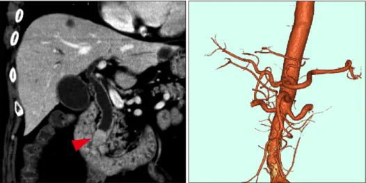
Abdominal computed tomography (CT) revealed a tu- mor in the lower bile duct with biliary dilatation (Fig. 1), and no evidence of metastasis in other organs was noted.
The tumor was low intensity at T1WI, iso-high intensity at T2WI, and high intensity at the diffusion-weighted im-
Fig. 1. Computed tomography findings showing tumor of the lower bile duct with biliary di- latation (arrow).
Fig. 2. Magnetic resonance imaging showed that the tumor was low intensity at T1WI, iso- high intensity at T2WI and high intensity at diffusion-weighted image (DWI) (arrows).
age in magnetic resonance imaging (MRI) findings (Fig.
2). Biliary dilatation and filling defect of the lower bile duct were found in magnetic resonance chol- angiopancreatography (MRCP) and endoscopic retrograde cholangiopancreatography (ERCP) (Fig. 3). MRCP and ERCP revealed no abnormal findings in the pancreatic duct.
Cytology of the bile juice was malignant cell-negative.
Therefore, we performed subtotal stomach-preserving pan- creatoduodenectomy (SSPPD, Pancreaticogastrostomy, Billroth I) (Fig. 4). The proximal 2 cm of the pancreatic
remnant is dissected from the retroperitoneum and anasto- mosed end-to-side to the posterior wall of the stomach.
With the suturing and tying, 1 cm of the pancreas was invaginated into the stomach. We sutured the pancreatic parenchyma and the seromuscular layer of the stomach at 2 cm radius from the pancreatic duct. The pathological findings revealed bile duct cancer of T2N0M0 StageⅡA (TNM staging by the International Union Against Cancer [UICC] 7th edition), and there was no evidence of cancer invasion into the pancreatic duct. In addition, the peri-
Fig. 3. Magnetic resonance chol- angiopancreatography findings.
(A) Biliary dilatation and filling defect of lower bile duct were observed. (B) No abnormality in the pancreatic duct was observed.
Fig. 4. Operative finding of the first operation. (A) Surgical find- ings of the pancreaticogastric anastomosis in the first operation (white arrow). (B) Transgastric pancreaticogastric anastomosis was performed. The pan- creaticogastric anastomosis was made using full-thickness su- tures of the stomach to the pancreas. Subsequently, gastro- jejunectomy was performed us- ing Billroth I method.
Fig. 5. Gastrointestinal endoscopy finding after 13 months of first operation: the recurrence at pancreaticogastrostomy site was noted.
neural/lymphatic invasion was not observed.
On the 16th day after the operation, hematemesis occurred. Gastrointestinal endoscopy revealed blood coag-
ulation in the stomach, but there was no obvious bleeding in the stomach. In angiography, development of pseudoa- neurysm of the stump of the gastroduodenal artery was observed and extravasation was confirmed. Therefore, coil embolization of the common hepatic artery was performed. After the treatment, the patient recovered and was discharged from the hospital.
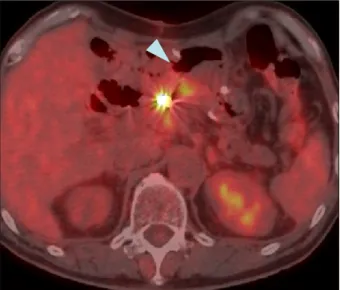
At 13 months after the operation, gastrointestinal en- doscopy revealed an irregular shaped tumor in the pan- creaticogastrostomy site and endoscopic biopsy revealed a well-differentiated tubular adenocarcinoma (Fig. 5). The lesion was low enhanced in the pancreaticogastrostomy site in CT scan. The lesion also showed the accumulation of fluorodeoxyglucose in positron emission tomography CT (Fig. 6). Although no changes in the level of serum tumor marker were noted. There was no evidence of other distant metastasis. The patient was diagnosed as having a recurrence of cholangiocarcinoma and a second surgery was performed. Partial pancreatectomy was performed from the opened anterior wall of the stomach (Fig. 7). The
Fig. 6. Positron emission tomography computed tomography show- ing hypermetabolic uptake indicating recurrence (arrow).
Fig. 8. Comparison of the pathological findings. (A) Pathological finding from the first operation. (B) Pathological finding of the recurrent lesion. Both the lesions revealed similar findings.
Fig. 7. Surgical findings of the second operation.
to that of primary biliary carcinoma. Consequently, the patient was diagnosed with recurrence of the bile duct cancer as well differentiated tubular adenocarcinoma (Fig.
8). The patient underwent chemotherapy of Tegafur/
Gimeracil/Oteracil (TS-1) as adjuvant chemotherapy for bile duct cancer for one year after the second operation.
For 40 months after the second operation, there has been no evidence of recurrence of cancer.
DISCUSSION
Biliary tract cancer is a rare malignancy associated with poor prognosis and complete surgical resection is the only curable treatment. However, biliary tract cancer patients are often diagnosed with advanced stages and treated em- ploying systemic chemotherapy or palliative treatment set- tings rather than curative surgery.
In general, patients with recurrent biliary cancer are treated using various chemotherapies. Some clinical trials provided a certain degree of clinical benefit to patients;
however, the results are not satisfactory.2,3 Regarding sur- gical treatment for the recurrence of extrahepatic biliary carcinoma, few reports have reported surgical indication.
If it is considered in resectable, surgery appears to be fea- sible for recurrent extrahepatic biliary carcinoma and of- fers longer survival.4,5
Pancreaticogastrostomy is one of the methods of re- construction after pancreaticoduodenectomy. When com- pared with pancreaticojejunostomy, pancreaticogas- trostomy did not show any significant differences in the overall postoperative complication rate or reduction of pancreatic fistula. However, biliary fistula, postoperative collections, and delayed gastric emptying were sig- nificantly reduced in patients treated with pancreaticogastrostomy. In addition, pancreaticogas- trostomy is associated with a significantly lower fre- quency of multiple surgical complications.6-10 There exist several reports on pancreatic fistula, however, only a few reports have dealt with cancer recurrence after pancreaticogastrostomy. In our case, recurrence occurred in the anastomosed site of pancreaticogastrostomy. As the resected pancreatic duct was normal, it was hypothesized that the recurrent lesion was disseminated via the pancre- atic duct. This type of metastasis is a very rare case. As far as we examined, no similar case was not found. The lesion was localized, and no evidence of other metastasis was observed, therefore, we performed the second operation. It was considered that curative resection was succeeded by partial pancreatectomy, but total pan- createctomy was difficult due to adhesion by the first
operation. Pancreaticogastrostomy was identified as a use- ful method due to ease of observation of the anastomosed site by endoscopy and easy approach to the anastomosed site during the operation.
We report a rare case of advanced bile duct cancer with long-term survival by repeated resection of recurrence in the pancreaticogastrostomy site after SSPPD. Reoperation should be considered for accurate diagnosis for location and format of recurrence.
REFERENCES
1. Miyakawa S, Ishihara S, Horiguchi A, Takada T, Miyazaki M, Nagakawa T. Biliary tract cancer treatment: 5,584 results from the Biliary Tract Cancer Statistics Registry from 1998 to 2004 in Japan. J Hepatobiliary Pancreat Surg 2009;16:1-7.
2. Takashima A, Morizane C, Ishii H, Nakamura K, Fukuda H, Okusaka T, et al. Randomized phase II study of gemcitabine plus S-1 combination therapy vs. S-1 in advanced biliary tract cancer:
Japan Clinical Oncology Group Study (JCOG0805). Jpn J Clin Oncol 2010;40:1189-1191.
3. Valle JW. Advances in the treatment of metastatic or un- resectable biliary tract cancer. Ann Oncol 2010;21 Suppl 7:vii345-vii348.
4. Song SC, Heo JS, Choi DW, Choi SH, Kim WS, Kim MJ.
Survival benefits of surgical resection in recurrent cholangiocarcinoma. J Korean Surg Soc 2011;81:187-194.
5. Noji T, Tsuchikawa T, Mizota T, Okamura K, Nakamura T, Tamoto E, et al. Surgery for recurrent biliary carcinoma: results for 27 recurrent cases. World J Surg Oncol 2015;13:82.
6. Ishikawa O, Ohigashi H, Eguchi H, Yokoyama S, Yamada T, Takachi K, et al. Long-term follow-up of glucose tolerance func- tion after pancreaticoduodenectomy: comparison between pan- creaticogastrostomy and pancreaticojejunostomy. Surgery 2004;
136:617-623.
7. Que W, Fang H, Yan B, Li J, Guo W, Zhai W, et al.
Pancreaticogastrostomy versus pancreaticojejunostomy after pan- creaticoduodenectomy: a meta-analysis of randomized controlled trials. Am J Surg 2015;209:1074-1082.
8. Bassi C, Falconi M, Molinari E, Salvia R, Butturini G, Sartori N, et al. Reconstruction by pancreaticojejunostomy versus pan- creaticogastrostomy following pancreatectomy: results of a com- parative study. Ann Surg 2005;242:767-771, discussion 771-773.
9. Fernández-Cruz L, Cosa R, Blanco L, López-Boado MA, Astudillo E. Pancreatogastrostomy with gastric partition after py- lorus-preserving pancreatoduodenectomy versus conventional pancreatojejunostomy: a prospective randomized study. Ann Surg 2008;248:930-938.
10. Wada H, Numata A, Fukunaga A, Sasamura Y, Takeyama S, Nenohi M. A case of recurrent intraductal papillary mucinous carcinoma at the pancreaticogastrostomy portion. Pancreas 2012;
27:206-211.