https://doi.org/10.4174/astr.2020.98.3.116 Annals of Surgical Treatment and Research
Prognostic factors predicting survival rate over 10 years of patients with intrahepatic cholangiocarcinoma after hepatic resection
Chung Hyeun Ma1, Dae Wook Hwang2, Ki Byung Song2, Song Cheol Kim2, Sang Hyun Shin3, Jae Hoon Lee2
1Department of Surgery, Gangneung Asan Hospital, University of Ulsan College of Medicine, Gangneung, Korea
2Division of Hepatobiliary and Pancreatic Surgery, Department of Surgery, Asan Medical Center, University of Ulsan College of Medicine, Seoul, Korea
3Department of Surgery, Samsung Medical Center, Sungkyunkwan University School of Medicine, Seoul, Korea
INTRODUCTION
Among the primary carcinomas in the liver, intrahepatic cholangiocarcinoma (IHCC) is the second most common cancer after hepatocellular carcinoma (HCC), and the incidence is increasing worldwide [1,2]. However, the volume of literature on IHCC is scarce compared to that for the widely studied HCC [3,4]. Only 10%–20% of IHCCs are deemed resectable at the
time of presentation [5], and the median survival time ranges from 6–9 months for patients with unresectable disease [6]. It is generally believed that IHCC is primarily a surgical disease, and surgical resection offers the only prospect for long-term survival [7]. Unfortunately, even after curative-intent surgery, the clinical outcomes of patients undergoing liver resection are disappointing, with a 5-year survival rate of 20% to 35%
[8]. Furthermore, the efficacy of adjuvant therapies, including
Received August 20, 2019, Revised November 27, 2019, Accepted January 11, 2020
Corresponding Author: Jae Hoon Lee
Division of Hepatobiliary and Pancreatic Surgery, Department of Surgery, Asan Medical Center, University of Ulsan College of Medicine, 88 Olympic-ro 43-gil, Songpa-gu, Seoul 05505, Korea
Tel: +82-2-3010-1521, Fax: +82-2-3010-6701 E-mail: [email protected]
ORCID: https://orcid.org/0000-0002-6170-8729
Copyright ⓒ 2020, the Korean Surgical Society
cc Annals of Surgical Treatment and Research is an Open Access Journal. All articles are distributed under the terms of the Creative Commons Attribution Non- Commercial License (http://creativecommons.org/licenses/by-nc/4.0/) which permits unrestricted non-commercial use, distribution, and reproduction in any medium, provided the original work is properly cited.
Purpose: Hepatic resection is considered as the optimal treatment for intrahepatic cholangiocarcinoma (IHCC); however, the survival rate after resection is low and the analysis of long-term (≥10 years) survivors is rare. This study aims to analyze the clinicopathological factors affecting the long-term survival of patients with IHCC.
Methods: Between January 2003 and December 2012, a single-institution cohort of 429 patients who underwent hepatic resection for IHCC were reviewed retrospectively. Surgical results, recurrence, and survival rates were investigated, and multivariate analyses were performed to identify prognostic factors.
Results: The overall 1- , 3- , 5- and 10-year survival rates of patients were 76.5%, 44.1%, 33.3%, and 25.1%, respectively.
Multivariate analysis showed that the serum CA 19-9 level (≥38 U/mL) (P < 0.001), lymph node (LN) metastasis (P = 0.001), and lymphovascular invasion (LVI) (P = 0.012) were independent factors associated with overall survival. In particular, CA 19-9 level and histologic type were determined to be independent factors affecting survival for more than 10 years.
Conclusion: CA 19-9 (≥38 U/mL), LN metastasis, and LVI were identified as independent risk factors for survival after resection of IHCC. CA 19-9 (<38 U/mL) and histologic type were independent factors predicting survival for more than 10 years.
[Ann Surg Treat Res 2020;98(3):116-123]
Key Words: Bile ducts, Cholangiocarcinoma, Prognosis, Survival analysis
systemic chemotherapy and radiotherapy, is poorly understood, and there are no standard criteria [9]. Many factors have been found to predict prognosis after surgical resection for IHCC, but a consensus has not yet been reached regarding the factors that could significantly and independently influence the survival rates [10-12]. Furthermore, because of the low survival rate of IHCC patients who undergo surgical treatment, it may be difficult to accurately assess the prognostic factors of long-term survival. Therefore, the purpose of this study is to identify the prognostic factors of IHCC, analyze the patients who have survived for more than 10 years after surgery, and to identify the prognostic factors associated with long-term survival in such patients.
METHODS
Data sources and study population
From January 2003 to December 2012, 429 single-institution patients who underwent hepatic resection for pathologically proven IHCC at Asan Medical Center were enrolled in this study. The cohort of patients did not have metastatic lesions at the time of diagnosis and had no preoperative chemotherapy;
subsequently, they underwent hepatic resection for therapeutic purposes and achieved R0 or R1 resection. Preoperative variables included age, sex, underlying liver cirrhosis, serum CA 19-9 levels, and antigens of Clonorchis sinensis. All patients underwent preoperative contrast-enhanced CT or magnetic resonance imaging (MRI) of the chest and abdomen. A biopsy was not routinely performed before surgery. Pathological data for tumors were examined, including data on size, number of tumors, the grade of differentiation, presence and extent of vascular invasion, perineural invasion (PNI), and lymph node (LN) metastases. Margin and nodal status were identified based on the final pathologic review by a dedicated pathologist.
The tumors were staged using the TNM classification, and the patients were regularly followed up after surgery. CT or MRI was used to monitor the abdomen and chest radiographs prospectively for up to 2 years every 3–4 months, and every 6 months for up to 5 years, after which the surveillance was performed annually. The date of the last follow-up, vital status, and recurrence-related information was collected for all patients, with recurrence defined as histologically confirmed or strongly-suspected recurrence in imaging studies. Additionally, the duration from the initial date of surgery to the development of recurrent disease was recorded. Data collection and analysis were performed according to the institutional guidelines, which conformed to the ethical standards of the Declaration of Helsinki. This retrospective study was approved by the Institu tional Review Board of Asan Medical Center (No. S2019- 0525). Patient informed consent was waived because of the retrospective nature of the study.
Statistical analyses
All statistical analyses were performed using IBM SPSS Statistics ver. 21.0 (IBM Co., Armonk, NY, USA). Summary statistics were obtained using established methods and presented as percentages, mean, or median values. Recurrence and survival curves were estimated by the Kaplan-Meier method. Predictive analysis of variables associated with the factor-specific hazard of recurrence and survival was performed using the multivariate Cox proportional hazard model. Hazard ratios (HRs) and 95% confidence intervals (CIs) were estimated.
All reported P-values were two-sided, and P < 0.05 was considered statistically significant.
RESULTS
Baseline characteristics
The clinicopathologic characteristics of patients in this study are shown in Table 1. In the cohort, the median age was 60.5 years (range, 31–83 years) and males were dominant (69.5%). Preoperatively, 27 patients (6.3%) had intrahepatic cholangitis, and 65 (15.2%) and 17 patients (4.0%) had HBV and HCV infection, respectively. Additionally, 49 patients (11.4%) had preoperative cirrhosis, and 204 patients (47.6%) had high preoperative CA 19-9. Subsequently, anatomical resection was performed on 92.3% patients, and R0 resection was performed on 79.3% of patients. Laparoscopic surgery was performed in 7 patients: 5 laparoscopic left lateral sectionectomy, 1 laparoscopic S6 monosegmentectomy, and 1 laparoscopic S6 partial hepatectomy. Postoperative complications occurred in 27.1% patients and Clavien-Dindo classification grade III/IV complications were noted in 14.2%. According to postoperative clinico-pathological factors and patient conditions, 204 patients (47.6%) underwent adjuvant chemo-radiation therapy.
Pathological report
The average tumor size was 5.42 cm, and 10.5% had more than one tumor. On macroscopy, the mass-forming type was the most common in 296 patients (69.0%), followed by the intraductal growth type (12.8%), and periductal infiltrating type (10.3%). Furthermore, the histopathologic analysis revealed that moderate differentiation was the most prevalent (n = 255 [59.4%]) among patients, while 10% had a vascular invasion, 25%
had LVI, and 37% had PNI.
Risk factor analysis for patient survival and tumor recurrence
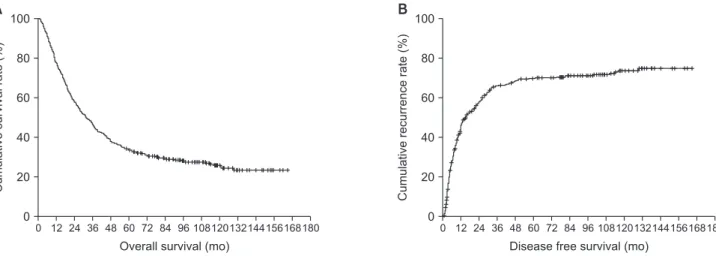
The overall 1-, 3-, and 5-year survival rates of patients were 76.5%, 44.1%, and 33.3%, respectively, while the disease-free 1-, 3-, and 5-year survival rates were 51.1%, 31.0%, and 28.3%, respectively (Fig. 1). Multivariate analysis showed that high CA 19-9 (≥38 U/mL) (HR, 2.191; 95% CI, 1.601–2.998; P < 0.001), LN
metastasis (HR, 1.750; 95% CI, 1.281–2.391; P = 0.001), and LVI (HR, 1.519; 95% CI, 1.097–2.104; P = 0.012) were independent factors associated with overall survival. Of these, high CA 19-9 (≥38 U/mL) (HR, 1.549; 95% CI, 1.126–2.133; P = 0.007) and LN metastasis (HR, 1.426; 95% CI, 1.022–1.990; P = 0.037) were also analyzed as independent factors affecting recurrence;
additionally, adjuvant therapy (HR, 1.743; 95% CI, 1.242–2.447;
P = 0.001) was another independent factor that affected recurrence (Table 2).
Comparison of clinicopathologic data between patients who survived ≥10 and <10 years
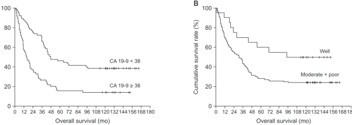
The results of the univariate analysis of patients who survived for over 10 years and less than 10 years are demonstrated in Table 3. Lower levels of CA 19-9 (<38 U/mL) were more frequently observed in patients who survived for more than 10 years (P = 0.001). Additionally, the prevalence macroscopic type (P = 0.008), histological type (P = 0.042), LVI (P = 0.040), PNI (P = 0.032), and adjuvant therapy (P = 0.030) were statistically different between both groups. Multivariate analysis also showed that low CA 19-9 (<38 U/mL) (HR, 3.755;
95% CI, 1.545–9.124; P = 0.003) and well-differentiated types (HR, 3.879; 95% CI, 1.282–11.735) were independent factors affecting the ≥10-year survival rates (Fig. 2).
Comparison of clinicopathologic data between patients who survived >5 and <1 year
Of the patients who died within 1 year (group 1) and those who survived for more than 5 years (group 2), the proportions of each of the following factors showed significant differences in univariable analysis: (1) CA 19-9 > 38 U/mL (group 1: 74.7%
vs. group 2: 34.7%, P = 0.001), (2) tumor size > 5 cm (group 1: 57.7% vs. group 2: 43.0%, P = 0.035), (3) intraductal growth type (group 1: 3.1% vs. group 2: 22.4%, P = 0.001), (4) well differentiation type (group 1: 6.2% vs. group 2: 27.1%, P = 0.003), (5) PNI (group 1: 54.2% vs. group 2: 19.6%; P < 0.001), (6) vascular invasion (group 1: 76.2% vs. group 2: 4.7%, P < 0.001), (7) LN metastasis (group 1: 52.5% vs. group 2: 16.7%; P < 0.001).
Table 1. Patient’s characteristics (n = 429)
Characteristic Value
Age (yr) 60.5 ± 9.8
Sex
Male 298 (69.5)
Female 131 (30.5)
Preoperative hepatolithiasis
Yes 27 (6.3)
No 402 (93.7)
Preoperative hepatitis
No 347 (90.9)
HBV 65 (15.2)
HCV 17 (4.0)
Cirrhosis
Yes 49 (11.4)
No 379 (88.3)
Preoperative CA 19-9 (U/mL) 901.2 (0.6–101,974.0) Operative methods
Anatomic resection 396 (92.3)
Nonanatomical resection 33 (7.7)
Postop complication
Yes 117 (27.1)
No 310 (72.9)
Clavien-Dindo classification grade
I/II 56 (13.0)
III/IV 61 (14.2)
Size of tumor (cm) 5.4 ± 3.0
Tumor multiplicity
Yes 45 (10.5)
No 384 (89.5)
Macroscopic type
Mass forming 296 (69.0)
Periductal infiltration type 44 (10.3)
Intraductal growth type 55 (12.8)
Mixed type 34 (8.0)
Histopathologic type
Well 73 (17.1)
Moderate 255 (59.4)
Poor 101 (23.6)
LN metastasis
N0 144 (33.6)
N1 87 (20.3)
No harvest 198 (46.2)
Surgical margin status
R0 340 (79.3)
R1 87 (20.3)
R2 2 (0.5)
Vascular invasion
Yes 43 (10.0)
No 386 (90.0)
Lymphovascular invasion
Yes 109 (25.5)
No 319 (74.5)
Perineural invasion
Yes 160 (37.4)
No 268 (62.6)
Table 1. Continued
Characteristic Value
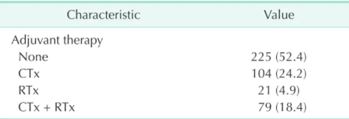
Adjuvant therapy
None 225 (52.4)
CTx 104 (24.2)
RTx 21 (4.9)
CTx + RTx 79 (18.4)
Values are presented as mean ± standard deviation, number (%), or median (range).
LN, lymph node; CTx, chemotherapy; RTx, radiation therapy.
DISCUSSION
It is widely acknowledged that the curative treatment of IHCC is surgical resection. Despite the surgical advances and risk factors for IHCC, the clinical outcome after resection is still dismal [13,14]. Consequently, the 5-year survival rate in our institution was 33.3%, which was comparable to the previously reported survival rates [11,13,15,16]. Although several studies have analyzed 5-year survival rates and recurrence rates, few studies have investigated the long-term survival for more than 10 years. This may be related to the low 5-year survival rate, and thus, the assumption that a small number of patients would survive for more than 10 years. To the best of our knowledge, only one retrospective single-institution study reported the results of an actual 10-year survival analysis [17].
Si et al. reported that 21 out of the 251 patients (8.4%) included in their study survived for more than 10 years; however, this value is not negligible. However, they also analyzed the clinicopathologic characteristics of patients who survived for more than 10 years and found that the prevalence of the following factors was higher in this cohort: time for the first recurrence, lower levels of alkaline phosphatase, tumor markers such as CEA and CA 19-9, single tumor, and smaller
tumor size. However, due to the small sample size, they did not obtain statistically string evidence that the factors mentioned above could affect long-term survival. In this study, 40 patients (25.1%) survived for more than 10 years after surgical resection, which is higher than the rate in the above-mentioned study.
Multivariate analysis demonstrated that low levels of CA 19-9 (<38 U/mL) and well-differentiated histologic types were predictors of survival for more than 10 years (Table 4). Among these factors, CA 19-9 is an easily and objectively measurable laboratory finding before surgery. These findings suggest that CA 19-9 may be used as a basis for the application of policies that consider more active surgical treatment in patients with low CA 19-9. In addition to curative resection in patients with high-level CA 19-9, further studies of adjuvant treatment may be needed to improve the poor survival rates.
Furthermore, LN metastasis appeared to be an independent factor affecting both survival and recurrence in this study. In a recently published meta-analysis, this has been shown to be the strongest predictor of survival (HR, 2.09; 95% CI, 1.80–
2.43; P < 0.001) [18]. The results were based on 5 previously published studies with the largest cohort. In only 2 of the 5 studies mentioned above, the frequencies of lymphadenectomy were described as 55.2% and 72.1%, respectively [8,11]. In these Table 2. Cox proportional hazard analysis of factor for survival and recurrence (n = 429)
Characteristic Survival Recurrence
HR (95% CI) P-value HR (95% CI) P-value
CA 19-9 (≥38 U/mL) 2.191 (1.601–2.998) 0.001 1.549 (1.126–2.133) 0.007
LN metastasis 1.750 (1.281–2.391) 0.001 1.426 (1.022–1.990) 0.037
Lymph vascular invasion 1.519 (1.097–2.104) 0.012 - -
Adjuvant treatment - - 1.743 (1.242–2.447) 0.001
HR, hazard ratio; CI, confidence interval; LN, lymph node.
Fig. 1. Kaplan-Meier survival curves. (A) Cumulative survival rate after curative resection. (B) Cumulative recurrence rate after curative resection.
0 180
Cumulativesurvivalrate(%)
Overall survival (mo) 100
80
60
40
20
0
12 24 36 48 60 72 84 96 108120132144156168 0 180
Cumulativerecurrencerate(%)
Disease free survival (mo) 100
80
60
40
20
0
12 24 36 48 60 72 84 96 108120132144156168
A B
Table 3. Clinicopathological finding in patients with IHCC who underwent curative resection: univariate analysis of patients with < or
≥10-year survival from 2003 to 2007 (n = 144)
Characteristic Survival < 10 years (n = 104) Survival ≥ 10 years (n = 40) P-value
Age (yr) 57.8 ± 9.3 57.7 ± 10.7 0.703
Sex 0.663
Male 67 (64.4) 28 (70.0)
Female 37 (35.6) 12 (30.0)
Preoperative hepatolithiasis >0.999
Yes 8 (7.7) 3 (7.5)
No 96 (92.3) 37 (92.5)
Preoperative hepatitis 0.184
No 90 (86.5) 31 (77.5)
HBV 11 (10.6) 5 (12.5)
HCV 3 (2.9) 4 (10.0)
Cirrhosis 0.220
Yes 7 (6.7) 6 (15.0)
No 97 (93.3) 34 (85.0)
Preoperative CA 19-9 (U/mL) 577.4 ± 1,329.9 46.6 ± 135.1 0.001
Operative methods 0.442
Anatomic resection 99 (95.2) 36 (90.0)
Nonanatomical resection 5 (4.8) 4 (10.0)
Postoperative complication 0.278
Yes 32 (30.8) 8 (20.0)
No 72 (69.2) 32 (80.0)
Size of tumor (cm) 5.9 ± 3.5 5.1 ± 2.5 0.110
Tumor multiplicity >0.999
Yes 12 (11.5) 5 (12.5)
No 92 (88.5) 35 (87.5)
Macroscopic type 0.008
Mass forming 71 (68.3) 24 (60.0)
Periductal infiltration type 11 (10.6) 3 (7.5)
Intraductal growth type 12 (11.5) 13 (32.5)
Mixed type 10 (9.6) 0 (0.0)
Histopathologic type 0.042
Well 10 (9.6) 10 (25.0)
Moderate 67 (64.4) 19 (47.5)
Poor 27 (26.0) 11 (27.5)
LN metastasis 0.348
N0 38 (36.5) 17 (42.5)
N1 21 (20.2) 4 (10.0)
No harvest 45 (43.3) 19 (47.5)
Surgical margin status 0.133
R0 78 (75.0) 36 (90.0)
R1 25 (24.0) 4 (10.0)
R2 1 (1.0) 0 (0.0)
Vascular invasion 0.821
Yes 5 (4.8) 3 (7.5)
No 99 (95.2) 37 (92.5)
Lymphovascular invasion 0.065
Yes 34 (32.0) 6 (15.0)
No 70 (68.0) 34 (85.0)
Perineural invasion 0.032
Yes 37 (35.0) 6 (15.0)
No 67 (65.0) 34 (85.0)
Adjuvant therapy 0.030
None 60 (57.7) 32 (80.0)
CTx 19 (18.3) 2 (5.0)
RTx 2 (1.9) 2 (5.0)
CTx + RTx 23 (22.1) 4 (10.0)
Values are presented as mean ± standard deviation or number (%).
LN, lymph node; CTx, chemotherapy; RTx, radiation therapy.
studies, LN metastasis was found in more than one-quarter of all patients undergoing LN dissection. Conversely, we performed lymphadenectomy in more than half of the patients, with one-third of these patients presenting with metastatic LN, which is comparable to the previous 2 studies. These results indicate that LN metastasis was found in at least 30% of patients who underwent lymphadenectomy, and this should not be ignored. Furthermore, LN metastasis is essential for accurate staging and subsequent prognosis prediction. Therefore, routine lymphadenectomy of IHCC should be considered. However, since this analysis is derived from a retrospective study, it is necessary to verify this proposal through a randomized control study.
One of the interesting findings of this study is that LVI is an independent prognostic factor for survival. In fact, LVI has been reported to be associated with LN metastasis in breast cancer [19], endometrial cancer [20], and colon cancer [21]. It has also been identified as an independent risk factor for survival in urinary tract carcinoma [22,23]. In a previous multi-institutional
analysis of IHCC, LVI proved to be an indicator of poor tumor biology and was suggested for use as a screening criterion for adjuvant treatment [24]. Although the oncologic effect of the adjuvant treatment was not statistically analyzed in this study, it is meaningful that the concept of LVI is applied to patients who undergo surgical treatment for IHCC. Therefore, future studies will need to demonstrate that adjuvant treatment can achieve a good oncologic outcome in patients with LVI.
There are several limitations to this study. First, by analyzing cohorts collected over a long period of time, it is possible that changes in surgical methods or treatment policies may have introduced some bias. Second, we did not routinely perform lymphadenectomy, and therefore, the influence of LN metastasis may have been reduced, which may have influenced multivariate analysis of risk factors. Additionally, the extent of lymphadenectomy may be determined by the subjective judgment of the surgeon. Finally, contrary to our expectation, adjuvant treatment appears to be a factor that increased the recurrence rate. This may be attributed to the limitations inherent to retrospective studies, and may also result from the more aggressive adjuvant treatment that is applied to patients with advanced-stage tumor or poor tumor biology. In the future, large-scale prospective studies should be conducted to establish a more detailed guideline to decide if adjuvant treatment should be recommended.
In conclusion, an elevated level of CA 19-9 (≥38 U/mL), LN metastasis, and LVI were identified as independent risk factors for survival after resection of IHCC. Furthermore, CA 19-9 (<38 U/mL) and the histologic type were independent factors predicting the survival for more than 10 years. Despite the poor outcome of surgical treatment for IHCC, patients with these factors are expected to survive for a relatively long time and they should be actively considered for surgical treatment.
Table 4. Multivariate analysis of patients with < or ≥10-year survival from 2003 to 2007 (n = 144)
Characteristics Multivariate
HR 95% CI P-value
CA 19-9 (<38 U/mL) 3.755 1.545–9.124 0.003 Macroscopic typea) 1.588a) 0.491–1.588 0.440 Histologic type 3.879 1.282–11.735 0.016 Lymphovascular invasion 0.831 0.255–2.705 0.831 Perineural invasion 0.472 0.162–1.376 0.169 Adjuvant therapy 0.675 0.252–1.803 0.433 HR, hazard ratio; CI, confidence interval.
a)HR of intraductal growth type compared to mass-forming type.
Fig. 2. (A) The results of Kaplan-Meier analysis of patients who survived over 10 years and below 10 years according to the CA 19-9 level. (B) The results of Kaplan-Meier analysis of patients who survived over 10 years and below 10 years according to the histopathological type.
0 180
Cumulativesurvivalrate(%)
Overall survival (mo) 100
80
60
40
20
0
12 24 36 48 60 72 84 96 108120132144156168 0 180
Cumulativerate(%)survival
Overall survival (mo) 100
80
60
40
20
0
12 24 36 48 60 72 84 96 108120132144156168
A B
CA 19-9 < 38
CA 19-9 > 38
Well
Moderate + poor
CONFLICTS OF INTEREST
No potential conflict of interest relevant to this article was reported.
ACKNOWLEDGEMENTS
The authors would like to thank Young Hun You (Department of Surgery, Samsung Medical Center, Sungkyunkwan University School of Medicine) for help with data analysis.
REFERENCES
1. Dodson RM, Weiss MJ, Cosgrove D, Herman JM, Kamel I, Anders R, et al. Intrahepatic cholangiocarcinoma:
management options and emerging therapies. J Am Coll Surg 2013;217:736-50.
e4.
2. Khan SA, Toledano MB, Taylor-Robinson SD. Epidemiology, risk factors, and pathogenesis of cholangiocarcinoma. HPB (Oxford) 2008;10:77-82.
3. Poultsides GA, Zhu AX, Choti MA, Pawlik TM. Intrahepatic cholangiocarcinoma.
Surg Clin North Am 2010;90:817-37.
4. Sempoux C, Jibara G, Ward SC, Fan C, Qin L, Roayaie S, et al. Intrahepatic cholangiocarcinoma: new insights in pathology. Semin Liver Dis 2011;31:49-60.
5. Tan JC, Coburn NG, Baxter NN, Kiss A, Law CH. Surgical management of intrahepatic cholangiocarcinoma--a population-based study. Ann Surg Oncol 2008;15:600-8.
6. Weimann A, Varnholt H, Schlitt HJ, Lang H, Flemming P, Hustedt C, et al. Retrospective analysis of prognostic factors after liver resection and transplantation for cholangiocellular carcinoma. Br J Surg 2000;87:1182-7.
7. Jonas S, Thelen A, Benckert C, Biskup W, Neumann U, Rudolph B, et al.
Extended liver resection for intrahepatic cholangiocarcinoma: a comparison of the prognostic accuracy of the fifth and sixth editions of the TNM classification. Ann Surg 2009;249:303-9.
8. Ribero D, Pinna AD, Guglielmi A, Ponti A, Nuzzo G, Giulini SM, et al. Surgical approach for long-term survival of patients with intrahepatic cholangiocarcinoma:
a multi-institutional analysis of 434
patients. Arch Surg 2012;147:1107-13.
9. Farges O, Fuks D, Boleslawski E, Le Treut YP, Castaing D, Laurent A, et al. Influence of surgical margins on outcome in patients with intrahepatic cholangiocarcinoma: a multicenter study by the AFC-IHCC-2009 study group. Ann Surg 2011;254:824-29.
10. Dhanasekaran R, Hemming AW, Zendejas I, George T, Nelson DR, Soldevila-Pico C, et al. Treatment outcomes and prognostic factors of intrahepatic cholangiocarcinoma.
Oncol Rep 2013;29:1259-67.
11. de Jong MC, Nathan H, Sotiropoulos GC, Paul A, Alexandrescu S, Marques H, et al. Intrahepatic cholangiocarcinoma: an international multi-institutional analysis of prognostic factors and lymph node assessment. J Clin Oncol 2011;29:3140-5.
12. Guglielmi A, Ruzzenente A, Campagnaro T, Pachera S, Valdegamberi A, Nicoli P, et al. Intrahepatic cholangiocarcinoma:
prognostic factors after surgical resection.
World J Surg 2009;33:1247-54.
13. Yamashita Y, Taketomi A, Morita K, Fukuhara T, Ueda S, Sanefuji K, et al.
The impact of surgical treatment and poor prognostic factors for patients with intrahepatic cholangiocarcinoma:
retrospective analysis of 60 patients.
Anticancer Res 2008;28(4C):2353-9.
14. Yamashita YI, Wang H, Kurihara T, Tsujita E, Nishie A, Imai K, et al. Clinical significances of preoperative classification of intrahepatic cholangiocarcinoma:
different characteristics of perihilar vs.
peripheral ICC. Anticancer Res 2016;36:
6563-9.
15. Nuzzo G, Giuliante F, Ardito F, De Rose AM, Vellone M, Clemente G, et
al. Intrahepatic cholangiocarcinoma:
prognostic factors after liver resection.
Updates Surg 2010;62:11-9.
16. Spolverato G, Kim Y, Alexandrescu S, Marques HP, Lamelas J, Aldrighetti L, et al. Management and outcomes of patients with recurrent intrahepatic cholangiocarcinoma following previous curative-intent surgical resection. Ann Surg Oncol 2016;23:235-43.
17. Si A, Li J, Xiang H, Zhang S, Bai S, Yang P, et al. Actual over 10-year survival after liver resection for patients with intrahepatic cholangiocarcinoma.
Oncotarget 2017;8:44521-32.
18. Mavros MN, Economopoulos K P, Alexiou VG, Pawlik TM. Treatment and prognosis for patients with intrahepatic c hol a ng io c a rc i nom a: s ystem at ic review and meta-analysis. JAMA Surg 2014;149:565-74.
19. Cornwell LB, McMasters KM, Chagpar AB.
The impact of lymphovascular invasion on lymph node status in patients with breast cancer. Am Surg 2011;77:874-7.
20. Guntupalli SR, Zighelboim I, Kizer NT, Zhang Q, Powell MA, Thaker PH, et al.
Lymphovascular space invasion is an independent risk factor for nodal disease and poor outcomes in endometrioid endometrial cancer. Gynecol Oncol 2012;
124:31-5.
21. Royston D, Jackson DG. Mechanisms of lymphatic metastasis in human colorectal adenocarcinoma. J Pathol 2009;217:608- 19.
22. Brunocilla E, Pernetti R, Martorana G.
The prognostic role of lymphovascular invasion in urothelial-cell carcinoma of upper and lower urinary tract. Anticancer
Res 2011;31:3503-6.
23. Gondo T, Nakashima J, Ozu C, Ohno Y, Horiguchi Y, Namiki K, et al. Risk stratification of survival by lympho- vascular invasion, pathological stage, and
surgical margin in patients with bladder cancer treated with radical cystectomy.
Int J Clin Oncol 2012;17:456-61.
24. Fisher SB, Patel SH, Kooby DA, Weber S, Bloomston M, Cho C, et al. Lympho-
vascular and perineural invasion as selection criteria for adjuvant therapy in intrahepatic cholangiocarcinoma: a multi- institution analysis. HPB (Oxford) 2012;
14:514-22.