Fenestration of liver cysts in polycystic liver disease to improve quality of life: a case report and literature review
Dong-Hwan Jung, Shin Hwang, Chul-Soo Ahn, Deok-Bog Moon, Gi-Won Song, Ki-Hun Kim, Tae-Yong Ha, Gil-Chun Park, and Sung-Gyu Lee
Division of Hepatobiliary Surgery and Liver Transplantation, Departments of Surgery, Asan Medical Center, University of Ulsan College of Medicine, Seoul, Korea
Polycystic liver disease (PCLD) is characterized by a large number of liver cysts scattered throughout the liver parenchyma. We herein intend to present the beneficial effect of palliative fenestration treatment on quality of life in a patient with symptomatic PCLD. A 48-year-old female patient had been followed up for 5 years for both polycystic liver and kidney diseases at another institution. During follow-up for last 1 year, we recognized that she had barely maintained her ability of function in daily activities due to progressive worsening of fatigue and dyspnea on exertion.
The patient finally underwent surgical fenestration treatment. Multiple cysts in the enlarged liver were opened and the cyst walls were excised with electrocautery. No surgical complication occurred and the patient was discharged 10 days after the open fenestration surgery. The total liver volume was 3,870 ml before surgery and 3,125 ml at 1 week after surgery, showing a volume reduction of 19.3%. After surgery, her performance status improved significantly. In the present case, significant improvement in quality of life and daily activity performance was achieved after open fenestra- tion treatment over 18 months of follow-up without disease recurrence. (Korean J Hepatobiliary Pancreat Surg 2015;19:
40-46)
Key Words: Fenestration; Polycystic liver disease; Quality of life; Liver transplantation
Received: January 11 2015; Revised: January 30 2015; Accepted: February 19, 2015 Corresponding author: Shin Hwang
Department of Surgery, Asan Medical Center, University of Ulsan College of Medicine, 88, Olympic-ro 43-gil, Songpa-gu, Seoul, 138-736, Korea
Tel: +82-2-3010-3930, Fax: +82-2-3010-6701, E-mail: [email protected].
Copyright Ⓒ 2015 by The Korean Association of Hepato-Biliary-Pancreatic Surgery
This is an Open Access article distributed under the terms of the Creative Commons Attribution Non-Commercial License (http://creativecommons.org/
licenses/by-nc/3.0) which permits unrestricted non-commercial use, distribution, and reproduction in any medium, provided the original work is properly cited.
Korean Journal of Hepato-Biliary-Pancreatic Surgery ∙ pISSN: 1738-6349ㆍeISSN: 2288-9213
INTRODUCTION
Polycystic liver disease (PCLD) is characterized by a large number of liver cysts scattered throughout the liver parenchyma. This disease entity is usually associated with autosomal dominant polycystic kidney disease. Patients with autosomal dominant polycystic kidney disease have polycystic kidneys, a feature that is absent in patients with autosomal dominant PCLD.1 Symptoms are often absent in patients with PCLD until there is a significant increase in cyst size. They mainly include abdominal pain, early satiety, dyspnea, nausea, and vomiting.2
Treatment is indicated if the symptom becomes severe or intolerable, and the therapeutic intention is to decrease the total liver volume leading to relief of compression-re- lated symptoms.3,4 Most of the PCLD patients present with only disturbing symptoms that deteriorate health
quality and limit daily activities rather than causing seri- ous liver function-related complications.
There are several therapeutic options, including aspira- tion-sclerotherapy, cyst fenestration, partial liver resection, and liver transplantation. The choice of treatment usually depends on the number, size, and location of the liver cysts.5 The technique of fenestration of liver cysts was first described in 1968,6 which was reported to be a safe and effective treatment for the management of sympto- matic non-parasitic cysts of the liver.7 In this study, we intend to present the beneficial effect of palliative fenes- tration treatment on quality of life in a patient with symp- tomatic PCLD.
CASE
A 48-year-old female patient had been followed up for
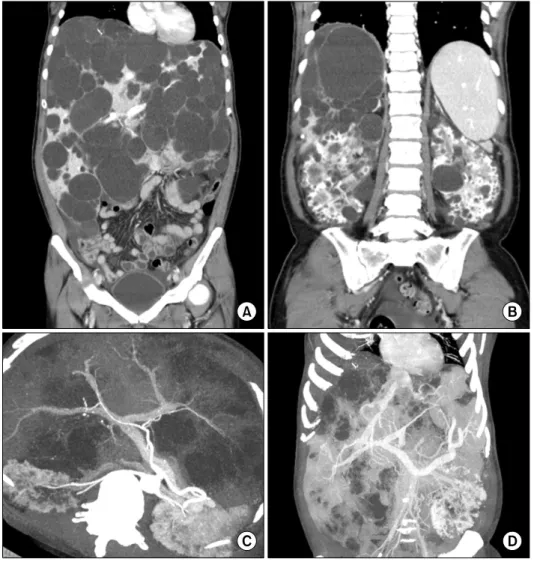
Fig. 1. Computed tomography images of the abdomen. Multiple cysts occupied the liver (A) and both kidneys (B), but the hepatic arterial and portal venous flow was well preserved (C) and hep- atic veins were extrinsically com- pressed (D).
Fig. 2. Gross morphology of polycystic liver disease according to Gigot’s classification. Type I: presence of less than 10 large hepatic cysts measuring more than 10 cm in maximum diameter. Type II: diffuse involvement of liver parenchyma by multiple cysts with remaining large areas of non-cystic liver parenchyma. Type III: presence of diffuse involvement of liver parenchyma by small- and medium-sized liver cysts with only a few areas of normal liver parenchyma.
5 years for both polycystic liver and kidney diseases at other institution. She was referred to us for identification of effective treatment. At the first visit, she complained of progressive distension of the abdomen and abdominal fullness leading to gradual decrease in body weight with- out shortness of breath. Her liver function was quite nor-
mal; hence, we decided to observe the patient regularly without considering palliative surgery or liver transplan- tation. Medical treatment with somatostatin analogue was not considered primarily due to the high medical cost as well as the undetermined therapeutic efficacy.
During the outpatient clinic follow-up for 1 year, we
Table 1. Qian’s classification according to the number of cysts and the presence of symptomatic hepatomegaly
Grade Number of cysts Hepatomegaly 1
2 3 4
0 1 to 10 11 to 20
>20
Asymptomatic Asymptomatic Asymptomatic Symptomatic
Fig. 3. Magnetic resonance imag- ing study of the abdomen. Water- filled multiple liver and kidney cysts were visible (A). The gall- bladder was collapsed by the ad- jacent liver cysts but the bile duct was not dilated (B).
Table 2. Schnelldorfer’s classification for polycystic liver disease
Type A Type B Type C Type D
Symptoms
Cyst characteristics Areas of relative normal liver parenchyma Presence of portal vein or hepatic vein occlusion in the preserved hepatic sections
Recommended therapy
Absent or mild or mild Any
Any
Any
Observation or medical therapy
Moderate or severe Limited No. large cysts
≥2 sections
Absent
Cyst fenestration
Severe (or moderate) Any
≥1 section
Absent
Partial hepatectomy with possible fenestration of remnant cysts
Severe (or moderate) Any
<1 section
Present
Liver transplantation recognized that she had barely maintained her ability of
function in daily activities due to progressive worsening of fatigue and dyspnea on exertion. Finally, she com- plained of shortness of breath even in a resting state.
Eastern Cooperative Oncology Group (ECOG) perform- ance status worsened from 2 to 3 during observation for 1 year.7 Gastrointestinal Quality of Life Index (GIQLI) score also deteriorated from 75 to 44 during observation for 1 year.8 Physical examination revealed that the liver was prominently bulged out of the abdomen, but leg ede- ma was absent. Liver and kidney functions were still quite normal. Abdominal computed tomography (CT) showed
progressive enlargement of multiple liver cysts with smooth and regular walls (Fig. 1), which was regarded as type III according to Gigot’s classification (Fig. 2),9 grade 4 according to Qian’s classification (Table 1),10 and type C according to Schnelldorfer’s classification (Table 2).11 Magnetic resonance imaging study revealed water-filled multiple liver and kidney cysts (Fig. 3).
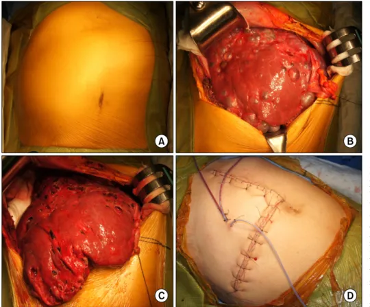
After obtaining consent of the patient on the uncertain effect of fenestration treatment as well as high risk of dis- ease recurrence, the patient underwent surgical fenestra- tion treatment. Multiple cysts in the enlarged liver were opened and the cyst walls were excised with electro- cautery (Fig. 4). To avoid bleeding and bile leakage, the thin membranous portions were meticulously fenestrated and none of the viable normal liver parenchyma was resected. The majority of accessible liver cysts were opened. At the end of the fenestration procedure, the liver appeared to be markedly shrunken. Three Jackson-Pratt type drains were inserted to evacuate the ascitic fluid: the abdominal drainage output was about 500 ml/day at post-
Fig. 4. Operative findings of the patient undergoing open fenes- tration surgery. Enlarged liver was bulged out of the abdomen (A); There were numerous liver cysts, but the majority of liver parenchyma was preserved (B);
Fenestration of the liver cyst led to moderate reduction of the whole liver volume (C); Bulging mass in the abdomen dis- appeared at the time of abdomi- nal wall closure (D).
operative day 1 and then it gradually decreased to less than 150 ml/day after 5 days. After performing follow-up CT at 1 week, the drains were removed. No surgical com- plication occurred and the patient was discharged 10 days after the open fenestration surgery. The total liver volume by using CT volumetry was 3,870 ml before surgery and 3,125 ml at 1 week after surgery, showing a volume re- duction of 19.3%.
At 1 month after surgery, ECOG performance status improved to 1 and the GIQLI score was significantly in- creased to 122. During regular follow-up at an interval of 6 months, follow-up CT showed no progression in cyst size and flat abdomen was observed (Fig. 5). After 1 year, ECOG performance status improved to 0 but the GIQLI score was slightly decreased to 114. The patient is doing well and she is performing her normal activities at 18 months after surgery with no evidence of disease recurrence.
DISCUSSION
The therapeutic intention of fenestration treatment for PCLD is to achieve relief of symptoms through liver vol-
ume reduction by rupture of liver cysts. In 1997, Gigot et al.9 evaluated 10 patients who had been treated by an aggressive attempt to reduce liver volume, mainly by open liver cyst fenestration. Deep-seated cysts were also opened with the aid of intraoperative ultrasonography, due to which the average liver volume decreased from 7,761 ml to 4,596 ml, resulting in a reduction of 43%. Both pre- operative liver volume and volume reduction were greater than that obtained in the present case probably due to more extensive fenestration of deep-seated cysts. Such an aggressive approach is associated with a higher prevalence of intra- and postoperative complications such as intra- operative massive hemorrhage and a biliary tear, post- operative biliary leakage and ascites, and obstruction of the inferior vena cava. Thus, the extent of cyst fenestra- tion should be prudently adjusted to minimize surgical complications. One study using laparoscopic fenestration showed a median liver volume reduction of 12.5%, im- plicating the limit of laparoscopic fenestration approach.4 Probably because high recurrence rate may be associated with low efficacy of liver volume reduction, laparoscopic fenestration for PCLD has not yet been accepted as a de- finitive treatment.12-15
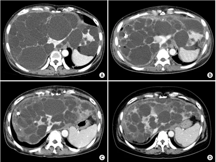
Fig. 5. Computed tomography follow-up of the abdomen. Multiple distended cysts occupied the liver just before surgery (A);
Cyst size was significantly reduced 1 week after fenestration surgery (B); No increase in the size of liver cysts was observed in the 6-month (C) and 18-month (D) images. Because of improvement in the nutritional status, subcutaneous fat was thickened in the 18-month image (D).
As shown in many other studies, liver cysts are prone to becoming enlarged with the passage of time. In view of the lack of an explicit definition of recurrence, it is dif- ficult to assess the recurrence rate after fenestration treat- ment in the literature.4 Some authors defined recurrence as recurrence of cysts on imaging studies, other authors defined recurrence as recurrence of symptoms, while some authors considered both symptomatic and radiological recurrence. Published series describing the results of open and laparoscopic fenestration has been summarized in the literature.16 Immediate symptom relief is achieved in 92%
of the patients, whereas up to 25% experience recurrence of the cysts or symptoms.17 Complication rate after fenes- tration is in the range of 23% while mortality is about 2%.
Factors that predict failure of fenestration are previous ab- dominal procedures, deep-seated cysts, incomplete unroof-
ing, cysts in segments VII and VIII, and the presence of diffuse PCLD.17
Hepatic resection is usually reserved for highly sympto- matic patients who are incapacitated by their disease due to the massive expansion of their livers.9 In such patients, fenestration alone is rarely successful because the liver pa- renchyma is rigid and it does not collapse.17 Symptom re- lief is achieved in 86% of the patients although cyst re- currence is expected in one third of the patients.18 The morbidity rate associated with this procedure can be up to 50% and includes ascites, pleural effusions, biliary leak- age, and hemorrhage. One of the reasons for these compli- cations is the fact that there is a significant distortion of the intra-hepatic vasculature and biliary tree which makes these procedures technically very challenging.
Orthotopic liver transplantation is the only curative
Fig. 6. Operative findings of a 52-year-old female patient un- dergoing liver transplantation with a MELD score of 18.
Markedly enlarged liver was bulged out of the abdomen (A);
There were numerous liver cysts that occupied the majority of liv- er parenchyma (B); Rupture of the liver cysts led to reduction of the liver volume (C), which fa- cilitated handling of the native liver for liver transplantation (D).
treatment for patients with severe PCLD.19 It is indicated in those patients with disabling symptoms that lead to de- creased performance status and quality of life. Patients with PCLD usually have normal liver function and the or- gan allocation system based on the Model for End-Stage Liver Disease (MELD) or Korean Network for Organ Sharing (KONOS) is often unable to assist this group of patients. For these patients, MELD exception criteria seem to be necessary.20,21 Because of the shortage of available grafts, the need for life-long immunosuppression, and the perioperative risks, liver transplantation is indicated only in symptomatic patients (Fig. 6). In the current Korean setting in which the majority of deceased donor liver grafts are allocated to urgent patients, most patients with PCLD have a very low chance of receiving deceased do- nor liver grafts; therefore, some of them have undergone living donor liver transplantation.22,23
In patients with PCLD, patient selection, timing and choice of treatments can be very challenging. In sympto- matic patients, treatment strategies should be based on the degree and progression of symptoms and the severity of other medical conditions. Symptomatic patients with large cysts or limited hepatic involvement might benefit from
fenestration or sclerotherapy. Hepatic resection with or without fenestration should be favored in patients with diffuse involvement of the liver but with sufficiently pre- served parenchyma. Finally, in the patients with diffuse disease, liver transplantation is a valid option and should be pursued as primary therapy prior to the development of debilitating disease that can significantly increase the risks of perioperative adverse events.16
In the present case, significant improvement in quality of life and daily activity performance was achieved after open fenestration treatment over 18 months of follow-up without disease recurrence.
REFERENCES
1. Qian Q, Li A, King BF, Kamath PS, Lager DJ, Huston J 3rd, et al. Clinical profile of autosomal dominant polycystic liver disease. Hepatology 2003;37:164-171.
2. Arnold HL, Harrison SA. New advances in evaluation and man- agement of patients with polycystic liver disease. Am J Gastroenterol 2005;100:2569-2582.
3. Szabó LS, Takács I, Arkosy P, Sápy P, Szentkereszty Z.
Laparoscopic treatment of nonparasitic hepatic cysts. Surg Endosc 2006;20:595-597.
4. van Keimpema L, Ruurda JP, Ernst MF, van Geffen HJ, Drenth JP. Laparoscopic fenestration of liver cysts in polycystic liver
disease results in a median volume reduction of 12.5%. J Gastrointest Surg 2008;12:477-482.
5. Everson GT, Taylor MR, Doctor RB. Polycystic disease of the liver. Hepatology 2004;40:774-782.
6. Lin TY, Chen CC, Wang SM. Treatment of non-parasitic cystic disease of the liver: a new approach to therapy with polycystic liver. Ann Surg 1968;168:921-927.
7. Oken MM, Creech RH, Tormey DC, Horton J, Davis TE, McFadden ET, et al. Toxicity and response criteria of the Eastern Cooperative Oncology Group. Am J Clin Oncol 1982;5:649-655.
8. Eypasch E, Williams JI, Wood-Dauphinee S, Ure BM, Schmülling C, Neugebauer E, et al. Gastrointestinal Quality of Life Index: development, validation and application of a new instrument. Br J Surg 1995;82:216-222.
9. Gigot JF, Jadoul P, Que F, Van Beers BE, Etienne J, Horsmans Y, et al. Adult polycystic liver disease: is fenestration the most adequate operation for long-term management? Ann Surg 1997;225:286-294.
10. Qian Q, Li A, King BF, Kamath PS, Lager DJ, Huston J 3rd, et al. Clinical profile of autosomal dominant polycystic liver disease. Hepatology 2003;37:164-171.
11. Schnelldorfer T, Torres VE, Zakaria S, Rosen CB, Nagorney DM. Polycystic liver disease: a critical appraisal of hepatic re- section, cyst fenestration, and liver transplantation. Ann Surg 2009;250:112-118.
12. Gigot JF, Legrand M, Hubens G, de Canniere L, Wibin E, Deweer F, et al. Laparoscopic treatment of nonparasitic liver cysts: adequate selection of patients and surgical technique.
World J Surg 1996;20:556-561.
13. Giuliante F, D'Acapito F, Vellone M, Giovannini I, Nuzzo G.
Risk for laparoscopic fenestration of liver cysts. Surg Endosc 2003;17:1735-1738.
14. Konstadoulakis MM, Gomatos IP, Albanopoulos K, Alexakis N, Leandros E. Laparoscopic fenestration for the treatment of pa- tients with severe adult polycystic liver disease. Am J Surg 2005;189:71-75.
15. Robinson TN, Stiegmann GV, Everson GT. Laparoscopic pallia- tion of polycystic liver disease. Surg Endosc 2005;19:130-132.
16. Abu-Wasel B, Walsh C, Keough V, Molinari M. Pathophysiology, epidemiology, classification and treatment options for polycystic liver diseases. World J Gastroenterol 2013;19:5775-5786.
17. Drenth JP, Chrispijn M, Nagorney DM, Kamath PS, Torres VE.
Medical and surgical treatment options for polycystic liver disease. Hepatology 2010;52:2223-2230.
18. Que F, Nagorney DM, Gross JB Jr, Torres VE. Liver resection and cyst fenestration in the treatment of severe polycystic liver disease. Gastroenterology 1995;108:487-494.
19. Everson GT, Taylor MR, Doctor RB. Polycystic disease of the liver. Hepatology 2004;40:774-782.
20. Freeman RB Jr, Gish RG, Harper A, Davis GL, Vierling J, Lieblein L, et al. Model for end-stage liver disease (MELD) ex- ception guidelines: results and recommendations from the MELD Exception Study Group and Conference (MESSAGE) for the ap- proval of patients who need liver transplantation with diseases not considered by the standard MELD formula. Liver Transpl 2006;12:S128-S136.
21. Arrazola L, Moonka D, Gish RG, Everson GT. Model for end-stage liver disease (MELD) exception for polycystic liver disease. Liver Transpl 2006;12:S110-S111.
22. Moon DB, Lee SG, Hwang S, Kim KH, Ahn CS, Ha TY, et al. More than 300 consecutive living donor liver transplants a year at a single center. Transplant Proc 2013;45:1942-1947.
23. Moon DB, Lee SG, Hwang S, Kim KH, Ahn CS, Ha TY, et al. Toward more than 400 liver transplantations a year at a single center. Transplant Proc 2013;45:1937-1941.