J Korean Surg Soc 2012;83:227-236 http://dx.doi.org/10.4174/jkss.2012.83.4.227
ORIGINAL ARTICLE
Journal of the Korean Surgical Society
JKSS
pISSN 2233-7903ㆍeISSN 2093-0488
Received May 31, 2012, Revised August 8, 2012, Accepted August 23, 2012 Correspondence to: Young Hoon Kim
Department of Surgery, Dong-A University Medical Center, Dong-A University College of Medicine, 26 Daesingongwon-ro, Seo-gu, Busan 602-715, Korea
Tel: +82-51-240-5146, Fax: +82-51-247-9316, E-mail: yhkim1@dau.ac.kr
cc Journal of the Korean Surgical Society is an Open Access Journal. All articles are distributed under the terms of the Creative Commons Attribution Non-Commercial License (http://creativecommons.org/licenses/by-nc/3.0/) which permits unrestricted non-commercial use, distribution, and reproduction in any medium, provided the original work is properly cited.
Prognostic factors for gallbladder cancer in the laparoscopy era
Hak Youn Lee, Young Hoon Kim, Ghap Joong Jung, Young Hoon Roh, Si Young Park, Nam Uk Kang, Soon Hwa Yoon, Jin Han Cho
1, Myung Hwan Roh
2, Sang Young Han
2, Sung Wook Lee
2, Yang Hyun Baek
2, Jin Sook Jeong
3Departments of Surgery, 1Radiology, 2Internal Medicine, and 3Pathology, Dong-A University College of Medicine, Busan, Korea
Purpose: Hepatobiliary surgery has changed dramatically in recent decades with the advent of laparoscopic techniques. The aim of this retrospective study was to compare survival rates according to stages, adjusting for important prognostic factors.
Methods: A retrospective study of a 17-year period from January 1994 to April 2011 was carried out. The cases studied were divided into two time period cohorts, those treated in the first 9-years (n = 109) and those treated in the last 7-years (n = 109).
Results: An operation with curative intent was performed on 218 patients. The 5-year survival rates according to the depth of invasion were 86% (T1), 56% (T2), 45% (T3), and 5% (T4). The number of cases of incidental gallbladder cancer found during 3,919 laparoscopic cholecystectomies was 96 (2.4%). Incidental gallbladder cancer revealed a better survival rate (P = 0.003).
Iatrogenic bile spillage was found in 20 perforations of the gallbladder during laparoscopic cholecystectomies, 16 pre- operative percutaneous transhepatic gallbladder drainages and 16 percutaneous transhepatic biliary drainages; only percu- taneous transhepatic biliary drainage patients showed a significantly lower survival rate than patients without iatrogenic bile spillage (P < 0.034). Chemoradiation appeared to improve overall survival (P < 0.001). Multivariate analysis also re- vealed that time period, type of surgery, surgical margin, lymphovascular invasion, lymph node involvement, and chemo- radiation therapy had significant effects. Conclusion: This study found that the prognosis of gallbladder cancer is still de- termined by the stage at presentation due to the aggressive biology of this tumor. Early diagnosis, radical resection and ap- propriate adjuvant therapy can increase overall survival.
Key Words: Gallbladder cancer, Laparoscopy, Prognosis
INTRODUCTION
The incidence of gallbladder cancer (GBC) shows wide racial and geographical variation, and some genetic and environmental factors have been suggested with regards
to etiologic association in the development of GBC. These factors include gallstones, presence of chronic inflam- mation, adenoma, anomalous pancreatobiliary duct un- ion, sex, age, obesity, and parity. The incidence of GBC is high in Korea, Japan, and Central and Eastern European
countries [1]. GBC is an uncommon cancer that has tradi- tionally been associated with a poor prognosis. This poor prognosis is considered to be related to advanced stage at diagnosis, which is due to both the anatomic position of the gallbladder, and the vagueness and non-specificity of symptoms. In the era of laparoscopic cholecystectomy (LC), incidental GBC has dramatically increased and is now the major way patients present with GBC [2,3]. In many cases, the diagnosis is made after a cholecystectomy has been performed and an incidental tumor is identified in the specimen. Frozen section diagnosis is often chal- lenging and has its own limitations because of sampling problems and freezing artifacts [4]. The risk of peritoneal seeding caused by inadvertent spillage of cancer con- taminated bile during percutaneous transhepatic gall- bladder drainage (PTGBD) and percutaneous transhepa- tic biliary drainage (PTBD) may be high, but has rarely been reported [5]. GBC is a fatal disease that can only be cured by radical surgical resection. The goal of radical re- section should be cholecystectomy with en bloc resection of the invaded organs (most commonly the liver) around the tumor to provide reasonable margins and to resect region- al lymph nodes. However, there is still a debate about the extent of hepatic resection and the extent of regional lym- phadenectomy [6,7].
The aim of this retrospective study was to compare sur- vival rates according to tumor stages, using Cox re- gression comparison of survival by stage adjusting for im- portant prognostic factors.
METHODS
A retrospective study spanning a 17-year period from January 1994 to April 2011 was performed, and 218 con- secutive inpatients were identified. Cases were divided in- to two time-period cohorts: those treated in the first 9 years (n = 109) and those treated in the last 7 years (n = 109).
Survival analysis was performed considering patient vari- ables (age and gender), operative variables (LC only, LC with conversion open cholecystectomy, radical second re- section, primary open cholecystectomy, and hemi-hep- atectomy), incidentality, iatrogenic bile spillage, chemo-
radiation, and pathologic variables (T-stage, histologic type, degree of differentiation, surgical margin status, lympho vascular invasion, perineural invasion, and lymph node involvement). Our standard radical operative procedure was extended cholecystectomy (cholecystecto- my plus partial resection of approximately 2 cm from the gallbladder bed) with lymph node dissection in the hep- atoduodenal ligament, skeletonizing the portal vein and hepatic artery. Hemi-hepatectomy has also been described by the Couinaud segments resected. After exploratory surgery, opening the gallbladder on the back table and identifying the type of disease that is present, as well as in- traoperative frozen sections, were also performed in all ex- cept four cases. Clinical staging of the disease was accord- ing to the American Joint Committee on Cancer (AJCC) staging system (7th edition). We recommended additional surgery when the tumor stage was over T2 (tumor invades the perivascular connective tissue). Adjuvant therapy af- ter surgical resection was not strictly protocol driven and was administered at the discretion of surgeons and oncol- ogists during the treatment of patients. When the resection margin was positive for tumor infiltration and the patient refused an additional second operation, chemoradiation therapy to the tumor bed was performed. Patients were regularly followed up in the outpatient clinic every 3 to 6 months.
Statistical analyses
Data were summarized using descriptive statistics that included frequency and percentage for categorical varia- bles and mean ± standard deviation for continuous vari- ables. Differences in patient demographic and clinical characteristics were compared across subgroups using the chi-square test for categorical variables and analysis of variance for continuous variables. Overall survival (OS;
defined as the time from date of surgery to death) was esti- mated using Kaplan-Meier curves. Survival curves were compared between groups using the log-rank test.
Univariate analyses of prognostic factors were performed using Cox regression to examine differences with respect to OS. The potential prognostic factors considered are shown in Tables 1 and 2. Multivariate analyses using Cox regression were performed to identify prognostic factors
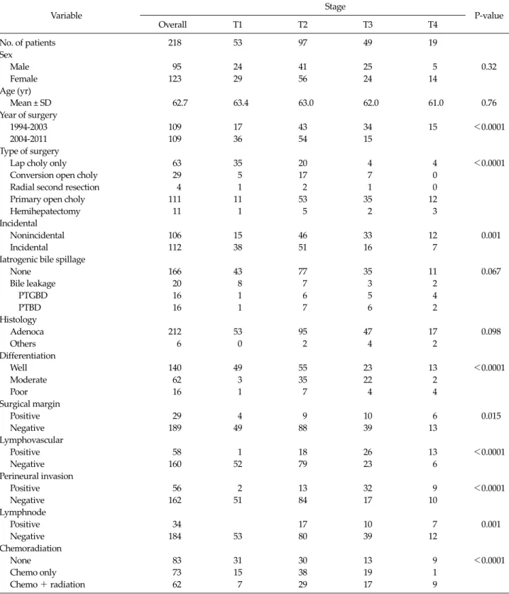
Variable Stage
P-value
Overall T1 T2 T3 T4
No. of patients 218 53 97 49 19
Sex
Male 95 24 41 25 5 0.32
Female 123 29 56 24 14
Age (yr)
Mean ± SD 62.7 63.4 63.0 62.0 61.0 0.76
Year of surgery
1994-2003 109 17 43 34 15 <0.0001
2004-2011 109 36 54 15
Type of surgery
Lap choly only 63 35 20 4 4 <0.0001
Conversion open choly 29 5 17 7 0
Radial second resection 4 1 2 1 0
Primary open choly 111 11 53 35 12
Hemihepatectomy 11 1 5 2 3
Incidental
Nonincidental 106 15 46 33 12 0.001
Incidental 112 38 51 16 7
Iatrogenic bile spillage
None 166 43 77 35 11 0.067
Bile leakage 20 8 7 3 2
PTGBD 16 1 6 5 4
PTBD 16 1 7 6 2
Histology
Adenoca 212 53 95 47 17 0.098
Others 6 0 2 4 2
Differentiation
Well 140 49 55 23 13 <0.0001
Moderate 62 3 35 22 2
Poor 16 1 7 4 4
Surgical margin
Positive 29 4 9 10 6 0.015
Negative 189 49 88 39 13
Lymphovascular
Positive 58 1 18 26 13 <0.0001
Negative 160 52 79 23 6
Perineural invasion
Positive 56 2 13 32 9 <0.0001
Negative 162 51 84 17 10
Lymphnode
Positive 34 17 10 7 0.001
Negative 184 53 80 39 12
Chemoradiation
None 83 31 30 13 9 <0.0001
Chemo only 73 15 38 19 1
Chemo + radiation 62 7 29 17 9
Lap choly, laparoscopic cholecystectomy; PTGBD, percutaneous transhepatic gallbladder drainage; PTBD, percutaneous transhepatic bile duct drainage.
Table 1. Patient baseline characteristics
independently related to OS. Cox regression was used to assess the difference between groups adjusting for sig-
nificant prognostic factors. In particular, the main analysis addressing the primary objective was a Cox regression
Fig. 1. Incidence by stage and group.
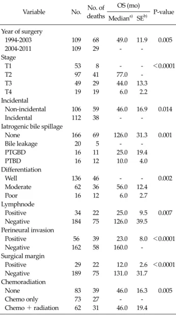
Variable No. No. of deaths
OS (mo)
P-value Mediana) SEb)
Year of surgery
1994-2003 109 68 49.0 11.9 0.005
2004-2011 109 29 - -
Stage
T1 53 8 - - <0.0001
T2 97 41 77.0 -
T3 49 29 44.0 13.3
T4 19 19 6.0 2.2
Incidental
Non-incidental 106 59 46.0 16.9 0.014
Incidental 112 38 - -
Iatrogenic bile spillage
None 166 69 126.0 31.3 0.001
Bile leakage 20 5 - -
PTGBD 16 11 25.0 19.4
PTBD 16 12 10.0 4.0
Differentiation
Well 136 46 - - 0.002
Moderate 62 36 56.0 12.4
Poor 16 12 6.0 2.7
Lymphnode
Positive 34 22 25.0 9.5 0.007
Negative 184 75 126.0 39.5
Perineural invasion
Positive 56 39 23.0 8.0 <0.0001
Negative 162 58 160.0 -
Surgical margin
Positive 29 22 12.0 2.6 <0.0001
Negative 189 75 131.0 31.7
Chemoradiation
None 83 39 46.0 16.3 0.005
Chemo only 73 27 - -
Chemo + radiation 62 31 46.0 19.4
OS, overall survival; SE, standard error; PTGBD, percutaneous transhepatic gallbladder drainage; PTBD, percutaneous transhe- patic bile duct drainage.
a)Median survival was not estimate since Kaplan-Meier curve did not reach 50% of the survival probability. b)Five-year OS estimate.
Table 2. Results from log-rank test
comparison of OS by stage, adjusting for important prog- nostic factors. All P-values < 0.05 were considered stat- ically significant. This study was explorative and therefore no adjustment for multiple testing was applied. All stat- istical analyses were carried out using SAS 9.1.3 (SAS Institute Inc., Cary, NC, USA) and R 2.9.2 (R Foundation for Statistical Computing, Vienna, Austria) statistical software.
RESULTS
Disease stage
Between January 1994 and April 2011, 218 patients un- derwent evaluation and surgical treatment for adenocar- cinoma of gallbladder. There was no postoperative death.
Median age was 63 years (range, 35 to 90 years) and 123 pa- tients were female (56%). The stage of the GBC was T1 in 53 (24%) patients, T2 in 97 (45%) patients, T3 in 49 (23%), and T4 in 19 (9%) patients.
The stage of disease at presentation affected the survival in all time periods. The 5-year estimated survival rates of patients according to the depth of invasion were: stage I (estimated survival rates limited to mucosa, muscularis, 86%; 95% confidence interval [CI], 0.73 to 0.94; P < 0.0001), stage II (subserosa, 56%; 95% CI, 0.44 to 0.66), stage III (serosa, 45%; 95% CI, 0.31 to 0.6), and stage IV (serosa with invasion to adjacent organs, 5%; 95% CI, 0.01 to 0.29).
Table 1 shows the distribution of patients by stage and time period. The notable change in tumor stage during the late period increased in the frequency of patients diag- nosed with stage I and II and decreased in stage III and IV disease (Fig. 1). Overall median survival improved com- paring early and late periods (Fig. 2). LC operations were more frequently performed in the late period and ap- peared to result in the earlier discovery of GBC, resulting in increased probability of survival.
Incidentality
During the 17-year period evaluated in this study, 3,919
Fig. 2. Comparison of overall survival according to groups.
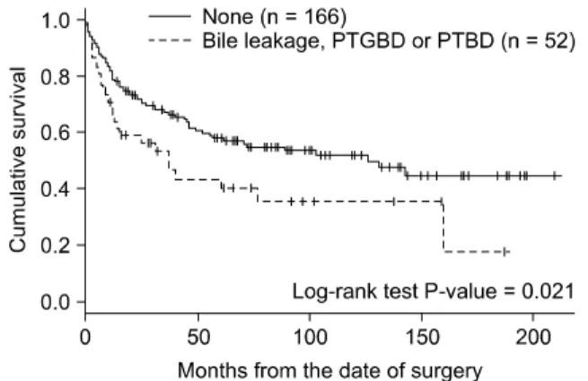
Fig. 4. Comparison of overall survival according to iatrogenic bile spillage. PTGBD, percutaneous transhepatic gallbladder drainage;
PTBD, percutaneous transhepatic bile duct drainage.
Fig. 3. Comparison of overall survival according to incidentality.
LCs were performed, of which 96 (2.4%) cases of GBC were incidentally discovered, and GBC was found incidentally in 112 of the 218 patients (50%) by permanent histological examination. Eighty nine (89%) of those patients whose GBC was found incidentally were stage T1 or T2. Patients who were found to have GBC incidentally at LC had a sig- nificant increase in survival when compared with those who were admitted electively with a known diagnosis (P = 0.014) (Fig. 3).
Intervention prior to definitive therapy and gall- bladder perforation during LC
Due to biliary colic, acute cholecystitis, cholangitis, choledocholithiasis, and gallstone pancreatitis, 16 patients had PTGBD, and 16 had PTBD with or without endoscopic sphincterotomy for the clearance of common bile duct stones prior to surgery. Gallbladder perforation during LC was found in 20 out of 218 patients (9%). Comparison of
OS according to incidence of bile leakage, iatrogenic bile spillage group (20 intraoperative bile leakage, 16 PTGBD, and 16 PTBD) demonstrated significantly worse survival (P = 0.021) (Fig. 4).
Surgical treatment (type of surgery)
The procedures performed as resection with curative intent included 11 (5%) major liver resection (≥hemi- hepatectomy) and 111 (51%) primary open cholecy- stectomy. Twenty nine patients (13%) were noted to have GBC during the laparoscopic procedures and were con- verted to open surgery. Four patients (2%) with histologi- cally proven GBC after LC underwent potentially curative resection, whereas 63 (29%) underwent LC only. In the LC only group, 55 patients (87%) were early stage (T1, T2) and, in the primary open cholecystectomy group, 47 pa- tients (42%) were late stage (T3, T4). In T1 and T2 compar- ison of 5-year OS according to type of surgery (LC only vs.
radical LC), there were no statistically significant differ- ences (T1, P = 0.502; T2, P = 0.216). In comparison of 5-year survival according to type of surgery (five categories), bet- ter survival was seen in re-open after LC (radical second resection) and LC plus conversion during the laparoscopic procedures (conversion open cholecystectomy). On the other hand, primary open cholecystectomy and hemi- hepatectomy showed poor survival (P = 0.0001).
Pathology
The distribution of histologic subtypes in the current re-
Variable Univariate Multivariate
HR 95% CI P-value HR 95% CI P-value
Stage (late vs. early) 2.61 1.75-3.89 <0.0001
Sex (male vs. female) 1.00 0.82-1.23 0.98
Age (≥63 yr vs.<63 yr) 1.29 0.86-1.93 0.21
Year of surgery (1994-2003 vs. 2004-2011) 1.86 1.19-2.90 0.006 1.78 1.09-2.89 0.021
Type of surgery (extended vs. LC only) 2.33 1.34-4.04 0.003 1.78 0.95-3.35 0.072
Incidental (yes vs. no) 1.66 1.10-2.50 0.016
Iatrogenic bile spillage
Bile leakage vs. none 0.72 0.29-1.78 0.476 0.50 0.20-1.23 0.14
PTGBD vs. none 1.96 1.04-3.71 0.039 1.12 0.56-2.23 0.75
PTBD vs. none 2.85 1.53-5.29 0.001 2.15 1.06-4.35 0.034
Adenoca
Poor vs. well 2.84 1.50-5.38 0.001
Moderate vs. well 1.57 1.02-2.43 0.042
Margin (positive vs. negative) 1.87 1.44-2.34 <0.0001 1.78 1.39-2.29 <0.0001
Lymphovascular (positive vs. negative) 1.69 1.38-2.07 <0.0001 1.41 1.12-1.77 0.003
Perineural invasion (positive vs. negative) 2.62 1.70-3.96 <0.0001
Lymphnode (positive vs. negative) 1.90 1.18-3.07 0.008 1.78 1.01-3.12 0.045
Chemo therapy (yes vs. no) 1.26 1.03-1.55 0.028 1.47 1.17-1.84 0.001
HR, hazard ratio; CI, confidence interval; LC, laparoscopic cholecystectomy; PTGBD, percutaneous transhepatic gallbladder drainage;
PTBD, percutaneous transhepatic bile duct drainage.
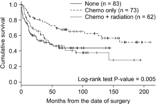
Table 3. Univariate and multivariate Cox regression analyses Fig. 5. Comparison of overall survival according to chemoradiation therapy.
port was similar to that reported from other centers, in which adenocarcinoma predominated (adenocarcinoma, 97%; adenosquamous, 1%; squamous, 1%). The majority of tumors were well differentiated (64%), 28% were mod- erately differentiated, and the remaining 7% were poorly differentiated. The T-stage of disease correlated with the likelihood of lymph node involvement, lymphovascular and perineural invasion, and surgical margin status. Of note, 34 cases (16%) involved regional lymph nodes, 56
(26%) exhibited perineural invasion, and 58 (27%) lym- phovascular invasion, whereas 29 cases (13%) were surgi- cal margin positive histologically.
Chemoradiation therapy
A total of 73 patients (34%) underwent adjuvant chemo- therapy and 62 patients (28%) underwent adjuvant che- motherapy with radiation, whereas the remaining 83 (38%) did not receive adjuvant therapy. Patients received a variety of doses and number of cycles of chemotherapy regimens and chemoradiation with fluorouracil and gem- citabine hydrochloride, which were the standard of care given to most patients. The chemotherapy alone group demonstrated a statistically significant improvement in survival than the no therapy or chemoradiation groups (P
= 0.005) (Fig. 5).
Survival
We examined the prognostic significance of the follow- ing factors: sex, age, T-stage, year of surgery, surgery type, incidentality, iatrogenic bile spillage, histologic type, dif- ferentiation of tumor, lymphovascular invasion, peri-
neural invasion, lymph node involvement, surgical mar- gin status, and postoperative chemoradiation therapy for patients who underwent resection with curative intent.
These clinical and pathologic variables are summarized in Tables 1 and 2. The results of univariative and multi- variative analysis for the factors that significantly influ- enced the survival are summarized in Table 3. In uni- variate analyses, stage, year of surgery performed, type of surgery, incidentality, intervention, differentiation, surgi- cal margin, lymphovascular, perineural invasion, lymph- node, and chemotherapy statistically affected OS. The hazard ratio (HR) for OS in the late stage patients com- pared with that in the early stage patients was 2.61 (P < 0.0001; 95% CI, 1.75 to 3.89). Comparing male and female patients, there was no significant difference on survival (P
= 0.98). The multivariate model was created using a back- ward elimination method, and the probability was set at 0.10 for removal. HRs and 95% CIs were calculated for death. In a multivariate analysis for OS, Cox models in- cluding all main effects were considered. A Cox model in- cluding year of surgery performed, type of surgery, surgi- cal margin, lymphovascular, lymph node, and chemo- therapy showed significant main effects.
DISCUSSION
The advent of LC has lowered the threshold for sympto- matic patients with gallbladder disease. It appears possi- ble that if gallbladder disease in general is operated on ear- lier, incidental GBCs may also be detected at an earlier stage, increasing the chance for survival.
As increasing numbers of LCs are being performed, their role in the management of potentially malignant dis- ease must be carefully examined. LC should not be per- formed when GBC has been diagnosed or is suspected preoperatively [8,9]. On the other hand, LC is now per- formed by surgeons with a reasonable degree of con- fidence, even in cases of possible malignant lesions [2,10].
GBC has one of the poorest OS rates of all the gastro- intestinal malignancies. The dismal results are due to the aggressive biology of this tumor [11].
In a review of the literature, GBC was suspected pre-
operatively in only 30% of patients and, in the remaining 70% of cases, GBC was incidentally discovered by the at- tending pathologist [3]. In our series, 80% of the patients that were found incidentally were stage I or II, and OS im- proved in the later period (2004 to 2011). Patients found to have GBC incidentally at LC had significant increased sur- vival when compared with those who were admitted elec- tively with a known diagnosis. Large quantities of viable tumor cells in the bile that spill out during the operation or perioperatively undoubtedly indicate the actual threat of implantation metastasis by the spilled bile. Spillage of tu- mor-laden bile may be a cause of port site/peritoneal re- currence following LC. Bile spillage and injuries to the bili- ary system occur more often during the laparoscopic pro- cedure than during the conventional open method, and can convert potentially curable GBC to an incurable dis- ease [12]. Thus, patients with preoperative findings suspi- cious for GBC should undergo open exploration with an intent to perform a radical cancer operation as a primary procedure if the diagnosis is confirmed intraoperatively [8,12].
Increasing numbers of reports have documented that mean survival is statistically shorter when complications (gallbladder perforation, injury to common bile duct, or tumor violation) occur during LC for GBC. It is thought that in addition to perioperative bile leak, factors such as the biological properties of the tumor, tumor stage, oper- ative maneuvers and impact of pneumoperitoneum pres- sure may be involved in the onset of peritoneal and port site recurrence [9].
Recently, the single port LC technique was reported to also increase the risk of bile leakage into the abdomen be- cause of “critical view of safety” [13]. It is undoubtedly a consequence of cancer-contaminated bile spillage through the liver pores created by PTGBD or PTBD, and sub- sequent cancer growth on the peritoneum [5]. In the cur- rent study, 20 biliary leakage incidences were reported during LC, 16 during PTGBD, and 16 during PTBD, only PTBD patients showed significantly worse survival.
Complete surgical resection is the only potential cura- tive treatment of GBC. If GBC is suspected preoperatively, an open cholecystectomy should be performed to enable a complete evaluation and radical resection [14].
Controversy still remains as to whether laparotomy and local additional excision should be performed when a di- agnosis of GBC is made during or after LC, and some groups recommended radical surgery [15-17], while oth- ers have come to the conclusion that further surgical inter- vention offers no advantage [18].
At our institution, early tumors (T1s or T1a) that are rec- ognized incidentally are curable with simple cholecystec- tomy alone. Also, extended cholecystectomy is not evi- dence-based in patients with T1b GBC. After postopera- tive diagnosis of incidental over T2 GBC, there is a need for a second radical procedure. In advanced GBC, radical sur- gery can cure only a small subset of patients. In our series, radical second resection showed statistically better surviv- al, whereas hemihepatectomy showed worse prognosis.
It is impossible to diagnosis the T stage of GBC precisely before histological confirmation, even if the best diag- nostic modalities are employed. Also, frozen biopsies have limited accuracy for the T staging of GBC because of sampling error and freezing artifacts. Sometimes frozen tissue diagnosis and the final diagnosis are not identical [4,14].
Patients found to have GBC incidentally during LC ap- pear to have a survival advantage if resected with curative intent. There have been suggestions that prognosis after two operations is less favorable than for patients treated with a single procedure [19]. Also, if GBC is diagnosed postoperatively after LC, only about one-third of patients eventually undergo a second procedure after LC for GBC [7,20].
Recently, some reports showed the feasibility and safety of total laparoscopic completion radical cholecystectomy for incidentally detected early GBC [21,22]. The magni- tude of partial hepatectomy in terms of number of excised nodes and integrity of the specimen did not differ between the open and laparoscopic approach [23]. Larger and pro- spective studies are needed to support the oncologic safety and efficacy of laparoscopic completion radical surgery in early GBC.
Features of GBC are characterized by a wide range of tu- mor extent, cancer stage-dependent survival and extent of standard resection are not yet established. Tumor-node- metastasis stage was found to be a significant prognostic
factor. The prognosis worsened with increasing disease stage and the survival rate decreased with increasing age, especially after the age of 75 [24]. Our study demonstrated a similar result, but the age factor did not influence surviv- al rate.
No consensus regarding the optimal extent of liver re- section has been established. In our series, we obtained 2 to 3 cm of negative margin for the standard resection.
Perineural invasion also has been reported as a factor re- sponsible for poor prognosis [9,25]. Also, lymph node mi- crometastasis has a significant survival impact in patients with GBC [26]. The results of the current study are similar to previous publication from other centers [27,28].
The effectiveness of a radical surgical approach for GBC has been the subject of a number of reports. When consid- ering the optimal operation for GBC, there are two major issues: the extent of hepatic resection and the extent of re- gional lymphadenectomy. Extended lymphadenectomy may prolong survival in selected patients with GBC, but the extent of lymph node resection for the optimal treat- ment of GBC has not been clearly established [15,27]. The AJCC suggests a minimum of three lymph nodes need to be assessed for appropriate pathologic nodal staging of GBC, and there are no established standards. Investigators from Western centers rely on limited nodal dissection in- volving hepato-duodenal ligament, while those from the East recommend extended lymphadenectomy including pericholedochal, periportal, common hepatic, peripancre- atic, and paraaortic lymph nodes, even if overt nodal metastasis is absent.
This difference in surgical approaches has led to higher lymph nodal yield reported in Eastern studies compared to Western studies [26,28]. Lymph node dissection at our institution involves complete portal dissection, skeletoni- zation of the biliary tree, hepatic artery and portal vein, and the pericholedochal and retropancreatic lymph nodes those are the most frequently involved nodal basins.
Optimal extent of lymph node dissection for the GBC should be decided keeping in mind various factors includ- ing the patients’ general condition and tumor stage.
Complete surgical resection is the only potential cura- tive treatment. The role of chemotherapy and radiation therapy in the management of GBC remains undefined. A
better understanding of the pathogenesis of the disease is needed to develop a more effective targeted adjuvant ther- apy [29,30]. The heterogenecity of the patient and the regi- men used makes it difficult to extrapolate any conclusion.
In our series, after adjusting for the stage parameters, the data supported the view that adjuvant chemoradiotherpy might improve OS for patients with GBC. The main limi- tation of this study was its retrospective, nonrandomized, short follow-up period, which prevented survival curve analysis. On the other hand, by comparing outcomes in the later 7 years to those in the first 9 years, we used histor- ical controls as a reference point for the new surgical approach. The main drawback to this type of comparison is that it is possible that the overall management of these patients has changed over time and is variable among sur- geons, irrespective of surgical factors.
In conclusion, the stage of disease at presentation af- fected the survival in all time periods. It is most likely that tumor biology and tumor stage, rather than extent of hep- atectomy, determine long-term prognosis. Complete sur- gical resection is the only potentially curative treatment.
Heightened awareness of the possibility of GBC and the knowledge of appropriate management are important for surgeons practicing LC. Early diagnosis, appropriate sur- gical resection, and better adjuvant therapy will be key fac- tors in improving results in the future.
CONFLICTS OF INTEREST
No potential conflict of interest relevant to this article was reported.
ACKNOWLEDGEMENTS
This work was supported by the Dong-A University re- search fund.
REFERENCES
1. Konstantinidis IT, Deshpande V, Genevay M, Berger D,
Fernandez-del Castillo C, Tanabe KK, et al. Trends in pre- sentation and survival for gallbladder cancer during a pe- riod of more than 4 decades: a single-institution expe- rience. Arch Surg 2009;144:441-7.
2. Goetze T, Paolucci V. Does laparoscopy worsen the prog- nosis for incidental gallbladder cancer? Surg Endosc 2006;20:286-93.
3. Shih SP, Schulick RD, Cameron JL, Lillemoe KD, Pitt HA, Choti MA, et al. Gallbladder cancer: the role of laparo- scopy and radical resection. Ann Surg 2007;245:893-901.
4. Yokomizo H, Yamane T, Hirata T, Hifumi M, Kawaguchi T, Fukuda S. Surgical treatment of pT2 gallbladder carcino- ma: a reevaluation of the therapeutic effect of hepatectomy and extrahepatic bile duct resection based on the long- term outcome. Ann Surg Oncol 2007;14:1366-73.
5. Tanaka N, Nobori M, Suzuki Y. Does bile spillage during an operation present a risk for peritoneal metastasis in bile duct carcinoma? Surg Today 1997;27:1010-4.
6. D'Angelica M, Dalal KM, DeMatteo RP, Fong Y, Blumgart LH, Jarnagin WR. Analysis of the extent of resection for adenocarcinoma of the gallbladder. Ann Surg Oncol 2009;
16:806-16.
7. Goetze TO, Paolucci V. Adequate extent in radical re-re- section of incidental gallbladder carcinoma: analysis of the German Registry. Surg Endosc 2010;24:2156-64.
8. Weiland ST, Mahvi DM, Niederhuber JE, Heisey DM, Chicks DS, Rikkers LF. Should suspected early gallbladder cancer be treated laparoscopically? J Gastrointest Surg 2002;6:50-6.
9. Kondo S, Takada T, Miyazaki M, Miyakawa S, Tsukada K, Nagino M, et al. Guidelines for the management of biliary tract and ampullary carcinomas: surgical treatment. J Hepatobiliary Pancreat Surg 2008;15:41-54.
10. Ouchi K, Mikuni J, Kakugawa Y; Organizing Committee, The 30th Annual Congress of the Japanese Society of Biliary Surgery. Laparoscopic cholecystectomy for gall- bladder carcinoma: results of a Japanese survey of 498 patients. J Hepatobiliary Pancreat Surg 2002;9:256-60.
11. Vollmer CM, Drebin JA, Middleton WD, Teefey SA, Linehan DC, Soper NJ, et al. Utility of staging laparoscopy in subsets of peripancreatic and biliary malignancies. Ann Surg 2002;235:1-7.
12. Vollmer CM Jr. Unexpected identification of gallbladder carcinoma during cholecystectomy. J Gastrointest Surg 2009;13:2034-6.
13. Allemann P, Schafer M, Demartines N. Critical appraisal of single port access cholecystectomy. Br J Surg 2010;97:
1476-80.
14. Sikora SS, Singh RK. Surgical strategies in patients with gallbladder cancer: nihilism to optimism. J Surg Oncol 2006;93:670-81.
15. Lee BS, Kim DH, Chang YS, Kang JH, Lee TS, Han JG.
Radical reresection for T2 gallbladder cancer patients diag- nosed following laparoscopic cholecystectomy. J Korean Surg Soc 2010;78:398-404.
16. Goetze TO, Paolucci V. Benefits of reoperation of T2 and more advanced incidental gallbladder carcinoma: analysis
of the German registry. Ann Surg 2008;247:104-8.
17. Wakai T, Shirai Y, Hatakeyama K. Radical second resection provides survival benefit for patients with T2 gallbladder carcinoma first discovered after laparoscopic cholecys- tectomy. World J Surg 2002;26:867-71.
18. Toyonaga T, Chijiiwa K, Nakano K, Noshiro H, Yamaguchi K, Sada M, et al. Completion radical surgery after chol- ecystectomy for accidentally undiagnosed gallbladder carcinoma. World J Surg 2003;27:266-71.
19. Fong Y, Jarnagin W, Blumgart LH. Gallbladder cancer:
comparison of patients presenting initially for definitive operation with those presenting after prior noncurative intervention. Ann Surg 2000;232:557-69.
20. Pawlik TM, Gleisner AL, Vigano L, Kooby DA, Bauer TW, Frilling A, et al. Incidence of finding residual disease for in- cidental gallbladder carcinoma: implications for re-resec- tion. J Gastrointest Surg 2007;11:1478-86.
21. Belli G, Cioffi L, D'Agostino A, Limongelli P, Belli A, Russo G, et al. Revision surgery for incidentally detected early gallbladder cancer in laparoscopic era. J Laparoendosc Adv Surg Tech A 2011;21:531-4.
22. Gumbs AA, Hoffman JP. Laparoscopic radical cholecy- stectomy and Roux-en-Y choledochojejunostomy for gall- bladder cancer. Surg Endosc 2010;24:1766-8.
23. Cho JY, Han HS, Yoon YS, Ahn KS, Kim YH, Lee KH.
Laparoscopic approach for suspected early-stage gall- bladder carcinoma. Arch Surg 2010;145:128-33.
24. Kayahara M, Nagakawa T. Recent trends of gallbladder cancer in Japan: an analysis of 4,770 patients. Cancer 2007;110:572-80.
25. Yamaguchi R, Nagino M, Oda K, Kamiya J, Uesaka K, Nimura Y. Perineural invasion has a negative impact on survival of patients with gallbladder carcinoma. Br J Surg 2002;89:1130-6.
26. Sasaki E, Nagino M, Ebata T, Oda K, Arai T, Nishio H, et al.
Immunohistochemically demonstrated lymph node micro- metastasis and prognosis in patients with gallbladder carcinoma. Ann Surg 2006;244:99-105.
27. Jensen EH, Abraham A, Jarosek S, Habermann EB, Al-Refaie WB, Vickers SA, et al. Lymph node evaluation is associated with improved survival after surgery for early stage gallbladder cancer. Surgery 2009;146:706-11.
28. Negi SS, Singh A, Chaudhary A. Lymph nodal involve- ment as prognostic factor in gallbladder cancer: location, count or ratio? J Gastrointest Surg 2011;15:1017-25.
29. Andre T, Reyes-Vidal JM, Fartoux L, Ross P, Leslie M, Rosmorduc O, et al. Gemcitabine and oxaliplatin in ad- vanced biliary tract carcinoma: a phase II study. Br J Cancer 2008;99:862-7.
30. Gold DG, Miller RC, Haddock MG, Gunderson LL, Quevedo F, Donohue JH, et al. Adjuvant therapy for gall- bladder carcinoma: the Mayo Clinic Experience. Int J Radiat Oncol Biol Phys 2009;75:150-5.