Hepatocellular carcinoma (HCC) is the most common primary malignant tumor of the liver. Extrahepatic metastasis can occur through hematogeneous metasta- sis, lymphatic extension and direct invasion (1).
The incidence of spontaneous rupture of primary HCC has been reported to be up to 14.5% (2, 3).
However, rupture of the metastatic lesions of HCC is very rare. We report here on a case of massive retroperi- toneal hemorrhage due to spontaneous rupture of right adrenal gland metastasis that was secondary to invasive HCC. This was successfully controlled by transcatheter arterial embolization (TAE).
Case Report
A 45-year-old man was admitted to our hospital be- cause of severe right upper quadrant pain of an acute onset, abdominal distension and hypovolemic shock.
He was diagnosed with massive HCC in the right lobe of the liver and lung metastasis; the HCC was associated with hepatitis B virus (HBV)-positive hepatitis, which he had been diagnosed with 6 months previously at our hospital. The patient had received transcatheter arterial chemombolization (TACE) three times during the previ- ous 6 months.
The contrast enhanced abdominal CT obtained 4 days before the onset of symptoms shows partial retention of iodized oil in the mass in the posterior segment of the right lobe of the liver with direct invasion to the right adrenal gland (Fig. 1).
On the present admission, the patient’s initial blood pressure was 70/40 mmHg and a laboratory test showed a rapid drop of hemoglobin during 4 hours, from 13.2 g/dl to 11.0 g/dl
Unenhanced and enhanced abdomen CT was then performed. The abdominal CT showed a large retroperi- toneal hematoma involving the right perirenal and ante- rior pararenal spaces and it abutted the right adrenal metastasis of the HCC, suggesting rupture. As a result, the duodenum and IVC were displaced to the left side by hematoma (Fig. 2).
Angiograhy was performed to evaluate for tumor bleeding from an arterial vessel and then to eventually
J Korean Radiol Soc 2007;56:327-330
─ 327 ─
Endovascular Treatment of Massive Retroperitoneal Hemorrhage Due to Spontaneous Rupture of Right Adrenal Gland Metastasis that was Secondary to Invasive
Hepatocellular Carcinoma: A Case Report1
Youn Kyung Lee, M.D., Seung Kwon Kim, M.D., Byung Ik Kim, M.D.2
1Department of Radiology, Kangbuk Samsung Hospital, Sungkyunkwan University School of Medicine, Seoul, Korea
2Department of Internal Mediocine, Kangbuk Samsung Hospital, Sungkyunkwan University School of Medicine, Seoul, Korea
Received December 21, 2006 ; Accepted February 12, 2007
Address reprint requests to : Seung Kwon Kim, M.D., Department of Radiology, Kangbuk Samsung Hospital, Sungkyunkwan University School of Medicine, 108, Pyung-dong, Jongro-gu, Seoul 110-746, Korea Tel. 82-2-2001-2336 Fax. 82-2-2001-1030 E-mail: [email protected]
The reported incidence of spontaneous rupture of primary hepatocellular carcinoma (HCC) is up to 14.5%. However, rupture of the metastatic lesions of HCC is very rare.
We describe here a case of massive retroperitoneal hemorrhage due to spontaneous rupture of right adrenal gland metastasis that was secondary to invasive HCC. This was successfully controlled by performing transcatheter arterial embolization (TAE).
Index words :Liver neoplasms, metastases
Liver neoplasms, chemotherapeutic embolization Adrenal gland
Retroperitoneal space
embolize it. A 5F Yashiro type catheter (Glidecath, Terumo, Tokyo, Japan) was placed via the right femoral artery approach at the celiac trunk. The aortogram, celi- ac angiogram and right renal arteriogram showed no tu- mor staining or extravasations of contrast material. A 5F Yashiro catheter was then placed at the right phrenic artery. The right phrenic angiogram showed tumor stain- ing at the right adrenal gland. A 2F microcatheter (Progreat, Terumo, Tokyo, Japan) was coaxially passed through the 5F Yashiro catheter; the selective right
phrenic artery angiogram then showed tumor staining at the right adrenal gland (Fig. 3A). TACE was initially per- formed with an infusion of 5 mL of iodized oil (Lipiodol;
Laboratoire Andre Guerbet, Aulnay-sous-Bois, France) and 20 mg of doxorubicin hydrochloride (Adriamycin;
Pharmacia, Milan, Italy) emulsion into the right phrenic artery. Additional embolization was performed with us- ing gelatin sponge (Cutanplast; Mascia Brunelli, Milan, Italy), which we cut into small pieces and soaked in a so- lution of 10 mL of nonionic contrast medium (Visipaque;
Amersham, Cork, Ireland) and 3 mL of saline solution.
The right phrenic artery angiogram after embolization with an emulsion of iodized oil and doxorubicin hy- drochloride and gelatin sponge showed retention of the embolized emulsion in tumor cells and there was still residual tumor staining; embolization of the branch of the right phrenic artery was next performed with micro- coils (two 2 mm×2 cm hilar coils, two 3 mm×2 mm tor- nado coils and six 4 mm×2 mm tornado coils, Cook, Bloomington, IN, U.S.A.). The right phrenic artery an- giogram obtained after the embolization with additional microcoils showed that the branch of the right phrenic artery was completely occluded, and iodized oil was tak- en up to the right adrenal gland; tumor staining at the right adrenal gland was no longer seen (Fig. 3B).
After the embolization, the patient’s blood pressure was 100/60 mmHg and the vital signs were stable. Abdominal CT was performed again after 6 days and it showed the interval size decreases of the retroperitoneal hematoma
Youn Kyung Lee, et al : Endovascular Treatment of Massive Retroperitoneal Hemorrhage Due to Spontaneous Rupture of Right Adrenal Gland Metastasis that was Secondary to Invasive Hepatocellular Carcinoma
─ 328 ─
A B
Fig. 2. A, B. The coronal reformatted image (A) and the transverse image (B) of the contrast enhanced abdominal CT show a large retroperitoneal hematoma involving the right perirenal (arrows) and anterior pararenal spaces (arrows); this hematoma abuts the right adrenal metastasis (arrowhead) of the HCC, suggesting rupture. As a result, the duodenum (small arrow) and IVC (arrow- head) were displaced to the left side by the hematoma.
Fig. 1. Coronal reformatted image of the contrast enhanced ab- dominal CT obtained 4 days before onset of symptoms shows partial retention of iodized oil (arrows) by the mass in the pos- terior segment of the right lobe of the liver with direct invasion to the right adrenal gland (small arrows).
involving the right perirenal and right anterior pararenal spaces, and retention of the iodized oil in the right adrenal metastasis and the embolized microcoils (Fig. 4). The pa- tient was discharged 6 days after the CT scan.
Discussion
Although the adrenal gland is the second most com- mon site of metastasis from HCC in autopsy cases (4), there has been only one reported case in the Japanese article describing spontaneous rupture of the adrenal metastasis from HCC (5).
The adrenal metastasis in this case was not a hematogenous metastasis, but it was considered as the direct extension to (and engulfing) the right adrenal gland because the normal medial and lateral limbs of the adrenal gland were obliterated and an oval mass was formed (Fig. 1). It is difficult to differentiate rupture of HCC invading the right adrenal fossa from rupture of adrenal metastasis from invasive HCC. For our case, the massive retroperitoneal hemorrhage may have been caused by spontaneous rupture of the right adrenal gland metastasis from the invasive HCC because the retroperitoneal hemorrhage abutted the right adrenal
J Korean Radiol Soc 2007;56:327-330
─ 329 ─
A B
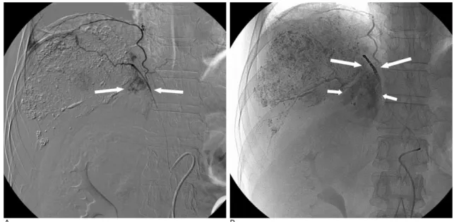
Fig. 3. (A) Selective right phrenic artery angiogram shows tumor staining (arrows) at the right adrenal gland. (B) The right phrenic artery angiogram obtained after transcatheter arterial embolization with an emulsion of iodized oil and doxorubicin hydrochloride, gelatin sponge and microcolis (arrows) shows that the right phrenic artery was completely occluded, iodized oil was taken up by the right adrenal gland (small arrows), and tumor staining at the right adrenal gland was no longer seen.
A B
Fig. 4. A, B. The coronal reformatted image (A) and transverse image (B) of the contrast enhanced abdominal CT obtained 6 days af- ter transcatheter arterial embolization shows interval size decreases of the retroperitoneal hematoma (arrows) involving the right perirenal and right anterior pararenal spaces and the uptake of iodized oil in the right adrenal metastasis (arrowheads).
metastasis of the HCC (Fig. 2A); further, the CT ob- tained 6 days after TACE showed uptake of iodized oil in the right adrenal metastasis (Fig. 4A)
Each adrenal gland has three sources of arterial blood supply from the inferior phrenic artery, the aorta and the renal arteries (6). Tumor staining was not seen on the aortogram and the right renal arteriogram, and the right phrenic artery was the main feeding artery in this case.
Unruptured adrenal metastasis can be treated by adrenalectomy, TACE or percutaneous ethanol injection (PEIT), according to the clinical features of each individ- ual; these features include the size of the metastatic tu- mor, whether there is invasion into the IVC, the func- tion of the remaining liver and the existence of intra- and/or nonadrenal extrahepatic lesions (7).
Spontaneous rupture of HCC or metastatic HCC is a critical and life-threatening condition and its prognosis is extremely poor. These patients usually present with a sudden onset of abdominal pain that’s accompanied by hypovolemic shock, abdominal distension and massive hemoperitoneum.
The mechanism of ruptured HCC is poorly understood.
The postulated mechanisms include rapid growth of tu- mor and necrosis, rupture by splitting of the overlying normal hepatic parenchyma or erosion of a vessel and oc- clusion of the hepatic veins by a tumor thrombus (3, 8).
Massive hemorrhage may be one of the causes of he- patic failure in cirrhotic patients with ruptured HCC.
Therefore, either emergency surgery or arterial em- bolization is necessary when faced with this condition.
Almost all of these patients are poor surgical candidates because of cirrhosis and extensive tumor replacement of the liver, which carries a high risk of morbidity and mortality. TAE has been reported to be highly effective to achieve hemostasis in patients with ruptured HCC,
and its immediate mortality rate is far less than that of surgery (9, 10). TAE may also be an appropriate treat- ment even for ruptured adrenal metastasis of HCC.
In summary, we describe here a case of massive retroperitoneal hemorrhage due to spontaneous rupture of right adrenal gland metastasis that was secondary to invasive HCC, and this was successfully controlled by performing TAE.
References
1. Nakashima T, Okuda K, Kojiro M, Jimi A, Yamaguchi R, Sakamoto K, et al. Pathology of hepatocellular carcinoma in Japan. 232 Consecutive cases autopsied in ten years. Cancer 1982;51:863-877 2. Miyamoto M, Sudo T, Kuyama T. Spontaneous rupture of hepato-
cellular carcinoma: a review of 172 Japanese cases. Am J Gastroenterol 1991;86:67-71
3. Ong GB, Taw JL. Spontaneous rupture of hepatocellular carcino- ma. Br Med J 1972;4:146-149
4. Edmondson HA, Steiner PE. Primary carcinoma of the liver. a study of 100 cases among 48,900 necropsies. Cancer 1954;7:462-503 5. Fukuoka K, Funatomi T, Ikegami F, Ito M, Shirai T, Tsuchiya H, et al. A case of hepatocellular carcinoma associated with multiple pulmonary tumor thrombi and rupture of its right adrenal metasta- sis. Gan No Rinsho 1987;33:199-204
6. Oglevie SB, Bookstein JJ. The roles of angiography in adrenal disease.
In Baum S. Abrams’ Angiography: Vascular and Interventional Radiology. 4th ed. Boston: Little, Brown and Company, 1997;1352- 1353
7. Momoi H, Shimahara Y, Terajima H, Iimuro Y, Yamamoto N, Yamamoto Y, et al. Management of adrenal metastasis from hepa- tocellular carcinoma. Surg Today 2002;32:1035-1041
8. Hermann RE, David TE. Spontaneous rupture of the liver caused by hepatomas. Surgery 1973;74:715-719
9. Hsieh JS, Huang CJ, Huang YS, Sheen PC, Huang TJ.
Intraperitoneal haemorrhage due to spontaneous rupture of hepa- tocellular carcinoma: treatment by hepatic artery embolization.
AJR Am J Roentgenol 1987;149:715-717
10. Okazaki M, Higashihara H, Koganemaru F, Nakamura T, Kitsuki H, Hoashi T ,et al. Intraperitoneal hemorrhage from hepatocellular carcinoma: emergency chemoembolization or embolization.
Radiology 1991;180:647-651
Youn Kyung Lee, et al : Endovascular Treatment of Massive Retroperitoneal Hemorrhage Due to Spontaneous Rupture of Right Adrenal Gland Metastasis that was Secondary to Invasive Hepatocellular Carcinoma
─ 330 ─
대한영상의학회지 2007;56:327-330
침습 간암의 우측 부신 전이의 자발 파열에 의한 대량 후복강출혈의 혈관 내 치료: 증례 보고1
1성균관대학교 의과대학 강북삼성병원 영상의학과
2성균관대학교 의과대학 강북삼성병원 내과
이윤경1・김승권1・김병익2
일차성 간암의 자발 파열은 14.5%까지 보고되어 있으나 간암 전이의 파열은 드물다. 저자들은 침습 간암의 우측 부신 전이의 자발 파열에 의한 대량 후복강출혈을 경관동맥색전술로 성공적으로 치료하여 보고하고자 한다.