Chronic pancreatitis can result in a number of vascu- lar complications including thromboses of the portal ve- nous system and the formation of a pseudoaneurysm.
Pseudoaneurysms occur in approximately 3.5-10.0% of patients with pancreatitis (1). The arteries most com- monly affected by pseudoaneurysms are (in decreasing percent occurrence), the splenic (40%), gastroduodenal (30%), pancreaticoduodenal (20%), gastric (5%), hepatic (2%), and others (superior mesenteric, jejunal, ileocecal, and aorta) (1-3%) (1, 2). Thrombosis of the portal ve- nous system is another important complication of chronic pancreatitis (1, 3). An isolated splenic vein thrombosis is more common than a portal or superior mesenteric vein thrombosis in patients with chronic pancreatitis (1).
However, as far as we know, a pseudoaneurysm with- in the thrombosed right portal vein of a patient with chronic pancreatitis has been not reported.
In this case report, we present the case of a right he- patic artery pseudoaneurysm which developed within the thrombosed right portal vein of a 35-year-old woman afflicted with chronic pancreatitis.
Case Report
A 35-year-old, heavy alcoholic woman was presented with sudden epigastric pain. The laboratory data re- vealed a normal range for WBC (7.1×10³), a markedly elevated CRP (92.7 mg/L), neutrophil (81.2%), lipase (332 U/L) and amylase (827 U/mL), in addition to de- creased hemoglobin (12.2 g/dL) and hematocrit (19.7%).
The patient underwent a dynamic pancreatic CT, which revealed intraductal stones in the pancreatic body (Fig. 1A), atrophy of the pancreas, and diffuse di- latation of the pancreatic duct (Fig. 1B). These CT find-
The Occurrence of a Pseudoaneurysm of the Hepatic Artery within the Thrombosed Portal Vein of a Patient
with Chronic Pancreatitis: A Case Report1
Eun Soo Kim, M.D., Kyung Mi Jang, M.D., Min-Jeong Kim, M.D., Hoi Soo Yoon, M.D., Hyun Lee, M.D., Eui Yong Jeon, M.D., Kwanseop Lee, M.D., Yul Lee, M.D.
1Department of Radiology, College of Medicine, Hallym University Received December 14, 2007 ; Accepted February 20, 2008
Address reprint requests to : Kyung Mi Jang, M.D., Department of Radiology, Hallym University, College of Medicine, 896 Pyungchon- dong, Dongan-gu, Anyang-city, Kyungki-do 431-070, Korea
Tel. 82-31-380-3885 Fax. 82-31-380-3878 E-mail: [email protected]
A pseudoaneurysm is an uncommon but important life threatening complication of chronic pancreatitis. The arteries most commonly affected by a pseudoaneurysm are (in decreasing percent occurrence), the splenic (40%), gastroduodenal (30%), pancre- aticoduodenal (20%), gastric (5%), hepatic (2%), and others (superior mesenteric, jeju- nal, ileocecal, and aorta) (1-3%). Thrombosis of the splenic or portal vein is another important complication of chronic pancreatitis. In this case report, we present a rare complication in the form of a right hepatic artery pseudoaneurysm which developed within the thrombosed right portal vein of a 35-year-old woman afflicted with chronic pancreatitis.
Index words :Pancreatitis, chronic Aneurysm, false Portal vein Chronic diseases
multiple collaterals were also demonstrated on CT (Fig.
1B, C). In addition, a small, well-enhanced nodular le- sion measuring about 1.5 cm in diameter, was identified at the thrombosed right posterior portal. These findings suggest the occurrence of a right hepatic artery pseudoa- neurysm (Fig. 1C). A three-dimensional volume render- ing CT image showed a pseudoaneurysm originating in the right hepatic artery. The neck portion of pseudoa-
ters below 5 cm were noted at the S8 site of the liver and surrounded the S8 thrombotic portal vein branch. The results were interpreted as liver abscesses (Fig. 1E).
Following this assessment, the patient underwent medical conservative treatment (antibiotics, NPO, for four weeks. A follow-up CT scan conducted two weeks after the onset of medical treatment illustrated the dis- appearance of the hepatic artery pseudoaneurysm (Fig.
A B
C D
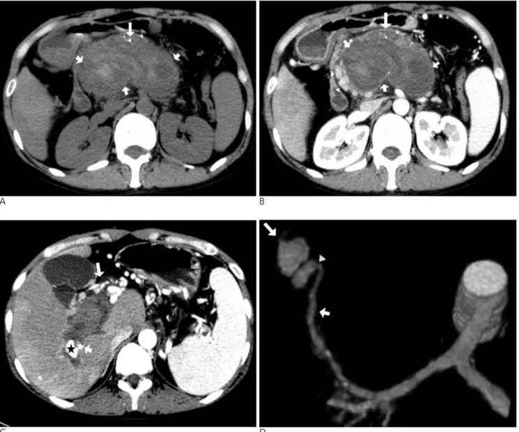
Fig. 1. A. The unenhanced axial CT scan reveals the intraductal stones (long arrow) in the pancreatic body and a high density thrombosis at the portal?splenic vein (short arrows).
B. The arterial phase CT scan shows the decreased pancreatic volume and duct dilatation, representing chronic pancreatitis (long arrow). The axial CT reveals thrombosis (short arrows) at the portal-splenic vein with collateral vessels (arrowheads).
C. An approximately 1.5 cm well-enhanced nodular lesion (asterisk) is observed within the thrombosed right posterior portal vein with collateral vessels (long arrows) on an arterial phase CT scan. This lesion originated from the right hepatic artery (short arrow), which suggests a hepatic artery pseudoaneurysm.
D. A three-dimensional volume rendering CT image shows the pseudoaneurysm (long arrow), which developed from the right he- patic artery (short arrow). The neck portion (arrowhead) of the pseudoaneurysm had a diameter of 1 mm.
1F), and the liquefaction of thrombosis at the portal- splenic vein (Fig. 1F, G). The last follow-up CT scan, conducted 4 weeks after medical treatment found an improvement in the previously identified liver abscesses (Fig. 1H).
Discussion
Chronic pancreatitis has a number of vascular compli- cations, including thromboses of the portal venous sys- tem and the formation of a pseudoaneurysm. Moreover,
patients afflicted with chronic pancreatitis were subject- ed to the possible erosion of the peripancreatic vessels to the cross-tissue planes and boundaries via the pancreat- ic enzymes, which resulted in formation of a pseudoa- neurysm (2). The hepatic artery is documented as being less commonly affected (2%) (1, 2). Two-thirds of hepat- ic artery pseudoaneurysm cases are extrahepatic and the right hepatic artery is involved more frequently than the left (4). The rupture of a pseudoaneurysm has been accompanied with mortality in greater than 90% of cas- es (1). Therefore, immediate surgery is the gold standard
E F
G H
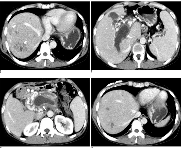
Fig. 1. E. Two clustered small low density lesions (arrows) are noted at the right hepatic lobe surrounding thrombosed S8 portal vein, which was interpreted as liver abscesses on a portal venous phase CT scan.
F. The portal venous phase CT scan conducted two weeks after medical treatment shows the disappeared hepatic artery pseudoa- neurysm at the thrombosed right portal vein (arrow).
G. The portal venous phase CT scan conducted two weeks after medical treatment indicates that the previously identified throm- bosis at the portal-splenic vein is liquefied and has a decreased diameter at the main portal vein (arrow).
H. The last follow-up portal venous phase CT scan conducted 4 weeks after medical treatment reveals an improvement in the pre- viously identified liver abscesses.
neurysm should undergo prompt initial angiographic evaluation and embolization if possible (5). On the other hand, patients who are hemodynamically stable and have angiographic evidence of bleeding can be treated by transcatheter embolization (1).
For patients with chronic pancreatitis, the occurrence of an isolated splenic vein thrombosis is more common than thrombosis of the portal and superior mesenteric vein, in relation to the close proximity of the splenic vein to the pancreas (1, 6).
In our case study, a hepatic artery pseudoaneurysm occurred within the thrombosed portal vein of a patient with chronic pancreatitis. Furthermore, liver abscesses were also associated. Until now, the association of por- tal?splenic vein thrombosis, intrahepatic pseudoa- neurysm, and liver abscesses has not been reported in patients with chronic pancreatitis.
A case of portal-splenic vein thrombosis accompanied with liver abscesses may suggest a case of pylephlebitis.
Portal-mesenteric vein thrombosis, pylephlebitis and liver abscesses have been reported as rare complications of inflammatory bowel disease and appendicitis (7, 8).
We believe that the portal vein thrombosis occurred as a complication of chronic pancreatitis, and the occurrence of pyephlebitis was probably due to a secondary infec- tion at the portal vein thrombosis site. Next, the propa- gation of inflammation to the hepatic artery may be the cause of the pseudoaneurysm.
In our case study, a hepatic artery pseudoaneurysm was improved only with medical treatment.
Spontaneous thrombosis of the visceral pseudoa- neurysm has rarely been reported (9, 10). The suggested medical factors that may have contributed to the aneurysm thrombosis include decreased flow and in- creased coagulability of blood due to factors such as hy- potension, dehydration, vasospasm, local damage to the arterial wall, and occult malignancy (11). However, none of these factors was present in our patient. The high ratio between aneurysmal size (maximum diame-
In aneurysms with a relatively small neck, intraluminal thrombosis cases have been documented (11, 12).
In conclusion, we experienced an extremely rare case of a hepatic artery pseudoaneurysm within the throm- bosed portal vein in a patient with chronic pancreatitis.
References
1. Ismail HR, Marc CW. Vascular complications of pancreatitis. J Pancreas 2004;5:328-337
2. Suzuki T, Ishida H, Komatsuda T, Oyake J, Miyauchi T, Heianna J, et al. Pseudoaneurysm of the gastroduodenal artery ruptured in- to the superior mesenteric vein in a patient with chronic pancreati- tis. J Clin Ultrasound 2003;31:278-282
3. Albertyn LE. Case report: Acute portal vein thrombosis. Clinical Radiology 1987;38:645-648
4. Kang M, Bapuraj JR, Khandelwal N, Kochhar R, Kalra N, Verma GR. Liver abscess associated with hepatic artery pseudoaneurysm with arteriovenous fistula: imaging and interventional manage- ment. Acta Radiol 2006;47:162-166
5. Udd M, Leppaniemi AK, Bidel S, Keto P, Roth WD, Haapiainen RK. Treatment of bleeding pseudoaneurysms in patients with chronic pancreatitis. World J Surg 2007;31:504-510
6. Weber SM, Rikkers LF. Splenic vein thrombosis and gastrointestinal bleeding in chronic pancreatitis. World J Surg 2003;27:1271-1274 7. Aguas M, Bastida G, Nos P, Beltran B, Grueso JL, Grueso J. Septic
thrombophlebitis of the superior mesenteric vein and multiple liv- er abscesses in a patient with Crohn’s disease at onset. BMC Gastroenterol 2007;12;7:22
8. Nishimori H, Ezoe E, Ura H, Imaizumi H, Meguro M, Furuhata T, et al. Septic thrombophlebitis of the portal and superior mesenteric veins as a complication of appendicitis: report of a case. Surg Today 2004;34:173-176
9. Vanlangenhove P, Defreyne L, Kunnen M. Spontaneous thrombo- sis of a pseudoaneurysm complicating pancreatitis. Abdom Imaging 1999;24:491-493
10. Tang LJ, Zipser S, Kang YS. Temporary spontaneous thrombosis of a splenic artery pseudoaneurysm in chronic pancreatitis during in- travenous octreotide administration. J Vasc Interv Radiol 2005;16:
863-866
11. Brownlee RD, Tranmer BI, Sevick RJ, Karmy G, Curry BJ.
Spontaneous thrombosis of an unruptured anterior communicat- ing artery aneurysm : an unusual cause of ischemic stroke. Stroke 1995;26:1945-1949
12. Black SPW, German WJ. Observation on the relationship between volume and size of the orifice of experimental aneurysms. J Neurosurg 1960;17:984-990
대한영상의학회지 2008;58:399-403
만성 췌장염과 합병된 간문맥 혈전 내에서 발생한 간동맥 가성동맥류: 증례 보고1
1한림대학교 성심병원 영상의학과
김은수・장경미・김민정・윤회수・이 현・전의용・이관섭・이 열
가성동맥류는 만성 췌장염에서 생길 수 있는 드물지만 중요하고 치명적인 합병증 중의 하나이다. 가성동맥류는 비 장동맥에서 가장 흔하게 발생하며 전체 40%에 이르며 그 다음으로 위 십이지장 동맥(20%), 위동맥(5%), 간동맥 (2%) 그리고 기타동맥(1-3%)에서 생길 수 있다. 만성 췌장염에서 비장정맥 또는 간문맥에서 혈전이 생기는 것 또 한 중요한 합병증이다. 이에 본 저자들은 만성 췌장염을 앓은 35세 여자 환자에서 오른쪽 간문맥 혈전 내에 생긴 오 른쪽 간동맥 가성동맥류를 경험하였기에 보고하고자 한다.