대한소화기학회지 2008;52:394-398
접수: 2008년 7월 31일, 승인: 2008년 10월 6일 연락처: 노명환, 602-103, 부산시 서구 동대신동 3가 1
동아대학교 의과대학 내과학교실 Tel: (051) 240-5627, Fax: (051) 240-2087 E-mail: [email protected]
Correspondence to: Myung-Hwan Roh, M.D.
Department of Internal Medicine, Dong-A University College of Medicine, 1, Dongdaesin-dong 3-ga, Seo-gu, Busan 602- 103, Korea
Tel: +82-51-240-5627, Fax: +82-51-240-2087 E-mail: [email protected]
위십이지장 동맥에서 분지한 우간동맥 압박에 의한 담관석을 동반한 폐쇄 황달
동아대학교 의과대학 내과학교실, 외과학교실*
백양현ㆍ최석렬ㆍ이종훈ㆍ김민지ㆍ김영훈*ㆍ노영훈*ㆍ노명환
Obstructive Jaundice due to Compression of the Common Bile Duct by Right Hepatic Artery Originated from Gastroduodenal Artery
Yang Hyun Baek, M.D., Suk Ryul Choi, M.D., Jong Hun Lee, M.D., Min Ji Kim, M.D., Young Hoon Kim, M.D.*, Young Hoon Roh, M.D.*, and Myung Hwan Roh, M.D.
Departments of Internal Medicine and Surgery*, Dong-A University College of Medicine, Busan, Korea
Obstructive jaundice by vascular compression is rare. The causative arteries were identified as the right hepatic artery, gastroduodenal artery, cystic artery, proper hepatic artery, and an unspecified branch of the common hep- atic artery. Also the venous system, such as enlarging collateral veins in cases of portal hypertension was a caus- ative vessel. Herein, we describe a case of a proximal choledocholithiasis due to compression of the common bile duct by right hepatic artery originated from gastroduodenal artery. Final diagnosis and treatment were achieved through an operation. (Korean J Gastroenterol 2008;52:394-398)
Key Words: Obstructive jaundice; Right hepatic artery; Choledocholithiasis
Introduction
Pulsatile vascular compression at the common bile duct is a rare cause of obstructive jaundice. Luttwak and Schwartz first described jaundice due to an obstruction of the common hepatic duct by an aberrant artery - celiac artery at 1961.1 Later, some cases of the biliary compression by hepatic artery aneurysm, gastroduodenal artery, and the right hepatic artery were reported.
Tsuchiya first described two cases of jaundice by compression due to right hepatic artery in English literature in 1984.2 Anatomically, the hepatic arteries are closely related to the extrahepatic bile duct. Particularly, the right hepatic artery has
many variations in its course,3 which make the intra- or extra- hepatic bile duct susceptible to extrinsic pulsatile arterial com- pression at the sites where these arteries cross the bile passages.4,5 We present here a rare case of biliary obstruction due to compression of the common bile duct by right hepatic artery originated from gastroduodenal artery combined with a large proximal choledocholithiasis.
Case report
A 49-year-old man with an unremarkable medical and family history was admitted to an emergency room, complaining
백양현 외 6인. 위십이지장 동맥에서 분지한 우간동맥 압박에 의한 담관석을 동반한 폐쇄 황달 395
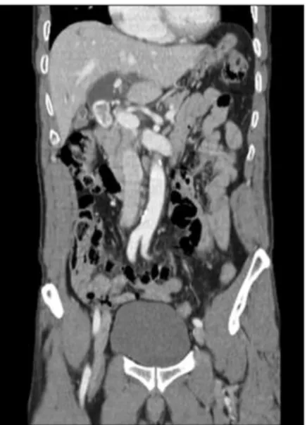
Fig. 1. Abdominal CT finding shows a large CBD stone with proximal ductal dilatation.
Fig. 2. Percutaneous transhepatic cholangiograpy shows a filling defect of round shape at the proximal part of common bile duct and the indentation of un- known cause below a filling de- fect (white arrow).
abdominal pain at right upper quadrant and vomiting.
On examination, he was sweaty with a temperature of 38.5oC, pulse rate of 95/min and the blood pressure 130/90 mmHg. He had an acute ill-looking appearance with icteric sclera. Abdominal examination revealed diffuse direct tender- ness at right upper quadrant. Laboratory examinations showed white cell count of 16,800/mm3 (seg 94.4%), hemoglobin 14.5 g/dL and the platelet count 172,000/mm3. A total bilirubin was
3.0 mg/dL, direct bilirubin 1.6 mg/dL, alkaline phosphatase 250 (104-338) IU/L, aspartate aminotransferase 372 (10-35) IU/L, alanine aminotransferase 172 (0-35) IU/L and gamma-glutamic transpeptidase 155 (8-53) IU/L. Electrolyte and renal function tests were normal. An abdominal computed tomography (CT) showed a 3 cm sized large proximal choledocholithiasis with upstream dilatation (Fig. 1).
Percutaneous transhepatic biliary drainage was performed, and a percutaneous transhepatic cholangiography (PTC) showed a partial obstruction of the extrahepatic bile duct at the level of upper common bile duct with a large 3 cm sized filling defect and the indentation below a filling defect (Fig. 2). And reconstructed CT images showed a variant right hepatic artery arising from gastroduodenal artery and this artery was seen at the level of upper bile duct (Fig. 3).
A patient had ERCP and failed standard stone extraction consisting of endoscopic sphincterotomy followed by basket extraction because of irremovable size of impacted stone. The presumptive diagnosis was a large impacted choledocholithiasis with cholangitis and an indentation of unknown cause. As a treatment, an operation was considered and performed. At laparotomy, a dilated proximal common bile duct containing a large stone was confirmed. Choledocholithotomy and T-tube choledochostomy were performed. At that time, the operator found that the right hepatic artery arising from the gastroduo-
396 The Korean Journal of Gastroenterology: Vol. 52, No. 6, 2008
Fig. 3. Reconstructed CT image. Reconstructed CT shows a var- iant right hepatic artery originated from gastroduodenal artery and this artery transverses the extrahepatic duct at the level of upper bile duct. (A) Common hepatic artery, (B) Left hepatic artery, (C) Right hepatic artery, (D) Gastroduodenal artery, (E) Variant right hepatic artery (dotted line).
Fig. 5. The cholaniography on the 6th POD shows that an in- dentation in the previous PTC is no longer found and maintains good passage.
Fig. 4. Operative findings. (A) Right hepatic artery from gastroduodenal artery (white arrow) crossing the common bile duct anteriorly and compressing the common bile duct is observed. (B) Operation is done by cholecystectomy and separation of a right hepatic artery (white arrow) from common bile duct.
denal artery was crossing the common bile duct anteriorly, and thus compressing the common bile duct (Fig. 4A). The right hepatic artery was separated from common bile duct and the common bile duct was free from compression (Fig. 4B). After the cholecystectomy, the right hepatic artery became even more mobile. The patient had a uneventful recovery and the cholaniography was performed (Fig. 5). Indentation showed in the previous PTC was not found any more and T-tube was removed on the 6th POD. At 6month follow-up after discharge, the patient had no complaints.
Discussion
Anatomic variations of biliary tract are found frequently.
Recognition of these anatomic variations is important because complications related to these variations are described in some reports and appropriate reconstruction of such variations is needed for surgical procedure. Among these variations, some reports have described anatomically variable vasculature of the hepatic artery.6-8 Koops represented that the finding of 604 selective angiographies showed normal anatomy of the hepatic artery in 79.1% and the anomalous arterial patterns in 20.9%.3
Baek YH, et al. Right Hepatic Artery Syndrome with Choledocholithiasis 397
Michels described that the incidence of the anterior crossing of the right hepatic artery to the extrahepatic bile duct was 12%9 and Tsuchiya presented that 11 (14%) among 79 patients receiving angiographys or operations due to intrahepatic stone showed the right hepatic artery anteriorly crossing to the bile duct.2 He presumed that the compression of the common bile duct resulted in prolonged bile stasis and this was an important role in the formation of hepatolithiasis. In 2005, Miyashita revealed the first case of bile duct obstruction due to the compression of the extrahepatic duct from dorsum by right hepatic artery10 in contrast with most cases with right hepatic artery which were crossing the extrahepatic duct anteriorly and compressing the common bile duct.11-14
In our case, the right hepatic artery showed anterior crossing to the bile duct and origin from gastroduodenal artery. Only a few authors mention hepatic arteries branching off the gastro- duodenal artery and two anomalies among 604 cases with hepatic artery arising from the gastroduodenal artery were reported by Koops in 2004.3
Assessment of extrahepatic duct obstruction often required the use of various imaging modalities to confirm the presence, level, and cause of obstruction, and to aid in treatment planning. Current techniques include transabdominal ultrasound (US), CT, ERCP, PTC, endoscopic ultrasound and magnetic resonance cholangiopancreatography (MRCP). Richard et al.
reviewed total of 103 consecutive patients with suspected biliary obstruction using CT and US to evaluate the relative accuracy of the methods.15 Their results reported that the precise level of obstruction was identified by CT in 88% and by US in 60%, and the cause of obstruction was accurately predicted by CT in 70% and by US in 38%. But, it is difficult to find the anatomic variations causing biliary obstruction by using non-operative typical imaging techniques, such as US, CT and PTC. Conventional or CT angiography could be helpful to determine more precisely on what variations exist and their hepatic artery collaterals. But, these can lead to duplication of testing with the possibility of increasing costs and delaying diagnosis. And indications of these tests have not been established yet.
In 2000, one study demonstrated that MRCP was helpful for diagnosis of biliary obstruction due to vascular compression.1 So, MRCP could be considered when typical imaging studies showed a large, proximal common bile duct stone without definite obstructive lesion or PTC showed a band-like filling defect with minimal or no upstream dilatation or vascular
structure transversing the extrahepatic duct was seen at the site of focal stenosis or obstruction on the coronal images. If biliary obstruction due to vascular compression was suggested via MRCP images, operation should be considered.12,16 Therapeutic surgical procedure is a cholecystectomy and separation of right hepatic artery from common hepatic duct. This surgical procedure is able to achieve the confirmation and treatment of biliary obstruction caused by compression of right hepatic artery. Post-surgical prognosis is very good and recurrence of obstructive jaundice have not been reported yet.2,10-14
In conclusion, our case present the biliary obstruction by compression of right hepatic artery originated from gastroduo- denal artery with large choledocholithiasis and surgical proce- dure is able to induce definite diagnosis and treatment.
References
1. Luttwak EM, Schwartz A. Jaundice due to obstruction of the common duct by aberrant artery: demonstration of celiac anomaly by translumbar aortography and simultaneous chol- edochogram. Ann Surg 1961;153:134-137.
2. Tsuchiya R, Eto T, Harada N, et al. Compression of the com- mon hepatic artery by the right hepatic artery in intrahepatic gallstones. World J Surg 1984;8:321-326.
3. Koops A, Wojciechowski B, Broering DC, Adam G, Krupski- Berdien G. Anatomic variations of the hepatic arteries in 604 selective celiac and superior mesenteric angiographies. Surg Radiol Anat 2004;26:239-244.
4. Hoizknecht N, Gauger J, Sackmann M, et al. Breath-hold MR cholangiography with snapshot techniques: prospective com- parison with endoscopic retrograde cholaniography. Radiology 1998;207:657-664.
5. Tabpga V, Braggion G, Marchesin G. Cholestasis caused by unnoticed anomaly of the right hepatic artery. Acta Chir Ital 1969;25:491-497.
6. Hiatt JR, Gabbay J, Busuttil RW. Surgical anatomy of the hepatic arteries in 1000 cases. Ann Surg 1994;220:50-52.
7. Michels NA. Newer anatomy of the liver and its variant blood supply and collateral circulation. Am J Surg 1966;112:
337-347.
8. Covey AM, Brody LA, Maluccio MA, Getrajdman GI, Brown KT. Variant hepatic arterial anatomy revisited: digital sub- traction angiography performed in 600 patients. Radiology 2002;224:542-547.
9. Michels NA. The hepatic, cystic and retroduodenal arteries and their relations to the biliary ducts. Ann Surg 1951;133:
503-524.
398 대한소화기학회지: 제52권 제6호, 2008
10. Myashita K, Shiraki K, Ito T, Taoka H, Nakano T. The right hepatic artery syndrome. World J Gastroenterol 2005;11:3008- 3009.
11. Ju JW, Kim MC, Kim YH, Oh JY, Nam KJ, Rho MH.
Hepatocholelithiasis due to compression of the common hep- atic duct by right hepatic artery. Korean J Hepatobiliary Pancreat Surg 2000;4:241-245.
12. Chung JP, Kim KW, Chi HS, et al. Obstructive jaundice due to compression of the common hepatic duct by right hepatic artery- a case associated with the absence of the lateral seg- ment of the left hepatic lobe. Younsei Med J 1994;35:231- 238.
13. Kim MH, Lee SK, Min YI, et al. Secondary intrahepatic
stones due to compression of common hepatic duct by right hepatic artery. Korean J Gastroenterol 1992;24:901-905.
14. Doi S, Yashimoto S, Hikita K, et al. Compression of the common hepatic duct by right hepatic artery in a case with cholecystolithiasis. Stom Intest 1979;14:687.
15. Baron RL, Stanley RJ, Lee JK, et al. A prospective compar- ison of the evaluation of biliary obstruction using computed tomography and ultrasonography. Radiology 1982;145:91-98.
16. Watanabe Y, Dohke M, Ishimori T, et al. Pseudo-obstruction of the extrahepatic bile duct due to artifact from arterial pul- satile compression: a diagnostic pitfall of MR cholangiopan- creatography. Radiology 2000;214:856-860.