Jun-Young Paeng
Department of Oral and Maxillofacial Surgery, Samsung Medical Center, 81 Irwon-ro, Gangnam-gu, Seoul 06351, Korea
TEL: +82-2-3410-2413 FAX: +82-2-3410-0038 E-mail: [email protected]
ORCID: https://orcid.org/0000-0002-0104-9338
This is an open-access article distributed under the terms of the Creative Commons Attribution Non-Commercial License (http://creativecommons.org/
licenses/by-nc/4.0/), which permits unrestricted non-commercial use, distribution, and reproduction in any medium, provided the original work is properly cited.
CC
Clinical analysis of neck node metastasis in oral cavity cancer
Aditi Sharma1, Jin-Wook Kim1, Jun-Young Paeng2
1Department of Oral and Maxillofacial Surgery, Kyungpook National University School of Dentistry, Daegu,
2Department of Oral and Maxillofacial Surgery, Samsung Medical Center, Seoul, Korea
Abstract(J Korean Assoc Oral Maxillofac Surg 2018;44:282-288)
Objectives: The purpose of this study was to evaluate the neck node metastasis pattern and related clinical factors in oral cavity cancer patients.
Materials and Methods: In total, 76 patients (47 males, 29 females) with oral squamous cell carcinoma (OSCC) who had no previous malignancies and were not undergoing neoadjuvant concomitant chemoradiotherapy or radiotherapy were selected for analysis.
Results: Occult metastases were found in 8 of 52 patients with clinically negative nodes (cN0, 15.4%). Neck node metastases were found in 17 pa- tients (22.4%). There was a statistically significant relationship between neck node metastasis and T stage (P=0.014) and between neck node metastasis and distant metastasis (Fisher’s exact test, P=0.019).
Conclusion: Neck node metastasis was significantly related to tumor size and distant metastasis during follow-up.
Key words: Mouth neoplasms, Lymph nodes, Metastasis, Squamous cell carcinoma
[paper submitted 2018. 1. 9 / revised 2018. 3. 12 / accepted 2018. 3. 22]
Copyright © 2018 The Korean Association of Oral and Maxillofacial Surgeons. All rights reserved.
I. Introduction
The oral cavity is the most common site of malignant tu- mors of the head and neck1. The most common malignant tu- mor type in the oral cavity is squamous cell carcinoma. Oral squamous cell carcinoma (OSCC) is frequently associated with poor prognosis2-4. Even when the tumors are small (T1 and T2), OSCC carries a high risk of cervical lymph node metastasis. Therefore, management of oral cancer remains controversial, especially for treatment of N0 neck patients.
More than 30% of OSCC patients with clinically N0 neck exhibit occult metastasis5,6. Cervical lymph node metastasis is the most significant independent prognostic factor, as it reduces the rate of survival by 50%7. Thus, appropriate treat- ment of cervical lymph nodes is essential for loco-regional control of the disease.
Various studies have revealed that elective neck dissection (END) is more beneficial than the “wait and see” approach in terms of survival rate8-10. END reduces the relapse rate and increases disease-free survival (DFS) and overall survival (OS)8,10-14. There are several radiological modalities for detec- tion of neck metastases. The sensitivity and specificity values range from 40% to 68% and 75% to 82%, respectively, for computed tomography (CT); from 50% to 58% and 75%
to 82% for ultrasonography; from 55% to 80% and 82% to 92% for magnetic resonance imaging (MRI); and from 57%
to 79% and 82% to 96% for positron emission tomography (PET)-CT. Thus, a single radiological modality cannot be used to confirm cervical lymph node metastasis12.
Surgical options for the neck include END at the time of primary tumor excision or observation with therapeutic neck dissection when neck node metastasis occurs during follow- up8-19. Some studies have demonstrated that the neck node (N) category; number, size, and location of positive lymph nodes;
and presence of extracapsular spread increase the risk of dis- tant metastasis16-20 and reduce DFS rate21-23. END at the time of primary tumor resection was found to reduce loco-regional spread by 93.8% compared with the observation approach24,25. On the other hand, some studies have found no significant difference in DFS and OS between the END group and the observation group26,27. In one study, nodal recurrence was re-
ported for 37% of patients in the observation group12.
Selective neck dissection alone is adequate treatment for oral cancer patients with N0 neck, even though nodal micro- metastases might be missed histopathologically. In patients with N+ neck, selective neck dissection and radiotherapy have been advised for better nodal control. Most patients in the “wait and see” group will require modified radical neck dissection (mRND) later when neck node metastasis occurs during follow-up, and mRND is associated with higher surgi- cal morbidity12. Therefore, END has been found to be a better treatment modality than the “wait and see” approach, which is usually associated with surgical morbidity. In the manage- ment of tongue carcinoma, especially stages I and II, late cervical lymph node metastasis is a major problem due to the high incidence of occult metastasis8. However, there is a lack of prospective studies demonstrating the benefits of END over therapeutic neck dissection28.
The purpose of this study was to evaluate the neck node metastasis pattern and related clinical factors in oral cavity cancer patients. The clinical factors that correlated with neck node metastasis in oral cavity cancer and the association of occult metastasis with different subsites were evaluated in this study.
II. Materials and Methods
This retrospective study included patients who were treated at the Department of Oral and Maxillofacial Surgery, Kyung- pook National University School of Dentistry, Daegu, South Korea, from January 2013 to August 2017. The institutional review board of Kyungpook National University Hospital ap- proved this study, and each patient signed an informed con- sent agreement (KNUH_06-1003).
The inclusion criteria were: (1) Patients between 18 and 75 years of age who had histopathologically proven invasive OSCC that met the staging criteria of the American Joint Committee on Cancer (AJCC)29 7th edition and who had un- dergone neck dissection at the time of primary tumor resec- tion. (2) Patients who had no history of head and neck cancer treatment.
Primary tumor size and location and lymph node involve- ment were evaluated through a physical examination, CT scan, MRI and PET-CT scan. Primary tumors were resected with a safety margin greater than 1 cm. Either supraomohy- oid neck dissection (SOHND; level I, II, III) or mRND (level I, II, III, IV, V) was performed, depending on neck involve- ment and tumor size.
Postoperatively, a specimen was sent for biopsy for evalu- ation of the resection margins or any invasion of the extra- capsular lymph nodes. The biopsy specimens were clearly labeled with neck levels and sublevels of dissection and were then immersed in 10% buffered formalin and sent for exami- nation. The clinical N0 or N+ status based on TNM staging (AJCC 7th edition29) and pathological pN0 or pN+ status were determined from the postoperative biopsy results. The predictor variables were oral cavity subsite, clinicopathologi- cal characteristics, T stage, primary tumor recurrence, and loco-regional nodal metastasis.
Adjuvant radiotherapy was performed in patients who had positive nodes, whose depth of invasion was greater than 10 mm, or whose safety margins were less than 4 mm. Radio- therapy was initiated within 4 to 8 weeks after surgery if nec- essary, according to the patient’s condition. A total dose of 60 to 70 Gy was delivered in 2 Gy per fraction.
The primary outcome variable was presence of occult me- tastasis. The secondary outcome variables were the relation between neck node metastasis and T stage and the relation between neck node metastasis and distant metastasis. Patients were followed once every 4 weeks for the first 6 months and every 6 weeks for the next 6 months.
Fisher’s exact test and the Cochrane Armitage test were used to analyze the categorical dichotomized variables and relationships. All tests were performed with the R software package (R Foundation for Statistical Computing, Vienna, Austria) on a personal computer, and P<0.05 was accepted as the level of statistical significance.
III. Results
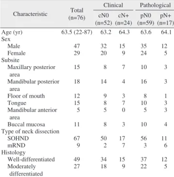
In total, 76 patients (47 males, 29 females; mean age, 63.5 years) with OSCC who had no previous malignancies and were not undergoing neoadjuvant concomitant chemoradio- therapy or radiotherapy were selected for this analysis.(Table 1) The mean follow-up period was 12.2 months, and the median was 12 months. The findings of neck node metastasis were cN0 (n=52, 68.4%), cN+ (n=24, 31.6%), pN0 (n=59, 77.6%), and pN+ (n=17, 22.4%).(Table 2) The mandibular and maxillary posterior areas were the predominant subsites.
(Fig. 1) All patients with clinically N0 or N+ neck were treat- ed with neck dissection (SOHND, n=67; mRND, n=9).(Fig.
2) The T stages of the pN0 necks were T1, n=29; T2, n=16;
T3, n=6; T4a, n=7; and T4b, n=1. In the pN+ necks, the tu- mor sizes were distributed as follows: T1, n=3; T2, n=7; T3, n=0; T4a, n=6; and T4b, n=1.(Table 3, Fig. 3) Based on the
AJCC 7th edition29 TNM classification, pN0 (pathologically negative lymph nodes) was noted in 29 patients in Stage I, 4 patients in Stage II, 12 patients in Stage III, and 14 patients in Stage IV (12 patients in IVa and 2 patient in IVb), while pN+ (with pathologically positive lymph nodes) was found in 5 patients in Stage I, 5 patients in Stage III, and 7 patients in Stage IV (6 patients in IVa and 1 patient in IVb).(Fig. 4)
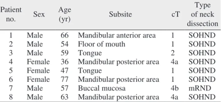
Occult metastases were found in 8 of the 52 cN0 patients
Table 1. Basic demographic data and treatments of the patients Characteristic Total
(n=76)
Clinical Pathological cN0
(n=52) cN+
(n=24) pN0 (n=59) pN+
(n=17)
Age (yr) 63.5 (22-87) 63.2 64.3 63.6 64.1
Sex Male 47 32 15 35 12
Female 29 20 9 24 5
Subsite
Maxillary posterior area
15 8 7 10 3
Mandibular posterior area
18 14 4 16 3
Floor of mouth 12 9 3 8 1
Tongue 15 8 7 10 3
Mandibular anterior
area 5 5 0 5 3
Buccal mucosa 11 8 3 10 4
Type of neck dissection
SOHND 67 50 17 56 11
mRND 9 2 7 3 6
Histology
Well-differentiated 49 34 15 37 12
Moderately differentiated
27 18 9 22 5
(SOHND: supraomohyoid neck dissection, mRND: modified radical neck dissection)
Values are presented as mean (range), mean only, or number only.
Aditi Sharma et al: Clinical analysis of neck node metastasis in oral cavity cancer. J Ko- rean Assoc Oral Maxillofac Surg 2018
Table 2. Correlations between clinical and pathological neck nodes
Clinical Pathological
pN0 pN+ Total
cN0 44 8 52
cN+ 15 9 24
Total 59 17
Values are presented as number of patients.
Aditi Sharma et al: Clinical analysis of neck node metastasis in oral cavity cancer. J Ko- rean Assoc Oral Maxillofac Surg 2018
Mandibular posterio
r area Floor
ofmout h
Tongue
Mandibular anterio
r area
Buccal mucosa
No.ofpatients
14 12 10 8 6 4 2 0
cN0 pN+
Maxillary posterio
r area 8
3 14
3 9
1
3 5
3
4
8 8
Fig. 1. Occult metastasis in relation to oral subsites.
Aditi Sharma et al: Clinical analysis of neck node metastasis in oral cavity cancer. J Ko- rean Assoc Oral Maxillofac Surg 2018
SOHND
No.ofpatients
60 50 40 30 20 10 0
pN0 pN+
11 56
mRND 3 6
Fig. 2. Type of neck dissection. (SOHND: supraomohyoid neck dissection, mRND: modified radical neck dissection)
Aditi Sharma et al: Clinical analysis of neck node metastasis in oral cavity cancer. J Ko- rean Assoc Oral Maxillofac Surg 2018
Table 3. Relation between neck node metastasis and T stage
T1 T2 T3 T4 Total
pN0 29 16 6 8 59
pN+ 3 7 0 7 17
Total 32 23 6 15 76
Values are presented as number of patients.
Cochrane Armitage trend test, P=0.014.
Aditi Sharma et al: Clinical analysis of neck node metastasis in oral cavity cancer. J Ko- rean Assoc Oral Maxillofac Surg 2018
T1
No.ofpatients
30 25 20 15 10 5 0
pN0 pN+
3 29
T2 7 16
T3 0 6
T4 8 7
Fig. 3. Tumor size (the American Joint Committee on Cancer [AJCC]
classification32).
Aditi Sharma et al: Clinical analysis of neck node metastasis in oral cavity cancer. J Ko- rean Assoc Oral Maxillofac Surg 2018
(15.4%). Neck node metastases were found in 17 patients (22.4%).(Table 4) We found no distant metastases in pN0 necks, while 4 of the 17 pN+ patients had distant metastases.
Regional LN metastases were found in one N0 neck and one N+ neck. Histopathological differentiation did not reveal any characteristics related to pathological lymph node metastasis (χ2 test, P>0.05).(Fig. 5) However, there was a statistically significant relationship between neck node metastasis and T stage (Cochrane Armitage trend test, P=0.014).(Table 3) Neck node metastasis and distant metastasis were also sig- nificantly related (Fisher’s exact test, P=0.019).(Table 5)
IV. Discussion
According to a meta-analysis of four well-designed pro- spective randomized controlled trials in oral cavity cancer patients, END significantly reduced the disease-specific death rates of OSCC patients with N0 neck30. The incidence of occult metastasis ranged from 6% to 30% in the END
group and from 37% to 58% in the observation group25. The reduced incidence of occult metastasis in the END group was most likely due to removal of fibro-fatty tissue in this group.
In the present study, histopathological diagnosis revealed oc- cult metastasis in 8 patients (15.4%). Our results were similar to those of Shimamoto et al.31, who found a 17.0% rate of cervical node metastasis; however, higher rates of occult me- tastasis have been found in other studies12,26,27,32. The tongue and mandibular posterior area were found to be common sub- sites associated with occult metastasis. This finding is in ac- cordance with the findings of Byers et al.5, who reported that the tongue was the predominant site for occult metastasis.
In various studies, END has been found to improve the regional control rate12,26,27. Observation of the neck tends to be associated with a greater number of regional recurrences33 and poor prognosis34. Small tumors (early stage) are poten- tially aggressive, and the incidence of nodal metastasis is high30. Weiss et al.35 suggested guidelines for N0 OSCC, rec- ommending END if the probability of occult cervical lymph node metastasis is greater than 20%. However, Okura et al.36 concluded that END should be recommended if the probabil- ity of occult metastasis is higher than 44.4%. The probability of occult metastasis has been reduced due to improvement
Table 4. Occult metastasis in 8 patients Pa tient
no. Sex Age
(yr) Subsite cT Type
of neck dissection 1 Male 66 Mandibular anterior area 1 SOHND
2 Male 54 Floor of mouth 1 SOHND
3 Male 59 Tongue 2 SOHND
4 Female 36 Mandibular posterior area 4a SOHND
5 Female 47 Tongue 1 SOHND
6 Female 77 Mandibular posterior area 1 SOHND
7 Male 57 Buccal mucosa 4b mRND
8 Male 63 Mandibular posterior area 4a SOHND (SOHND: supraomohyoid neck dissection, mRND: modified radical neck dissection)
Aditi Sharma et al: Clinical analysis of neck node metastasis in oral cavity cancer. J Ko- rean Assoc Oral Maxillofac Surg 2018
Stage I
No.ofpatients
30 25 20 15 10 5 0
pN0 pN+
5 29
0 4
Stage II Stage III 12
5
Stage IVb 2 1 Stage IVa
12
6
Fig. 4. Clinical staging of oral squamous cell carcinoma size.
Aditi Sharma et al: Clinical analysis of neck node metastasis in oral cavity cancer. J Ko- rean Assoc Oral Maxillofac Surg 2018
Well-differentiated
No.ofpatients
40
30
20
10
0
pN0 pN+
12 37
Moderately differentiated 5
22
Fig. 5. Histological classification of oral squamous cell carcinoma.
Aditi Sharma et al: Clinical analysis of neck node metastasis in oral cavity cancer. J Ko- rean Assoc Oral Maxillofac Surg 2018
Table 5. Relation between neck node metastasis and distant me- tastasis
Distant metastasis Pathological
pN0 pN+
– 59 13
+ 0 4
Values are presented as number of patients.
Fisher’s exact test, P=0.019.
Aditi Sharma et al: Clinical analysis of neck node metastasis in oral cavity cancer. J Ko- rean Assoc Oral Maxillofac Surg 2018
of radiological and treatment modalities. In our study, 15 of 24 clinically cN+ patients were found to be pathologically pN0 after the operation. Sentinel node biopsies were not performed in this study, and the discussion regarding this is beyond the scope of this article. The advantages of SOHND compared with mRND as a therapeutic procedure remain controversial due to a lack of prospective studies.
The most common subsites of oral cavity cancer are the tongue and floor of the mouth37,38. Tongue cancer metasta- sizes more often than floor-of-the-mouth cancer38,39. In addi- tion, these two subunits have a tendency to spread to the con- tralateral side12. In our study, the maxillary and mandibular posterior areas were found to be more common subsites than the tongue and floor of the mouth. The mandibular anterior area and buccal mucosa were predominant subsites for occult metastasis.
Nodal metastases may be missed in histological sections.
Yuen et al.12 detected nodal recurrence in 37% of patients in the observation neck group. The authors concluded that selective neck dissection alone is an adequate treatment for oral cancer patients with N0 neck, even though nodal micro- metastasis might be missed histopathologically. In patients with N+ neck, selective neck dissection and radiotherapy have been advised for better nodal control. Most studies have failed to demonstrate that survival outcomes differ signifi- cantly between the END group and the observation group40,41. Even fewer studies have demonstrated the significance of END in OSCC patients with clinically N0 neck42.
The major advantage of END in clinically N0 neck patients is that its surgical morbidity is lower than that of mRND for patients with nodal recurrence in the observation group.
Most patients in the observation group will need mRND and will thus have greater chances of surgical morbidity. On the other hand, the disadvantage of END is that 70% of N0 neck patients will undergo unnecessary neck dissections, incurring additional costs and surgical morbidity. The advantage of observation is that only 30% to 40% of patients report nodal metastasis requiring neck treatment. The major disadvantage of observation is that patients may need radical or modified neck dissection, which have reduced survival rates.
When metastasis occurs, close follow-up is a major deter- minant of survival outcome and nodal recurrence irrespective of the choice of treatment for N0 neck12. Kligerman et al.32 demonstrated that the survival rate was lower (low salvage rate) and the nodal recurrence rate was 27% in observed necks, while END was beneficial in terms of survival out- come in clinically N0 necks. Vandenbrouck et al.26 reported
a high salvage rate (around 84%), so the survival outcomes of END were not found to differ significantly from those of observation.
Kuntz and Weymuller42 noted that shoulder disabilities at 6 months were more common with mRND than with SOHND.
Rastogi et al.43 found statistically significant differences be- tween super-selective neck dissection (level I, IIa, III) and se- lective neck dissection (level I, IIa, IIb, III) groups, including less shoulder morbidity and a better quality of life in the su- per-selective neck dissection group. Giordano et al.44 reported that sublevel IIb dissection impaired nerve conduction and reduced the quality of life in OSCC patients. Therefore, clini- cal, radiological, and histopathological evaluations should play a major role in the decision to involve level IIb in neck dissection. Positive nodal metastasis in sublevel IIa strongly signifies an association of notal metastasis of sublevel IIb45. Still, there is no clear indication for neck dissection in cN0 necks. However, neck dissection in cN0 necks has been shown to be better than observation25-27,32.
The limitation of our study is its retrospective nature. Nev- ertheless, we found a statistically significant relationship between neck node metastasis and T stage (P=0.014) and be- tween neck node metastasis and distant metastasis (P=0.019).
V. Conclusion
Our results revealed occult metastasis in 15.4% of the final surgical neck node specimens, a relatively low percentage compared with those of other studies12,26,27,32. Neck node me- tastasis was significantly related to tumor size and distant me- tastasis during follow-up. Therefore, additional well-designed prospective randomized controlled trials are required to de- termine the appropriate treatment modalities for N0 necks.
ORCID
Aditi Sharma, https://orcid.org/0000-0002-5948-4818 Jin-Wook Kim, https://orcid.org/0000-0003-4074-877X Jun-Young Paeng, https://orcid.org/0000-0002-0104-9338
Authors’ Contributions
A.S. participated in data collection and wrote the manu- script. J.W.K. participated in patients operation. J.Y.P. partici- pated in the study design and revised the manuscript.
Ethics Approval and Consent to Participate
The institutional review board of Kyungpook National University Hospital approved this study, and each patient signed an informed consent agreement (KNUH_06-1003).
Conflict of Interest
No potential conflict of interest relevant to this article was reported.
References
1. Jemal A, Bray F, Center MM, Ferlay J, Ward E, Forman D. Global cancer statistics. CA Cancer J Clin 2011;61:69-90.
2. Ren ZH, Wu HJ, Wang K, Zhang S, Tan HY, Gong ZJ. Anterolat- eral thigh myocutaneous flaps as the preferred flaps for reconstruc- tion of oral and maxillofacial defects. J Craniomaxillofac Surg 2014;42:1583-9.
3. Ren ZH, Wu HJ, Tan HY, Wang K, Zhang S. Transfer of antero- lateral thigh flaps in elderly oral cancer patients: complications in oral and maxillofacial reconstruction. J Oral Maxillofac Surg 2015;73:534-40.
4. Ren ZH, Xu JL, Fan TF, Ji T, Wu HJ, Zhang CP. The harmonic scalpel versus conventional hemostasis for neck dissection: a meta-analysis of the randomized controlled trials. PLoS One 2015;10:e0132476.
5. Byers RM, El-Naggar AK, Lee YY, Rao B, Fornage B, Terry NH, et al. Can we detect or predict the presence of occult nodal metasta- ses in patients with squamous carcinoma of the oral tongue? Head Neck 1998;20:138-44.
6. Po Wing Yuen A, Lam KY, Lam LK, Ho CM, Wong A, Chow TL, et al. Prognostic factors of clinically stage I and II oral tongue car- cinoma-a comparative study of stage, thickness, shape, growth pat- tern, invasive front malignancy grading, Martinez-Gimeno score, and pathologic features. Head Neck 2002;24:513-20.
7. Woolgar JA, Triantafyllou A, Lewis JS Jr, Hunt J, Williams MD, Takes RP, et al. Prognostic biological features in neck dissection specimens. Eur Arch Otorhinolaryngol 2013;270:1581-92.
8. Haddadin KJ, Soutar DS, Oliver RJ, Webster MH, Robertson AG, MacDonald DG. Improved survival for patients with clinically T1/
T2, N0 tongue tumors undergoing a prophylactic neck dissection.
Head Neck 1999;21:517-25.
9. Lydiatt DD, Robbins KT, Byers RM, Wolf PF. Treatment of stage I and II oral tongue cancer. Head Neck 1993;15:308-12.
10. Yuen AP, Wei WI, Wong YM, Tang KC. Elective neck dissection versus observation in the treatment of early oral tongue carcinoma.
Head Neck 1997;19:583-8.
11. Keski-Säntti H, Atula T, Törnwall J, Koivunen P, Mäkitie A. Elec- tive neck treatment versus observation in patients with T1/T2 N0 squamous cell carcinoma of oral tongue. Oral Oncol 2006;42:96- 12. Yuen AP, Ho CM, Chow TL, Tang LC, Cheung WY, Ng RW, et al. 101.
Prospective randomized study of selective neck dissection versus observation for N0 neck of early tongue carcinoma. Head Neck 2009;31:765-72.
13. Capote A, Escorial V, Muñoz-Guerra MF, Rodríguez-Campo FJ, Gamallo C, Naval L. Elective neck dissection in early-stage oral squamous cell carcinoma--does it influence recurrence and sur- vival? Head Neck 2007;29:3-11.
14. Huang SF, Chang JT, Liao CT, Kang CJ, Lin CY, Fan KH, et al.
The role of elective neck dissection in early stage buccal cancer.
Laryngoscope 2015;125:128-33.
15. Kirita T, Omura K. Oral cancer: diagnosis and therapy. Tokyo:
Springer; 2015:221-30.
16. Mamelle G, Pampurik J, Luboinski B, Lancar R, Lusinchi A, Bosq J. Lymph node prognostic factors in head and neck squamous cell carcinomas. Am J Surg 1994;168:494-8.
17. León X, Quer M, Orús C, del Prado Venegas M, López M. Distant metastases in head and neck cancer patients who achieved loco- regional control. Head Neck 2000;22:680-6.
18. Lim JY, Lim YC, Kim SH, Kim JW, Jeong HM, Choi EC. Predic- tive factors of isolated distant metastasis after primary definitive surgery without systemic treatment for head and neck squamous cell carcinoma. Oral Oncol 2010;46:504-8.
19. Garavello W, Ciardo A, Spreafico R, Gaini RM. Risk factors for distant metastases in head and neck squamous cell carcinoma. Arch Otolaryngol Head Neck Surg 2006;132:762-6.
20. Liao CT, Wang HM, Chang JT, Ng SH, Hsueh C, Lee LY, et al.
Analysis of risk factors for distant metastases in squamous cell car- cinoma of the oral cavity. Cancer 2007;110:1501-8.
21. Reinisch S, Kruse A, Bredell M, Lübbers HT, Gander T, Lanzer M.
Is lymph-node ratio a superior predictor than lymph node status for recurrence-free and overall survival in patients with head and neck squamous cell carcinoma? Ann Surg Oncol 2014;21:1912-8.
22. Prabhu RS, Hanasoge S, Magliocca KR, Hall WA, Chen SA, Hig- gins KA, et al. Lymph node ratio influence on risk of head and neck cancer locoregional recurrence after initial surgical resection:
implications for adjuvant therapy. Head Neck 2015;37:777-82.
23. Gil Z, Carlson DL, Boyle JO, Kraus DH, Shah JP, Shaha AR, et al. Lymph node density is a significant predictor of outcome in pa- tients with oral cancer. Cancer 2009;115:5700-10.
24. Guo CB, Feng Z, Zhang JG, Peng X, Cai ZG, Mao C, et al. Su- praomohyoid neck dissection and modified radical neck dissec- tion for clinically node-negative oral squamous cell carcinoma: a prospective study of prognosis, complications and quality of life. J Craniomaxillofac Surg 2014;42:1885-90.
25. Fasunla AJ, Greene BH, Timmesfeld N, Wiegand S, Werner JA, Sesterhenn AM. A meta-analysis of the randomized controlled tri- als on elective neck dissection versus therapeutic neck dissection in oral cavity cancers with clinically node-negative neck. Oral Oncol 2011;47:320-4.
26. Vandenbrouck C, Sancho-Garnier H, Chassagne D, Saravane D, Cachin Y, Micheau C. Elective versus therapeutic radical neck dissection in epidermoid carcinoma of the oral cavity: results of a randomized clinical trial. Cancer 1980;46:386-90.
27. Fakih AR, Rao RS, Borges AM, Patel AR. Elective versus thera- peutic neck dissection in early carcinoma of the oral tongue. Am J Surg 1989;158:309-13.
28. Bryne M. Prognostic value of various molecular and cellular fea- tures in oral squamous cell carcinomas: a review. J Oral Pathol Med 1991;20:413-20.
29. Edge SB, Compton CC. The American Joint Committee on Cancer:
the 7th edition of the AJCC cancer staging manual and the future of TNM. Ann Surg Oncol 2010;17:1471-4.
30. Silverberg E. Cancer statistics. 1986. CA Cancer J Clin 1986;36:9- 31. Shimamoto H, Oikawa Y, Osako T, Hirai H, Mochizuki Y, Tanaka 25.
K, et al. Neck failure after elective neck dissection in patients with oral squamous cell carcinoma. Oral Surg Oral Med Oral Pathol Oral Radiol 2017;124:32-6.
32. Kligerman J, Lima RA, Soares JR, Prado L, Dias FL, Freitas EQ, et al. Supraomohyoid neck dissection in the treatment of T1/T2 squamous cell carcinoma of oral cavity. Am J Surg 1994;168:391- 33. Kowalski LP. Results of salvage treatment of the neck in patients 4.
with oral cancer. Arch Otolaryngol Head Neck Surg 2002;128:58- 62.
34. Byers RM, Weber RS, Andrews T, McGill D, Kare R, Wolf P.
Frequency and therapeutic implications of “skip metastases” in the neck from squamous carcinoma of the oral tongue. Head Neck 1997;19:14-9.
35. Weiss MH, Harrison LB, Isaacs RS. Use of decision analysis in planning a management strategy for the stage N0 neck. Arch Oto- laryngol Head Neck Surg 1994;120:699-702.
36. Okura M, Aikawa T, Sawai NY, Iida S, Kogo M. Decision analysis and treatment threshold in a management for the N0 neck of the oral cavity carcinoma. Oral Oncol 2009;45:908-11.
37. Dias FL, Lima RA, Kligerman J, Farias TP, Soares JR, Manfro G, Sa GM. Relevance of skip metastases for squamous cell carcinoma of the oral tongue and the floor of the mouth. Otolaryngol Head Neck Surg 2006;134:460-5.
38. Jerjes W, Upile T, Petrie A, Riskalla A, Hamdoon Z, Vourvachis M, et al. Clinicopathological parameters, recurrence, locoregional and distant metastasis in 115 T1-T2 oral squamous cell carcinoma patients. Head Neck Oncol 2010;2:9.
39. Duvvuri U, Simental AA Jr, D’Angelo G, Johnson JT, Ferris RL, Gooding W, et al. Elective neck dissection and survival in patients with squamous cell carcinoma of the oral cavity and oropharynx.
Laryngoscope 2004;114:2228-34.
40. Dias FL, Kligerman J, Matos de Sá G, Arcuri RA, Freitas EQ, Farias T, et al. Elective neck dissection versus observation in stage I squamous cell carcinomas of the tongue and floor of the mouth.
Otolaryngol Head Neck Surg 2001;125:23-9.
41. Marchioni DL, Fisberg RM, do Rosário M, Latorre DO, Wunsch V.
Diet and cancer of the oral cavity and pharynx: a case-control study in São Paulo, Brazil. IARC Sci Publ 2002;156:559-61.
42. Kuntz AL, Weymuller EA Jr. Impact of neck dissection on quality of life. Laryngoscope 1999;109:1334-8.
43. Rastogi S, Sharma A, Choudhury R, Tripathi S, Al Wayli H, Am- rithraj A, et al. Is superselective neck dissection safer than suprao- mohyoid neck dissection for oral carcinoma patients with N0 neck in terms of shoulder morbidity and recurrence rate? J Oral Maxil- lofac Surg 2018;76:647-55.
44. Giordano L, Sarandria D, Fabiano B, Del Carro U, Bussi M.
Shoulder function after selective and superselective neck dissec- tions: clinical and functional outcomes. Acta Otorhinolaryngol Ital 2012;32:376-9.
45. Mashberg A, Meyers H. Anatomical site and size of 222 early as- ymptomatic oral squamous cell carcinomas: a continuing prospec- tive study of oral cancer. II. Cancer 1976;37:2149-57.