대 한 생 식 의 학 회 지 : 제 35 권 제 4 호 2008
조기난소부전의 유전학적 이해
포천중문의과대학교 차병원 여성의학연구소 최 영 석*
Genetics of Non-Syndromic Premature Ovarian Failure
Youngsok Choi *Fertility Center of CHA General Hospital, Pochon CHA University College of Medicine
[Korean. J. Reprod. Med. 2008; 35(4): 239-245.]
I. 서 론
불임이란 일반적으로 결혼 후 원만한 부부관계 를 가져도 1~2년 이내에 임신이 되지 않는 경우로 엄밀히 말해 병이라고 단정 지을 수 없지만 일정 기간이 지나도록 임신을 못하는 상태를 말한다.1,2 불임의 40~60%가 여자 쪽에 원인이 있는 경우로 분류되며 배란장애, 난관이상, 자궁이상 등이 포함 된다.2 이들 장애 중에 조기난소부전 (Premature ovarian failure, POF)은 여성의 불임을 일으키는 한 원인이다. 미국에서 40세 이전 여성의 약 1% 정 도가 POF로 진단받고 있다.3 POF는 난소 내 난 포들이 사라지거나 감소되면서 영구불임으로 진 단받는 경우를 말하는데, POF 환자들은 에스트로 겐 (Estrogen) 분비저하로 혈액 내 난포자극호르몬 (Follicle stimulating Hormone, FSH) 분비가 (>40 IU/
liter) 높고 6개월 이상 월경주기가 없다. 현재까지 알려진 POF의 원인으로는 감염 (Infection), 화학치 료 (chemotherapy), 자궁경부 외과수술 (pelvic sur-
gery), 자가면역질병 (autoimmune diseases), 환경적 요인, 유전적요인 등 다양하게 존재한다. 그렇지만 POF의 정확한 유전적 원인은 밝혀져 있지 않다.
몇 해 전까지 만해도 POF는 성염색체 X (X chromosome)와 상염색체 (Autosome) 이상과 연관 이 있다고 보고되어 왔다. X chromosome의 일부인 POF (Premature Ovarian Failure)-1, POF-2, POF-3를 포함하고 있는 유전자위 (loci)의 손실 (deletion)이 나 재배열 (rearrangement)은 POF를 유발한다.4~8 POF1B의 점 돌연변이 (point mutation)는 POF1B가 nonmuscle actin filament 결합을 방해함으로써 이상 이 발생한다. 그리고 Turner 증후군 환자와 Fragile X 증후군 환자에게서도 난소부전이 관찰된다. Turner 증후군의 경우 X chromosome 특정지역에 존재하 는 유전자의 Haploinsufficiency에 기인하는 것으로 여겨지고 있다.9,10 Fragile X 증후군 환자의 경우는 FMR1 (Fragile X mental retardation 1) 돌연변이와 FMR2 micro-deletion에 의해 조기폐경이 유발된다.
11~13
그리고 X-linked된 유전자 중 하나인 BMP15 (Bone morphogenetic protein 15)의 유전자사슬 변이 (sequence variant) 도 난소이상 (Ovarian dysfunction) 과 연관되어 있다.14~16 그리고 상염색체 (Autosome) 상에 존재하는 유전자의 돌연변이들도 POF 환자와 연관이 있다. FSHR (FSH receptor), LHR (Luteinizing
주관책임자: 최영석, 우) 135-080 서울특별시 강남구 역삼동 606-5, 포천중문의과대학교 차병원 여성의학연구소
Tel: (02) 3468-3504, Fax: (02) 3468-3464 e-mail: youngsokchoi@gmail.com
*본 연구는 보건복지가족부 보건의료기술연구개발사업의 지원에 의해 이루어진 것임 (A080364).
종 설
Hormone receptor), GDF9 (Growth differentiation factor 9), INHA (Inhibin), GALT (Galactose-1-phosphate uridyltransferase), FOXL2 (Forkhead transcription factor 2) 등이 여기에 포함된다.17~24 그렇지만 최근까지 이러한 유전적 결함들이 어떻게 난소 내 난포형 성과 유지에 관계가 있는지 분자생물학적 접근이 이뤄지지 않았다. 본 고찰에서는 Non-syndromic ovarian failure의 유전학적 이해를 설명하기 위해 최 근에 발견된 난자특이 전사조절인자의 유전자 결 핍 생쥐모델과 임상적으로 보고된 연구 등을 살펴 보고 그 가능성에 대해 알아보고자 한다.
II. 본 론
1. Factor in the germline alpha (FIGLA)
FIGLA는 난포의 투명대 (zona pellucida, Zp) 단 백질 발현을 조절하는basic helix-loop-helix (bHLH) 전사조절인자이다.25 FIGLA는 생쥐에서 배발달 13.5 (E13.5)일째 발현되며, 난소에서 원시난포 (Primor- dial follicle) 형성에 중요한 역할을 한다.26 FIGLA 유전자 결핍생쥐 암컷은 불임이지만, 수컷은 정상 이다.26 FIGLA 유전자 결핍은 난소에서 primordial follicle 형성을 방해하며, 태어나면서 난소 내 난자 들이 빠른 속도로 사라지게 만든다.26 FIGLA 유전 자 결핍생쥐 난소에서 Pou5f1 (Known as Oct4), Zp2, Pad6 등 수 많은 난자특이 유전자들이 유의적으로 감소되거나 결핍된다.27 Zp2는 투명대 (Zona matrix) 를 구성하는 세 가지 인자인 Zp1, Zp2, Zp3 중 하 나로 Zp2 유전자 결핍생쥐도 난포형성이상으로 인 한 불임이다.28 Zp2 프로모터에 FIGLA 결합장소인 E-box (CANNTG)가 위치하고 있다. FIGLA가 직접 이 결합장소에 결합하고 프로모터 활성을 조절한 다.25 이것은 FIGLA 전사조절인자가 난자의 형태 및 형성에 중요한 유전자를 조절함으로써 난자의 생리적 역할을 조절한다는 것을 의미한다. Zhao 등 은 100명의 중국인 POF 환자의 염기서열 분석을 통해 FIGLA genomic DNA에서 세 개의 유전자 변 이 (Variant)을 발견하였고 두 명의 환자에서 유전자
돌연변이 (missence mutation, c.11C → A (p.A4E))을 발견하였다.29 이러한 돌연변이의 경우 프레임시 프트 (frame shift)를 일으켜 haploinsufficiency를 유 도한다. 그리고 한 명의 환자에서는 유전자 결손 (deletion, c.419-421 delACA (p.140 delN))이 발견된 다.29 이러한 missense 돌연변이와 유전자 결손은 FIGLA 전사조절인자의 활성에 영향을 미치는데, FIGLA의 heterodimer 파트너인 TCF3 bHLH 결합을 방해한다.25 이러한 연구는 FIGLA가 난포형성 초기 에 중요하며 난자특이의 유전자들을 직접적으로 조 절함으로써 난포의 성장과 유지에 관여한다는 것을 의미한다.
2. Newborn ovary homeobox gene (NOBOX)
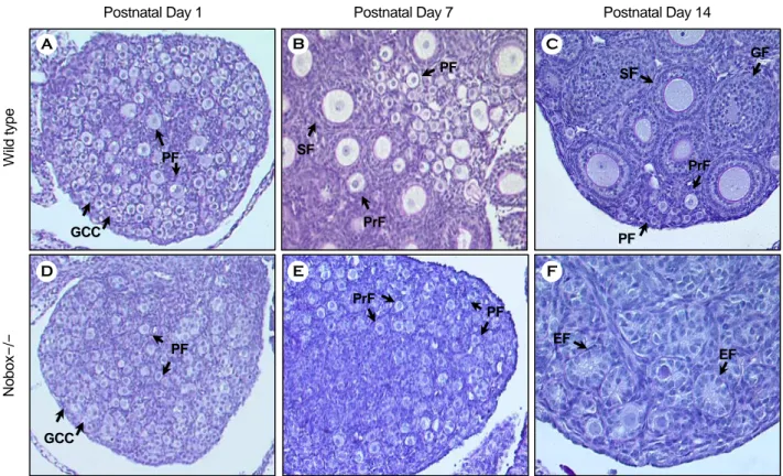
NOBOX는 호메오박스 (Homeobox) 도메인을 가 지고 있는 전사조절인자이다.30 NOBOX는 생쥐에 서 배발달 E15.5일째 발현되며, 난소에서 생식세포 및 원시난포 (Primordial follicle)를 포함한 모든 성 장난포의 난자 내에 존재한다.30,31 NOBOX 유전자 결핍생쥐 수컷은 정상 표현형을 보여주지만 암컷 은 불임이다.31 NOBOX 유전자 결핍생쥐는 생후 14일 이내에 난소에 존재하던 난자와 난포를 모두 잃어버린다.31 갓 태어난 NOBOX 유전자 결핍생쥐 암컷의 난소와 정상생쥐의 난소를 비교했을 때 조 직학적으로 차이가 없다. NOBOX 유전자 결핍생쥐 암컷의 난소에는 비슷한 숫자의 생식세포와 원시 난포를 포함하고 있다.31 그렇지만 생후 7일째가 되 면 NOBOX 유전자 결핍생쥐 난소에는 정상생쥐의 난소에서 발견되는 Secondary follicle를 볼 수 없 고 난포의 숫자도 현저히 감소됨을 보여주고 있다 (Figure 1).31 그리고 갓 태어났을 때 NOBOX 유전 자 결핍생쥐 암컷의 난소가 조직학적으로 문제가 없었지만 유전학적으로 많은 변화가 있다. NOBOX 유전자 결핍생쥐 암컷의 난소에서 Gdf9 (Growth differentiation factor 9), Bmp15 (Bone morphogenetic protein 15), Mos, Pou5f1와 같은 수많은 난자 및 생 식세포 특이적 유전자의 발현이 현저하게 감소된
다.31,32 NOBOX는 호메오 전사조절인자로 TAA/
GTTG/A에 결합하여 직접적으로 유전자 발현을 조 절한다.33 NOBOX 유전자 결핍생쥐 암컷의 난소에 서 현저하게 감소되는 유전자들 중 Gdf9은 프로 모터 부분에 3개의 NOBOX 결합장소가 존재한다.33 NOBOX는 이들 각각의 결합장소에 강한 결합력을 보여주고 있고 Gdf9 프로모터의 활성을 조절한다.33 특이할 점은 난자에서 Gdf9의 조절이다. Choi 등이 NOBOX가 Gdf9 유전자를 직접적으로 조절한다고 보고할 때까지 Gdf9의 분자생물학적 역할은 알려 져 있지 않았다. Gdf9은 난자에서 분비되며 난자주 변을 둘러싸고 있는 granulosa 세포들의 성장에 영 향을 미쳐 난포의 성장을 조절하는 매우 중요한 난 자특이 성장인자이다.34~37 Gdf9 유전자가 없다면 암 컷은 불임되고 난소 내 난포들은 primary 난포에서 성장을 멈춘다.38 이것은 NOBOX 유전자 결핍생쥐 암컷의 표현형과 거의 유사하다. 즉 NOBOX 전사 조절인자는 형태학적으로 초기 원시난포형성에는
영향을 미치지 않지만 생후 난포의 생존과 성장에 중요한 유전자의 발현을 조절함으로써 난자에 중요 한 역할을 한다는 것을 의미한다. Qin 등은 96명의 백인 POF 환자의 염기서열 분석을 통해 네 명의 환자에게서 NOBOX genomic DNA에서 네 개의 유 전자 변이를 발견하였고 NOBOX 호메오 도메인 부 분에서 두 개의 p.Arg355His와 p.Arg360Gln missense 돌연변이를 찾아내었다.39 이 중에서 p.Arg355His missense 돌연변이는 NOBOX의 DNA 결합능력을 현저히 감소시킨다.39 그리고 정상 NOBOX와 일대 일 반응에서도 돌연변이 NOBOX는 정상 NOBOX 의 DNA 결합능력을 절반으로 감소시켰다.39 이것 은 p.Arg355His missense 돌연변이가 haploinsuffi- ciency를 유도한다는 것을 의미한다.
3. LIM-homeobox gene 8 (LHX8)
LHX8은 호메오박스 (Homeobox) 도메인과 LIM
Postnatal Day 1 Postnatal Day 7 Postnatal Day 14
Wild type Nobox-
/-
GCC
PF
PF
PrF SF
PF SF
GF
PrF
GCC
PF
PF PrF
EF EF
A B C
D E F
Figure 1. Ovaries from wild type and Nobox null (Nobox-/-) mice at postnatal day 1, 7, and 14. GCC, germ cell cluster; PF, primordial follicle; PrF, primary follicle; SF, secondary follicle; GF, growing follicle; EF, empty follicle.
도메인을 가지고 있는 전사조절인자로서 뇌 발달에 관여하는 인자로 발견이 되었다.40 그 이후에 Choi 등은 LHX8가 생쥐난소에서 초기 배발달 E13.5일째 발현되며, 난소에서 생식세포 및 원시난포 (Primor- dial follicle)를 포함한 모든 성장난포의 난자 내에 특이적으로 존재한다고 보고하였다.41 LHX8 유전자 결핍생쥐 수컷은 정상이지만 암컷은 불임이다.41 LHX8 유전자 결핍생쥐는 생후 7일 이내에 난소에
존재하던 난자와 난포를 모두 잃어버린다.31 갓 태 어난 LHX8 유전자 결핍생쥐 암컷의 난소는 정상 생쥐와 비교했을 때 원시생식세포의 숫자는 차이 가 없었지만 원시난포의 숫자는 감소한다.41 그리고 생후 3일째가 되면 LHX8 유전자 결핍생쥐 난소에 는 정상생쥐의 난소에서 발견되는 primary follicle를 볼 수 없고 난포의 숫자도 현저히 줄어든다.41 갓 태어난 LHX8 유전자 결핍생쥐 암컷의 난소는 조 A
B
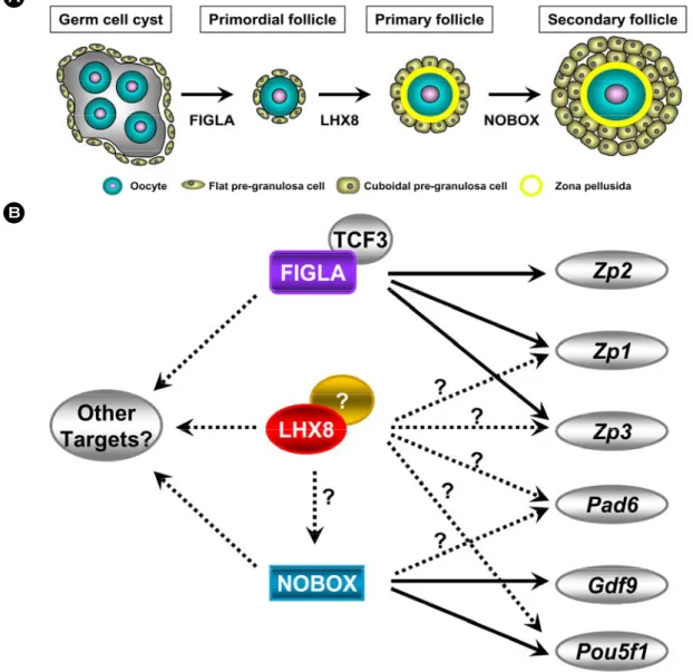
Figure 2. A) Transcription factors that are important in the formation of primordial and primary follicles at early stage of folliculogenesis. The follicle structures involving the oocyte, zona pellusida, and granulosa cells are shown.
Transcriptional regulations are controlled by FIGLA, LHX8 and NOBOX. B) Transcriptional network at early stage of folliculogenesis. Numerous oocyte-specific genes including Zp1, Zp2, Zp3, Pad6, Gdf9, and Pou5f1 are controlled by FIGLA, LHX8 or NOBOX.
직학적으로 차이가 거의 없지만 Pou5f1, Gdf9, Kitl, Zp3와 같은 난자 및 생식세포 특이적 유전자의 발 현이 유의적으로 감소한다.41 LHX8도 난자특이 호 메오 전사조절인자 NOBOX처럼 DNA에 결합하는 호메오 도메인을 가지고 있다. LHX8는 전사조절 인자의 DNA 결합장소는 TG/AATTA로 NOBOX의 DNA 결합장소인 TAA/GTTG/A와 약간 차이가 있 다.41 이것은 조직발달에 시간적으로 공간적으로 작 동하는 호메오 전사조절인자들이 조직특이적으로 작동하게 하는 원리이다. 특이한 점은 LHX8 유전 자 결핍난자에서 유의있게 감소 하는 유전자 중에 NOBOX가 포함된다.41 그리고 NOBOX 프로모터에 한 개의 LHX8 DNA 결합장소가 존재하며 LHX8 에 강한 결합력 가지고 있다 (Unpublised data, Choi 등, 2009). 이것은 LHX8가 난포성장과 생존에 필 요한 NOBOX 전사조절인자의 상위조절인자로서 NOBOX를 직접적으로 조절한다는 것을 의미한다.
그렇지만 아직까지 LHX8와 POF와 상관관계는 보 고된 바가 없다.
III. 맺음말
POF의 유전적 원인을 규명하고자 하는 연구가 계속적으로 진행 중 이었지만 난자에 특이적으로 발현되는 전사조절인자들이 발굴되기 전 까지 그 노력은 답보상태에 있었다 (Figure 2). FIGLA는 10 여 년 전에 발견되었지만 얼마 전 POF 환자군에 서 임상적 자료가 발표될 때까지 알려지지 않았다.
그리고 Rajkovic 그룹에서 NOBOX, LHX8와 같은 새로운 전사조절인자들을 발굴하면서 POF의 유전 학적 규명에 관한 연구는 탄력을 받기 시작했다.
현재 FIGLA, NOBOX, LHX8 이 세 가지 전사조절 인자들이 각각의 하위 조절 메커니즘을 갖는지 아 니면 서로 전사조절 네트워크를 형성하면서 난자 와 난포의 성장과 생존에 영향을 미치는지 명확하 지 않다. 그리고 이 종설에서는 언급하지 않았지 만 전사조절인자들 외에도 PTEN (Phosphatase and tensin homolog)과 같은 세포질인자들도 primordial
follicle pool를 조기에 활성화시킴으로 POF가 유발 한다고 보고되고 있다.42 PTEN은 Phosphatidylinositol 3-kinase (PI3K)의 주요 negative regulator로 이는 난 포 내 난자가 PTEN-PI3K pathway를 통하여 성장과 활성화를 조절하는 것으로 여겨지고 있다. 이처럼 난포형성과 생존 조절 메커니즘에 필수적인 인자 들을 발굴하고 이를 통하여 유전자이상으로 유발 된 POF의 원인을 분자수준에서 설명할 수 있다면 POF를 미리 진단 예측하고 치료 예방할 수 있는 실마리를 제공할 수 있을 것으로 생각된다.
참 고 문 헌
1. Habbema JD, Collins J, Leridon H, Evers JL, Lunenfeld B. te Velde ER. Towards less confusing terminology in reproductive medicine: a proposal. Hum Reprod 2004; 19: 1497-501.
2. Smith S, Pfeifer SM, Collins JA. Diagnosis and management of female infertility. JAMA 2003; 290: 1767-70.
3. Conway GS. Premature ovarian failure. Br Med Bull 2000;
56: 643-9.
4. Davison RM, Fox M, Conway GS. Mapping of the POF1 locus and identification of putative genes for premature ovarian failure. Mol Hum Reprod 2000; 6: 314-8.
5. Lacombe A, Lee H, Zahed L, Choucair M, Muller JM, Nelson SF, et al. Disruption of POF1B binding to nonmuscle actin filaments is associated with premature ovarian failure. Am J Hum Genet 2006; 79: 113-9.
6. Marozzi A, Manfredini E, Tibiletti MG, Furlan D, Villa N, Vegetti W, et al. Molecular definition of Xq common-deleted region in patients affected by premature ovarian failure. Hum Genet 2000; 107: 304-11.
7. Powell CM, Taggart RT, Drumheller TC, Wangsa D, Qian C, Nelson LM, et al. Molecular and cytogenetic studies of an X;autosome translocation in a patient with premature ovarian failure and review of the literature. Am J Med Genet 1994;
52: 19-26.
8. Tharapel AT, Anderson KP, Simpson JL, Martens PR, Wilroy RS Jr, Llerena JC Jr, et al., Deletion (X)(q26.1-->q28) in a proband and her mother: molecular characterization and phenotypic-karyotypic deductions. Am J Hum Genet 1993;
52: 463-71.
9.Elsheikh M, Dunger DB, Conway GS, Wass JA. Turner's syndrome in adulthood. Endocr Rev 2002; 23: 120-40.
10. Zinn AR, Ross JL. Turner syndrome and haploinsufficiency.
Curr Opin Genet Dev 1998; 8: 322-7.
11. Allingham-Hawkins DJ, Babul-Hirji R, Chitayat D, Holden JJ, Yang KT, Lee C, et al. Fragile X premutation is a significant risk factor for premature ovarian failure: the International Collaborative POF in Fragile X study--preliminary data. Am J Med Genet 1998; 83: 322-5.
12. Murray A, Webb J, Dennis N, Conway G, Morton N. Micro- deletions in FMR2 may be a significant cause of premature ovarian failure. J Med Genet 1999; 36: 767-70.
13. Murray A, Webb J, Grimley S, Conway G, Jacobs P. Studies of FRAXA and FRAXE in women with premature ovarian failure. J Med Genet 1998; 35: 637-40.
14. Dixit H, Rao LK, Padmalatha VV, Kanakavalli M, Deenadayal M, Gupta N, et al. Missense mutations in the BMP15 gene are associated with ovarian failure. Hum Genet 2006; 119: 408-15.
15. Laissue P, Christin-Maitre S, Touraine P, Kuttenn F, Ritvos O, Aittomaki K, et al. Mutations and sequence variants in GDF9 and BMP15 in patients with premature ovarian failure. Eur J Endocrinol 2006; 154: 739-44.
16. Laissue P, Vinci G, Veitia RA, Fellous M. Recent advances in the study of genes involved in non-syndromic premature ovarian failure. Mol Cell Endocrinol 2008; 282: 101-11.
17. Aittomaki K, Lucena JL, Pakarinen P, Sistonen P, Tapanainen J, Gromoll J, et al. Mutation in the follicle-stimulating hormone receptor gene causes hereditary hypergonadotropic ovarian failure. Cell 1995; 82: 959-68.
18. Crisponi L, Deiana M, Loi A, Chiappe F, Uda M, Amati P, et al. The putative forkhead transcription factor FOXL2 is mutated in blepharophimosis/ ptosis/ epicanthus inversus syndrome. Nat Genet 2001; 27: 159-66.
19. Dixit H, Deendayal M, Singh L. Mutational analysis of the mature peptide region of inhibin genes in Indian women with ovarian failure. Hum Reprod 2004; 19: 1760-4.
20. Dixit H, Rao KL, Padmalatha V, Kanakavalli M, Deenadayal M, Gupta N, et al. Expansion of the germline analysis for the INHA gene in Indian women with ovarian failure. Hum Reprod 2006; 21: 1643-4.
21. Dixit H, Rao LK, Padmalatha V, Kanakavalli M, Deenadayal M, Gupta N, et al. Mutational screening of the coding region of growth differentiation factor 9 gene in Indian women with
ovarian failure. Menopause 2005; 12: 749-54.
22. Guerrero NV, Singh RH, Manatunga A, Berry GT, Steiner RD, Elsas LJ 2nd. Risk factors for premature ovarian failure in females with galactosemia. J Pediatr 2000; 137: 833-41.
23. Latronico AC, Anasti J, Arnhold IJ, Rapaport R, Mendonca BB, Bloise W, et al. Brief report: testicular and ovarian resistance to luteinizing hormone caused by inactivating mutations of the luteinizing hormone-receptor gene. N Engl J Med 1996; 334: 507-12.
24. Shelling AN, Burton KA, Chand AL, van Ee CC, France JT, Farquhar CM, et al. Inhibin: a candidate gene for premature ovarian failure. Hum Reprod 2000; 15: 2644-9.
25. Liang L, Soyal SM, Dean J. FIGalpha, a germ cell specific transcription factor involved in the coordinate expression of the zona pellucida genes. Development 1997; 124: 4939-47.
26. Soyal SM, Amleh A, Dean J. FIGalpha, a germ cell-specific transcription factor required for ovarian follicle formation.
Development 2000; 127: 4645-54.
27. Joshi S, Davies H, Sims LP, Levy SE, Dean J. Ovarian gene expression in the absence of FIGLA, an oocyte-specific tran- scription factor. BMC Dev Biol 2007; 7: 67.
28.Rankin TL, O'Brien M, Lee E, Wigglesworth K, Eppig J, Dean J. Defective zonae pellucidae in Zp2-null mice disrupt folliculogenesis, fertility and development. Development 2001;
128: 1119-26.
29.Zhao H, Chen ZJ, Qin Y, Shi Y, Wang S, Choi Y, et al.
Transcription factor FIGLA is mutated in patients with pre- mature ovarian failure. Am J Hum Genet 2008; 82: 1342-8.
30.Suzumori N, Yan C, Matzuk MM, Rajkovic A. Nobox is a homeobox-encoding gene preferentially expressed in primor- dial and growing oocytes. Mech Dev 2002; 111: 137-41.
31. Rajkovic A, Pangas SA, Ballow D, Suzumori N, Matzuk MM.
NOBOX deficiency disrupts early folliculogenesis and oocyte- specific gene expression. Science 2004; 305: 1157-9.
32. Choi Y, Qin Y, Berger MF, Ballow DJ, Bulyk ML, Rajkovic A.
Microarray analyses of newborn mouse ovaries lacking Nobox.
Biol Reprod 2007; 77: 312-9.
33. Choi Y, Rajkovic A. Characterization of NOBOX DNA binding specificity and its regulation of Gdf9 and Pou5f1 promoters. J Biol Chem 2006; 281: 35747-56.
34. Elvin JA, Clark AT, Wang P, Wolfman NM, Matzuk MM.
Paracrine actions of growth differentiation factor-9 in the mammalian ovary. Mol Endocrinol 1999; 13: 1035-48.
35. Elvin JA, Yan C, Matzuk MM. Growth differentiation factor-9 stimulates progesterone synthesis in granulosa cells via a prostaglandin E2/EP2 receptor pathway. Proc Natl Acad Sci U S A 2000; 97: 10288-93.
36. Elvin JA, Yan C, Wang P, Nishimori K, Matzuk MM. Molecu- lar characterization of the follicle defects in the growth differentiation factor 9-deficient ovary. Mol Endocrinol 1999;
13: 1018-34.
37. McGrath SA, Esquela AF, Lee SJ. Oocyte-specific expression of growth/differentiation factor-9. Mol Endocrinol 1995; 9:
131-6.
38. Dong J, Albertini DF, Nishimori K, Kumar TR, Lu N, Matzuk MM Growth differentiation factor-9 is required during early ovarian folliculogenesis. Nature 1996; 383: 531-5.
39. Qin Y, Choi Y, Zhao H, Simpson JL, Chen ZJ, Rajkovic A.
NOBOX homeobox mutation causes premature ovarian failure.
Am J Hum Genet 2007; 81: 576-81.
40. Zhao Y, Guo YJ, Tomac AC, Taylor NR, Grinberg A, Lee EJ, et al. Isolated cleft palate in mice with a targeted mutation of the LIM homeobox gene lhx8. Proc Natl Acad Sci U S A 1999; 96: 15002-6.
41. Choi Y, Ballow DJ, Xin Y, Rajkovic A. Lim homeobox gene, lhx8, is essential for mouse oocyte differentiation and survival.
Biol Reprod 2008; 79: 442-9.
42. Reddy P, Liu L, Adhikari D, Jagarlamudi K, Rajareddy S, Shen Y, et al. Oocyte-specific deletion of Pten causes premature activation of the primordial follicle pool. Science 2008; 319:
611-3.