Corresponding author: Young-Kwon Kim
Department of Health Sciences, The Graduate School of Konyang University, 158 Gwanjeodong-ro, Seo-gu, Daejeon 35365, Korea
E-mail: [email protected]
ORCID: https://orcid.org/0000-0002-5029-4962
†
The first two authors contributed equally to this work.
ORIGINAL ARTICLE
Comparison of Quantitative Relationship between Real-Time PCR and Acid Fast Bacilli Staining for Diagnosis of Pulmonary Tuberculosis
Taewon Jung 1,† , Sang-Ha Kim 2,† , Sunghyun Kim 3 , Jae-Sun Choi 4 , Young-Kwon Kim 5
1
Department of Laboratory Medicine, Samsung Medical Center, Seoul, Korea
2
Department of Laboratory Medicine, Konyang University Hospital, Daejeon, Korea
3
Department of Clinical Laboratory Science, College of Health Sciences, Catholic University of Pusan, Busan, Korea
4
Department of Biomedical Laboratory Science, Far East University, Eumseong, Korea
5
Department of Health Sciences, The Graduate School of Konyang University, Daejeon, Korea
폐결핵 진단을 위한 실시간중합효소연쇄반응과 AFB 염색진단검사의 정량적 연관성 비교
정태원 1,† , 김상하 2,† , 김성현 3 , 최재선 4 , 김영권 5
1
삼성서울병원 진단검사의학과,
2건양대학교병원 진단검사의학과,
3부산가톨릭대학교 보건과학대학 임상병리학과,
4극동대학교 임상병리학과,
5
건양대학교 보건복지대학원 보건학과
ARTICLE INFO ABSTRACT
Received September 5, 2020 Revised 1
stSeptember 18, 2020 Revised 2
ndSeptember 24, 2020 Revised 3
rdSeptember 27, 2020 Revised 4
thSeptember 28, 2020 Accepted September 29, 2020
This study investigates the association of the AFB stain with the cycle threshold (C
t) value of the Cobas TaqMan MTB test (CTM test, Roche Diagnostics, Basel, Switzerland), and it establishes the base data for semi-quantitative identification of M . tuberculosis by the C
tvalue. CTM test were simultaneously conducted on 8,389 specimens submitted to the Samsung Medical Center from January 2015 to December 2015, and the results were analyzed and compared retrospectively investigates the association of the AFB stain with the C
tvalue of the CTM test, and it establishes the base data for semi-quantitative identification of M . tuberculosis by the C
tvalue. The C
tvalues for 135 positive specimens of the CTM were inversely correlated with the AFB stain (rs=−0.545, P <0.01).
When the C
tvalue of the CTM test and the time to positivity (TTP) of the mycobacteria cultures were verified based on the AFB stain, they were found to have a positive correlation (rs=0.136, P <0.01).
The negative correlation between the CTM test and the AFB stain grade was demonstrated. The clinical significance was verified by applying these criteria to the clinical results. The semi- quantitative criteria of this study can be used to facilitate the rapid isolation of patients with active tuberculosis and infection control in the hospital.
Copyright Ⓒ 2020 The Korean Society for Clinical Laboratory Science. All rights reserved.
Key words Cycle threshold value Mycobacterium tuberculosis Real-time PCR
Smear positivity grade
서 론
결핵은 Mycobacterium tuberculosis (MTB)균에 의해 감 염되는 질환으로 폐결핵 및 폐 외 결핵을 일으킬 수 있으며, 감염 된 사람의 기침을 통한 공기 전파로 타인에게 재감염될 수 있는 질환이다[1]. 세계보건기구에 의하면 2015년 한 해 동안 전 세 계적으로 약 1,040만 명의 결핵환자가 발생하고 있으며, 우리
Korean Society for Clinical Laboratory Science
나라에서는 같은 해 결핵 감염자 40,847명(80.2/100,000명 당)으로 경제협력개발기구(Organization for Economic Cor- poration and Development, OECD) 참여국 중 가장 높은 결 핵 발생률을 보이고 있다[2]. 병원에서 결핵균 감염관리를 위해 서는 초기에 무엇보다 정확하게 결핵균을 확인하고 조기에 환자 를 격리해야 하며, 다양한 임상 소견으로 진단이 어려운 경우가 많다[3]. 항산균 도말 검사의 민감도는 배양과 비교하였을 때 25∼80%로 낮은 편이나 역학적으로 주요한 전염력이 높은 환 자를 신속하게 검출할 수 있는 장점이 있으며[4] 활동성 결핵환 자의 30∼50%만이 양성의 소견을 보이며, 민감도가 낮고 결핵 균뿐만 아니라 비결핵 항산균도 양성으로 보고될 수 있는 단점 이 있다. 우리나라의 경우도 도말 양성 검체의 약 10%가 비결핵 항산균이라고 알려져 있다[5].
결핵균의 배양검사는 결핵을 확진하기 위한 가장 중요한 검 사로 결핵균을 분리, 동정함으로써 첫째 결핵을 확진할 수 있고, 둘째 항결핵제에 대한 약제감수성검사를 시행할 수 있고, 셋째 역학적 연구에 도움을 줄 수 있다는 장점이 있는 반면 다른 검사 방법에 소요시간이 길며, 검사자의 위험노출도와 함께 검사과 정이 복잡한 단점이 있다. 배양은 매우 적은 수의 세균도 검출할 수 있는 민감도가 높은 방법이다[6].
결핵균 핵산증폭검사는 결핵균에만 존재하는 핵산(DNA)을 중합효소연쇄반응(polymerase chain reaction, PCR)을 이 용하여 핵산을 증폭하여 확인할 수 있는 분자생물학적 검사이다 [1]. 핵산 증폭방법(nucleic acid amplification test, NAAT) 은 특이도와 양성 예측률이 높아서 항산균 도말 양성시 MTB로 확진하는데 도움이 된다[7].
최근에는 MTB의 확진에 핵산 증폭을 이용한 다른 방법으 로 실시간 중합효소연쇄반응(real-time polymerase chain reaction, real-time PCR)을 사용한다[8]. Real-time PCR은 증폭핵산과 형광체부착 probe를 접합시키고 thermal cyclers 내에서 형광신호를 측정하는 방법이다. 일반적으로 민감도 71∼
98%, 특이도 100% 정도로 보고되지만[9], 민감도는 결핵균 농 도가 균일한 배양액과 달리 결핵균 농도가 일정하지 않은 임상 검체에서는 초기 검체내 세균의 DNA 양에 따라 영향을 받을 수 있다[10]. 결핵균은 그 검출 여부뿐만 아니라 결핵균의 양 (mycobacterial load)을 파악하는 것이 결핵의 중증도를 평가 하고 치료과정을 모니터링 하는데 중요하다[11, 12].
Cobas
Ⓡ
TaqManⓇ
MTB (CTM)는 결핵균의 존재 유무만을 확인하기 위한 검사법으로 결과보고 과정에서 판독할 수 있는 임계값(cycle threshold, Ct
)을 기준으로 결핵균의 양(myco- bacterial load)을 측정하는 연구는 시행되지 않았다.따라서 본 연구에서는 환자로부터 채취한 가검물을 직접 처 리하여 항산균 도말검사(acid fast bacillus stain, AFB stain) 와 실시간 중합효소 연쇄반응(real-time PCR) C
t
값과의 정량 적 연관성을 증명하고, 결핵균 검출을 위한 각 검사법의 측정 한 계를 측정하고 그에 따른 CTM의 Ct
값과 AFB stain 등급을 기 준으로 반정량적인 진단의 판단에 도움을 주고자 하였다.재료 및 방법
1. 검사방법
결핵균 표준균주인 H37Rv를 6단계로 희석하여 AFB stain, 결핵 액체 배양 검사와 실시간 중합효소연쇄반응 Cobas
Ⓡ
TaqManⓇ
MTB (CTM; Roche Diagnostics, Basel, Switzer land)를 3회씩 진행하여 균의 검출 한계를 측정하고, 항산균 도 말 검사와 CTM 값의 연관성으로 반정량 기준을 정하였다.반정량 기준을 2015년 삼성서울병원에서 보고된 양성 검체 135건을 대상으로 항산균 도말검사 등급과 CTM과의 연관성을 후향적 분석방법으로 유용성을 확인하였다.
2. 항산균 도말 염색검사
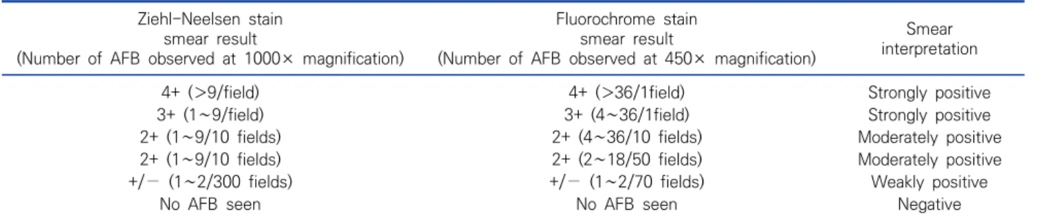
모든 임상 검체는 N-acetyl-L-cysteine-NaOH (NALC- NAOH) 방법을 이용하여 융화, 오염제거과정을 진행 후 3,000 G로 20분간 원심 분리하여 농축하였다. 농축된 침사는 항산균 도말 검사 및 결핵균 배양 검사, 실시간 중합효소연쇄반응 검사 에 사용하였다. 항산균 도말 검사는 형광염색(fluorochrome stain)으로 양성을 선별하고 Ziehl-Neelsen 염색법을 확인하 였다. 항산균 도말 염색 결과는 ATS/CDC guidelines 기준에 따라 negative, trace 및 양성은 1+에서 4+까지 구분하는 6 grade 기준으로 판정하였다(Table 1) [13].
3. 결핵균 배양 검사
결핵균 배양 검사는 고체배지 배양으로 3% Ogawa 배지 (Asan Pharmaceutical Co., Seoul, Korea)와 액체배지 배양 으로 Bactec MGIT (Mycobacterium Growth Indicator Tube) 960 system (Becton Dickinson Diagnostic Instrument Systems, Sparks, MD, USA)을 이용하여 최대 6주까지 배양하 였다.
배양과정에서 자란 균주는 Genedia MTB/NTM detection kit (Green Cross Medical Science Co., Yongin, Korea) 결 핵균과 비정형 항산균(nontuberculous mycobacteria, NTM) 으로 각각 확인하였다.
Table 1. AFB smear staining standards according to CDC guidelines Ziehl-Neelsen stain
smear result
(Number of AFB observed at 1000× magnification)
Fluorochrome stain smear result
(Number of AFB observed at 450× magnification)
Smear interpretation
4+ (>9/field) 4+ (>36/1field) Strongly positive
3+ (1∼9/field) 3+ (4∼36/1field) Strongly positive
2+ (1∼9/10 fields) 2+ (4∼36/10 fields) Moderately positive
2+ (1∼9/10 fields) 2+ (2∼18/50 fields) Moderately positive
+/− (1∼2/300 fields) +/− (1∼2/70 fields) Weakly positive
No AFB seen No AFB seen Negative
Abbreviation: AFB, acid-fast bacillus.
Table 2. Comparison of Cobas MTB test semi-quantitative ranges and AFB smear results for pelleted samples Cobas MTB test
(MTB C
trange)
AFB stain smear grade
Total
Negative Trace 1+ 2+ 3+ 4+
Low (40<Ct≤47) % 66.7 29.2 2.1 2.1 0 0 100
N 32 14 1 1 0 0 48
Medium (35<Ct≤40) % 18 39.3 24.6 13.1 3.3 1.6 100
N 11 24 15 8 2 1 61
High (Ct≤34) % 3.8 15.4 15.4 19.2 7.7 38.5 100
N 1 4 4 5 2 10 26
Abbreviations: AFB, acid-fast bacillus; MTB, Mycobacterium tuberculosis ; C
t, threshold value.
4. 실시간 중합효소 연쇄반응
항산균 도말검사 및 배양 검사 후 남은 침사를 CTM을 이용하 여 핵산을 추출 과정을 거친 후 Cobas
Ⓡ
TaqManⓇ
48 analyzer (Roche Diagnostic)를 이용하여 제조사의 지침에 따라 수행하였다. 양성 결과는 내부 정도관리 물질(internal quality control, IQC)의 양성표준물질의 Ct
값과 비교했을 때 그 이하의 값을 양성, 그 이상의 Ct
값인 경우 invalid로 판정하 고, 측정 증폭 대상이 없는 경우 음성으로 판정하였다.5. 결핵균 검사방법에 따른 세균의 양 측정
동일한 검사실내에서 항산균 도말검사의 grade에 대해 예측 되는 CTM의 Ct 값과 액체배양을 시행하여 세균의 양을 측정하 고자 하였다. Bactec MGIT 960 system 이용하여 M.
tuberculosis H37Rv를 액체배지에 100 μL를 분주하여 배양 시킨 후 배양되는 시점에서 결핵균 양을 10
6
CFU /mL의 기준 으로 10배율씩 5단으로 희석하여 1×101
, 1×10²
, 1×10³
, 1×10⁴
, 1×10⁵
와 1×10⁶
CFU/mL를 포함한 6단계의 세균 희 석액을 준비하여 항산균 도말검사를 각 단계별 10회 실시하고, 액체 배양과 CTM을 단계별 3회를 시행하여 양성 배양시간과 Ct값을 확인 하였다. 각 검사별 측정한계치는 50% 이상 검출된 시점에 cut-off를 적용하였다.6. 결핵균 양의 측정에 따른 반정량적 판정 기준
결핵균 양의 측정을 통하여 각 검사법에 대한 검출 한계를 확 인하고 항산균 도말검사의 검출 가능한 값과 CTM의 C
t
값으로 반정량적 판정 기준을 설정하였다. 판정기준에 따라 본 연구에 서 진행된 CTM 양성 검체의 Ct
값과 항산균 도말검사 grade에 적용하여 유용성을 파악하였다.결 과
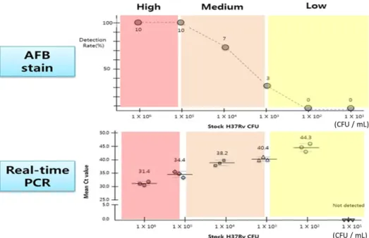
1. 표준균주를 단계별 희석하여 항산균 도말검사 분석 결과 M. tuberculosis H37Rv (CFU/mL)를 6단계로 희석하여 시행한 항산균 도말검사에서 각 단계별 10회씩 시행한 결과는 1×10
6
에서 10/10 (100%); 1×10⁵
에서 10/10 (100%);1×10
4
에서 7/10 (70%); 1×10³
에서 3/10 (30%); 1×10²
에 서 0/10 (0%); 1×101
에서 0/10 (0%)으로 검출되었다.Cut-off를 산정을 위한 기준으로 정성검사의 양성 기준값인 50%를 적용하였을 때 항산균 도말검사의 결핵균의 최소검출농 도가 1,000∼10,000 CFU/mL에서 검출됨을 확인하였다 (Table 2, Figure 1).
Figure 1. Mycobacterial load determination using AFB stain.
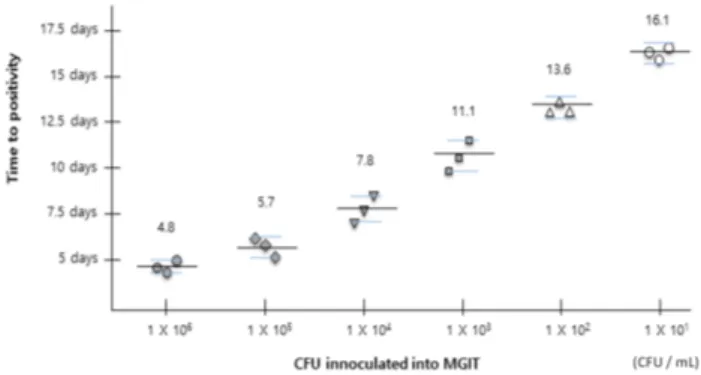
Figure 2. Automated liquid culture (using BACTEC MGIT 960) time to positivity.
Figure 3. PCR (COBAS MTB test) changes in cycle threshold (Ct).
2. 표준균주를 단계별 희석하여 액체배양 분석 결과
M. tuberculosis H37Rv (CFU/mL)를 6단계로 희석하여 액체배지에 각각 3회씩 배양한 세균 액은 모두 배양되었으며, 평균 배양 소요일은 1×10
6
에서 4.8일, 1×10⁵
에서 5.7일, 1×10⁴
에서 7.8일, 1×10³
에서 11.1일, 1×10²
에서 13.6일, 1×101
에서 16.1일의 결과를 얻었다. 가장 낮은 농도인 10 CFU/mL에서 배양됨을 확인하였고 최소검출농도가 10 CFU/mL 이하에서 배양될 수 있음을 확인하였다(Figure 2).3. 표준균주를 단계별 희석하여 Real-time PCR 분석 결과 M. tuberculosis H37Rv (CFU/mL)를 6단계 희석한 세균 액을 CTM test로 단계별 각 3회씩 진행하였으며, 모든 검사는 양성을 확인하였고, 그에 따른 Ct값(cycles)의 평균은 1×10
6
에서 31.4, 1×10⁵
에서 34.4, 1×10⁴
에서 38.2, 1×10³
에서 40.4, 1×10²
에서 44.3, 1×101
에서는 검출되지 않았다. 본원 기준 환자대상 검체의 결핵균 양성 Ct
판정 기준 25∼45 cycles 기준으로 비교했을 때 결핵균의 농도가 높을수록 Ct값이 낮음 을 확인하였으며, CTM의 검출 최소 농도는 100 CFU/mL 임을 확인하였다(Figure 3).4. 결핵균 양의 측정에 따른 반정량적 기준
결핵균의 양 측정을 위한 검사법으로 항산균 도말검사는 검 출 한계치가 1,000∼10,000 CFU/mL를 확인하였고, 액체배 지 배양법은 10 CFU/mL 이하에서도 가능하고, CTM test는 10∼100 CFU/mL의 검출한계를 확인하였다.
Low, medium, high의 설정에 대한 기준은 항산균 도말검 사의 결과가 양성일 때와 CTM test 결과가 동일한 기준으로 100% 일치하는 균수 농도인 1×10
1
농도의 Ct
값 ≤34에 대해 서는 “high”, 30% 미만인 1×10³
농도의 Ct 값 40.1 이상을“low”, 30∼70% 중간 값인 1×10
4∼5
에 해당하는 Ct
값은 35∼40으로 “medium”으로 설정하였다(Figure 4).
2015년 1년간 삼성서울병원에 의뢰된 CTM의 양성 검체 135건에 대한 항산균 도말 염색 결과는 ATS/CDC guidelines 기준에 따라 negative, trace 및 positive는 1+에서 4+까지 구 분하는 6 grade 기준으로 판정하였다. 첫째, low 40∼47 Ct값 기준에 해당하는 항산균 도말검사 grade은 − (66.7%), ± (29.2%), 1+ (2.1%), 2+ (2.1%)의 분포를 보였다. 둘째, medium 35∼40 Ct값 기준에 해당하는 AFB grade은 − (18.0%), ± (39.3%), 1+ (24.6%), 2+ (13.1%), 3+ (3.3%), 4+ (1.6%)의 분 포를 보였다. 셋째, high ≤34 C
t
값 기준에 해당하는 항산균 도 말검사 grade은 − (3.8%), ± (15.4%), 1+ (15.4%), 2+ (19.2%), 3+ (7.7%), 4+ (38.5%)의 분포를 나타냈다.고 찰
최근 분자생물학적 방법을 이용한 신속한 결핵진단법을 많이 사용하고 있다. 그들 중에 실시간 중합효소연쇄반응을 이용하 여 직접 결핵균을 검출하는 방법은 민감도, 특이도가 높아 널리 사용하고 있다[10]. 기존에 진행되었던 결핵 진단 방법의 대부 분은 결핵균의 존재 유무를 판단하는 검사법이 대부분이었다.
Figure 4. Setting the semi-quantitative criteria for diagnosis of tuberculosis.
그러나 결핵균의 균수의 측정은 감염성평가, 치료효과 판정과 함께 환자로부터 결핵의 병원 감염관리를 위한 판단을 위해서는 결핵균 양의 측정이 중요한 상황이다[9, 14].
국내 연구 개발된 Advansure TB/NTM real-time PCR kit (LG Life Science, Seoul, Korea)에 대한 Ct값과 항산균 도말 검사 grade에 대한 음의 상관관계(r
s
=−0.635)를 검증한 자료 도 있었다[8]. 본 연구에서의 음의 상관 관계(rs
=−0.545)는 PCR Cycle의 횟수가 많아 조금 높은 값을 나타내며, 해당 논문 양성건수(N=108)보다 양성 건수(N=135)가 더 많은 점에서 신 뢰도가 높다[8].본 연구에서 시행한 CTM은 검체에서 직접 결핵균을 검출하 는 검사실에서 약 10% [20] 사용하고 있는 검사법으로 임상적 효율성 검증 및 결과에 대한 신뢰성에 대한 선행연구는 많았지 만 검사결과물인 Ct 값에 대한 정량적 연구는 없었다[17, 21, 22].
이전 연구에서는 항산균 도말검사와 배양검사까지는 같이 진 행을 하지만, PCR 검사는 다른 검체로 진행하는 연구로 PCR 양 성 검체를 기준으로 비교 분석한 자료가 있었다[8, 21].
본 연구의 장점은 한 검체로 항산균 도말염색과 배양검사와 PCR검사까지 같은 검체로 진행하여 얻은 결과물로 신뢰 할 수 있는 결과라고 볼 수 있다.
항산균 도말검사의 검출한계 범위로 5,000∼10,000 CFU/mL 를 측정했던 연구[16, 24]와 비슷한 결과로 1,000∼10,000 CFU/mL의 값을 본 연구에서 확인하고, 또한 액체배지의 검출 한계는 <10 CFU/mL 이하에서 가능하였고, CTM 검사의 검출 한계는 46 CFU/mL의 연구 결과[23]에 비해 본 연구에서의 검
출 한계는 100 CFU/mL로 비슷한 결과를 얻을 수 있었다.
Bactec 960 MGIT를 사용한 자동화 액체 배양 검사의 검출 한계가 가장 적은 수의 결핵균을 검출할 수 있음을 다시 한 번 확 인하고, 결핵균의 수와 TTP는 상관관계가 있음을 확인하였다.
결핵균 양에 대한 반정량적으로 Ct 값을 very low 28∼38, low 22∼28, medium 16∼22, high <16 이하의 값으로 항산 균 도말검사와 비교하여 4단계 grade 정하여 진료에 도움을 주 고자 했던 GeneXpert system (Cepheid, Sunnyvale, CA)의 자료[9, 12]를 토대로 새로운 장비의 도입이 아닌 기존에 사용하 던 검사법을 반정량적으로 구분하여 진료에 도움을 주기 위한 방향을 모색하였다.
우선 결핵균 검출을 위한 CTM test검사의 Ct값과 항산균 도 말검사는 음의 상관관계(r
s
=−0.545)가 있음을 확인하였고, 결 핵균 검출을 위한 3가지 검사법으로 검출 할 수 있는 한계를 측 정하였으며, 항산균 도말검사와 PCR과의 균수의 검출기준으로 1×10⁵∼6
이상의 균수일 때 high (Ct<34), 1×10³∼4
medium (35∼40), 1×101∼2
low (40∼47)로 반정량적인 결과를 보고 에 대한 기준을 제시하였으며, Gene Xpert system 검사에서 항산균 도말검사 grade 빈도가 높은 기준으로 very low (negative 65.0%, scanty 20.6%), low (25.5%, scanty 19.3%), medium (1+ 21.9%, 2+ 22.7%, 3+ 49.1%), high (3+90.5%)의 보였고[8], 본 연구의 PCR 양성 대상 항산균 도말검 사 grade을 비교 했을 때 low (negative 66.7%, trace 29.2%), medium (trace 39.3%, 1+ 24.6%), high (trace 15.4, 1+
15.4%, 2+ 19.2%)의 grade별 분포기준을 확인했을 때 low,
medium의 분포는 비슷하나 high에서의 비율이 골고루 분포 가 되었으나 medium보다 높은 비율의 grade를 확인할 수 있 었다.
본 연구에서 결핵균의 양 측정 방법에 몇 가지 한계가 있다. 첫 째, Ogawa 배지에 배양된 M. tuberculosis H37Rv를 0.5 McFarland 농도로 측정을 시도하였으나 결핵균이 잘 균질화 되지 않아 정확한 농도의 측정이 어려웠다. Bactec 960 MGIT 장비를 이용하여 결핵균을 배양시킨 뒤 검출된 시점을 10
6
CFU/mL으로 진행을 하여 장비 검출 특성상 반비례적 측정으 로 결핵균의 수의 측정오차가 발생할 수 있었다. 둘째, 결핵균 양 의 측정을 위한 단계별 희석법에서 시행 횟수가 적어 전체를 대 표할 수는 없으며, 항산균 도말검사의 한계점인 검사자의 숙련 도와 검사실의 여러 가지 특성에 의한 변수는 배제할 수 없다. 셋 째, 임상환자 대상 결핵균 양성 검체 수가 작아(N=135), 낮은 양 성 예측도를 나타낼 수 있다.요 약
본 연구는 Cobas TaqMan MTB 검사(CTM test, Roche Diagnostics, Basel, Switzerland)와 항산균 도말염색검사의 연관성을 확인하고 그에 따른 반정량적 판정기준을 확립하고자 하였다. 2015년 1월부터 2015년 12월까지 삼성서울병원 진단 검사의학과에 의뢰된 8,389개의 검체에 대해 결핵균 도말 검사, 배양 검사 및 CTM 검사를 동시에 실시하였으며, 그 결과를 분석 하여 AFB 염색과주기의 연관성을 후향적으로 분석하였다.
CTM 검사의 임계 값(Ct)값으로, 결핵균 검출방법의 반정량적 판정기준을 설정 하였다. CTM 검사의 135개의 양성 표본에 대 한 Ct값은 항산균 도말염색과 반비례적 상관관계가 있었다(r
s
=−0.545, P<0.01). CTM 검사와 항산균 염색 등급의 음의 상관 관계가 입증되었으며, 임상 기준에 이러한 기준을 적용하여 임 상적 의의를 검증하였다. 이 연구의 반정량적 기준은 병원에서 활동성 결핵 및 감염을 일으킬 수 있는 환자의 빠른 진단의 판단 에 도움이 될 것으로 사료된다.
Acknowledgements: None Conflict of interest: None
Author’s information (Position): Jung T
1
, M.T.; Kim SH2
, M.T.; Kim S3
, Professor; Choi JS4
,Professor; Kim YK5
, Professor.REFERENCES