INTRODUCTION
Shoulder pain is one of the most common complaints en- countered in patients with rheumatoid arthritis (RA). Dur- ing the first 2 yr of RA, nearly 50% of patients have shoul- der symptoms, and 90% complain of shoulder pain at some time during the course of the disease (1). In addition to the synovitis of the glenohumeral (GH) joint, shoulder pain in RA arises from pathologies involving diverse periarticular soft tissues, and the involvement of more than one anatomi- cal structure is common. Because it is difficult to detect and identify the site of anatomical alterations with clinical exam- inations even in non-RA shoulders (2), correct diagnosis and management of painful RA shoulder by clinical examination alone is often problematic.
Radiographic assessment of peripheral joints has served as an objective standard for the evaluation of RA progression.
However, it is difficult to evaluate complex anatomical struc- tures such as the shoulder joint by conventional radiography alone. Ultrasonographic (US) evaluation is useful for diag- nosing a variety of regional pain syndrome and soft tissue
rheumatism and has been increasingly employed in the rheu- matologic practice (3). Given the great improvement in res- olution achieved by high frequency ultrasound, it is expect- ed to serve as an important tool for accurate evaluation of RA shoulders. In a study performed in 43 RA patients, US examination detected more erosions in the humeroscapular joint compared to conventional radiography (4). In addition, US examination detected synovitis, tenosynovitis, and bursi- tis in a significant number of patients, implicating its value for the dignosis of shoulder pain in RA patients (4).
The objectives of this study were: 1) to identify the US abnormalities and 2) to compare the physical examination with US findings, especially of the rotator cuff abnormali- ties in RA patients with shoulder pain.
MATERIALS AND METHODS
We studied 30 consecutive RA patients visiting a universi- ty affiliated rheumatology clinic. Sex, age, height, weight, body mass index (BMI), duration of RA, involved joint groups,
Hyun Ah Kim, Su Ho Kim, Young-Il Seo
Department of Internal Medicine, Hallym University Sacred Heart Hospital, Anyang, Korea
Address for correspondence Hyun Ah Kim, M.D.
Division of Rheumatology, Department of Internal Medicine, Hallym University Sacred Heart Hospital, 896 Pyongchondong, Dongan-gu, Anyang 431-070, Korea
Tel : +82.31-380-5928, Fax : +82.31-386-2269 E-mail : [email protected]
*This study was supported by a grant from the Korea Health 21 R & D project, Ministry of Health and Welfare (01-PJ3-PG6-01GN11-0002).
660
Ultrasonographic Findings of the Shoulder in Patients with Rheumatoid Arthritis and Comparison with Physical Examination
The objectives of this study were: 1) to identify the ultrasonographic (US) abnor- malities and 2) to compare the findings of physical examination with US findings in rheumatoid arthritis (RA) patients with shoulder pain. We studied 30 RA patients.
Physical examination was performed systemically as follows: 1) area of tender- ness; 2) range of passive and active shoulder motion; 3) impingement tests; 4) maneuvers for determining the location of the tendon lesions. US investigations included the biceps, the supraspinatus, infraspinatus, and subscapularis tendons;
the subacromial-subdeltoid bursa; and the glenohumeral and acromioclavicular joints. Thirty RA patients with 35 painful and 25 non-painful shoulders were exam- ined. The range of motion affected the most by shoulder pain was abduction. The most frequent US finding of shoulder joint was effusion in the long head of the biceps tendon. Among the rotator cuff tendons, subscapularis was the most frequently involved. Tendon tear was also common among non-painful shoulders. Physical examination used for the diagnosis of shoulder pain had low sensitivity and speci- ficity for detecting abnormalities in the rheumatoid shoulder joint. In conclusion, US abnormalities showed frequent tendon tears in our RA patients. Physical examina- tion had low sensitivity and specificity for detecting rotator cuff tear in the rheuma- toid shoulder joint.
Key Words : Rheumatoid Arthritis; Shoulder; Pain; Ultrasonography; Rotator Cuff; Osteoarthritis; Physical Examination
Received : 6 September 2006 Accepted : 8 December 2006
current RA medications, erythrocyte sedimentation rate (ESR), and C-reactive protein (CRP) were recorded. Detailed histo- ry of shoulder pain including duration, involved site, previ- ous diagnosis, and the category of previous treatment was obtained. Patients who developed shoulder pain after trau- ma in the shoulder area were excluded.
Physical examination
Physical examination of the shoulder was performed sys- temically by one blinded rheumatologist as follows: 1) area of tenderness in the GH joint, acromioclavicular (AC) joint, bicipital groove, and subacromial space; 2) range of passive and active motion for abduction, forward flexion, external rotation, and internal rotation measured with a goniometer;
3) Neer and Hawkins tests for shoulder impingement; 4) maneuvers for determining the location of the tendon lesions (Jobe’s test for supraspinatus, Patte’s test for infraspinatus, Gerber’s lift off test for subscapularis, and Yegarson’s test for the long head of the biceps brachii). For the impingement maneuver of Neer (5, 6), the examiner stands behind the seated patient and uses one hand to prevent rotation of the scapula while passively raising the patient’s arm with the other hand to produce both forward elevation and abduc- tion. In Hawkins’s test (7), the examiner stands facing the patient and after raising the patient’s arm to 90°of strict forward elevation with the elbow in 90°flexion, rotates the arm medially by lowering the forearm. These tests are posi- tive when patients experience pain during the maneuvers.
In Jobe’s maneuver (8), the patient places both arms in 90° abduction and 30°horizontal adduction, in the plane of the scapula; the examiner then pushes the patient’s arms down- ward while asking the patient to resist the pressure. For Patte’s maneuver (9), the examiner supports the patient’s elbow in 90°flexion while the patient is asked to rotate the arm laterally. Jobe’s and Patte’s maneuvers can produce three types of response: 1) absence of pain, indicating that the tested tendon is normal; 2) the ability to resist despite pain, denoting tendinitis; 3) the inability to resist with gradual lowering of the arm or forearm, indicating tendon rupture.
In Gerber’s lift off test (10), the patient is asked to place the hand against the back at the level of the waist with the elbow in 90°flexion. The examiner pulls the hand to about 5-10 cm from the back while maintaining the 90°bend in the elbow. In Yergason’s test (11), pain along the course of the biceps tendon produced by resisted supination of the fore- arm denotes bicipital tendinitis.
Ultrasonographic (US) examination
US examination was performed by a rheumatologist who was blinded to the clinical test. A linear array 7 MHz trans- ducer (HDI 5000, ATL ultrasound, Bothell, U.S.A.) was used. Investigations included transverse and longitudinal
planes from the long head of the biceps, the supraspinatus, the infraspinatus, and the subscapularis tendons; the subacro- mial-subdeltoid bursa; and the GH and AC joints. Tendon thickness, homogeneity of the fibrillar pattern, and the pres- ence of calcification were noted. In all patients, images of the bilateral shoulder were obtained in order to compare US findings between 2 shoulders in case of unilateral involve- ment. The US examination technique for the shoulder is widely described (5, 12). The biceps tendon was examined with the patient seated with the elbow flexed to 90°and the forearm half pronated on the lap. On the anterior aspect of the shoulder, the long head of biceps tendon is imaged as an oval-shaped echogenic structure. Anteromedial to the biceps tendon, the hyperechoic subscapularis tendon was identified with slight external rotation of the GH joint. The supras- pinatus tendon was examined with the patient’s shoulder in hyperextension and full internal rotation with the dorsum of the hand placed in the small of the back. These tendons appear as a hyperechoic fibrillar layer, convex, tapered and inserting at the greater tuberosity on longitudinal view. The subacromial-subdeltoid bursa was imaged as a hypoechoic line, between the deltoid muscle and the supraspinatus and infraspinatus tendons. The infraspinatus tendon and GH joint were examined with the patient's hand placed on the contralateral shoulder. The transducer was oriented in the axial plane until the head of the humerus was seen adjacent to the posterior glenoid labrum. The presence of GH joint effusion, pannus and humeral head irregularity was observed.
The infraspinatus tendon and its insertion were observed by moving the transducer laterally from the GH joint. The AC joint was examined with the transducer oritented along the coronal plane, and the presence of intra-articular fluid was noted. US diagnostic criteria of shoulder abnormalities are presented in Table 1.
Statistical analysis
Data are presented as mean±standard deviation for con- tinuous variables and as frequency (%) for categorical vari- ables. A p value was calculated by Student’s t-test for con- tinuous variables or by chi-squared test for categorical vari- ables. A p value less than 0.05 was considered statistically
Shoulder abnormality Diagnostic criteria
Biceps sheath effusion Thickness of the hypoechoic halo of fluid surrounding the biceps tendon >2 mm Glenohumeral effusion Distance from the posterior labrum to the posterior infraspinatus tendon >2 mm Full thickness tear Non-visualization of tendon or complete
fiber discontinuity Partial thickness tear Partial fiber discontinuity
Subdeltoid effusion Hypoechoic fluid filled bursa >2 mm Table 1.Ultrasonographic diagnostic criteria of shoulder abnor- malities
significant. All the statistical analyses were performed using SPSS for Windows (Version 10.0, Chicago, IL, U.S.A.).
RESULTS
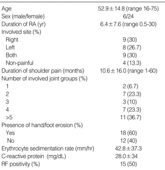
Table 2 shows the baseline demographic characteristics of the subjects. The majority of patients had active RA with a
mean ESR of 42.8 mm/hr. Sixteen (53.3%) patients were treated with oral corticosteroid. Twenty-seven (90%) patients were treated with disease-modifying anti-rheumatic drug (DMARD), and the median number of DMARDs used was 2. Simple radiography findings of the shoulder included 18 (60%) patients with normal findings, 6 (20%) with erosion, 5 (16.7%) with a degenerative change showing osteophytes and joint space narrowing, 2 (6.7%) with total destruction of shoulder bony architecture, and 1 (3.3%) with calcification.
Table 3 shows physical examination findings for the shoul- der joints of the study patients. Overall, painful shoulders showed more positive findings on physical examination com- pared to non-painful shoulders. The range of motion affected the most by shoulder pain in our patients was abduction, fol- lowed by forward flexion and internal rotation. For physical examination of individual tendons, we first tried to differenti- ate positive response as either pain or weakness. However, during the physical examination, we found that it was very difficult to differentiate the failure to resist due to pain or weakness in many of our patients; patients often refused to continue the test due to elicitation of severe pain by the ma- neuver. Therefore, this examination was recorded as either positive or negative, with positive result including inability to resist the examiner’s force, be it due to pain or weakness.
The majority of the shoulder joints exhibited abnormalities in Jobe’s test, indicating supraspinatus lesion, followed by
Age 52.9±14.8 (range 16-75)
Sex (male/female) 6/24
Duration of RA (yr) 6.4±7.6 (range 0.5-30) Involved site (%)
Right 9 (30)
Left 8 (26.7)
Both 9 (30)
Non-painful 4 (13.3)
Duration of shoulder pain (months) 10.6±16.0 (range 1-60) Number of involved joint groups (%)
1 2 (6.7)
2 7 (23.3)
3 3 (10)
4 7 (23.3)
>5 11 (36.7)
Presence of hand/foot erosion (%)
Yes 18 (60)
No 12 (40)
Erythrocyte sedimentation rate (mm/hr) 42.8±37.3 C-reactive protein (mg/dL) 28.0±34
RF positivity (%) 15 (50)
Table 2.Baseline characteristics of the 30 RA patients examined
Tenderness
Glenohumeral 3 (12)* 22 (62.9)
Subacromial 4 (16) 15 (42.9)
Bicipital 5 (20) 17 (48.6)
Acromioclavicular 4 (16) 8 (22.9)
Limitation of motion
Forward flexion 3 (12) 18 (51.4)
Abduction 4 (16) 19 (54.3)
External rotation 3 (12) 12 (34.3)
Internal rotation 4 (16) 16 (45.7)
Range of passive motion greater 4 (16) 14 (40) than active
Impingement test
Neer 1 (4) 13 (37.1)
Hawkins 2 (8) 15 (42.9)
Individual tendon test
Jobe’s test (supraspinatus) 5 (20) 21 (60) Patte’s test (infraspinatus) 1 (4) 11 (31.4) Gerber’s lift off test (subscapularis) 2 (8) 14 (40)
Yegarson’s test (Biceps) 3 (12) 6 (17.2)
Table 3.Positive physical examination findings of RA shoulders Painful (n=35) non-painful
(n=25)
*, % in parenthesis. RA, rheumatoid arthritis.
RA, rheumatoid arthritis.
Long head of the biceps tendon
Effusion 9 (36)* 13 (37.1)
Rupture 2 (8) 5 (14.3)
Subdeltoid effusion 1 (4) 5 (14.3)
Supraspinatus tendon
Thickening 2 (8) 6 (17.1)
Thinning 5 (20) 7 (20)
Tears
Partial thickness 4 (16) 6 (17.1)
Full thickness 3 (12) 6 (17.1)
Calcification 1 (4) 2 (5.7)
Infraspinatus
Thickening 0 (0) 3 (8.6)
Thinning 6 (24) 6 (17.1)
Tears
Partial thickness 2 (8) 4 (11.4)
Full thickness 1 (4) 0 (0)
Subscapularis
Thickening 0 (0) 2 (5.7)
Thinning 6 (24) 13 (37.1)
Tears
Partial thickness 5 (20) 8 (22.9)
Full thickness 3 (12) 5 (14.3)
GH effusion 5 (20) 12 (34.3)
Humeral bony irregularity 2 (8) 10 (28.6)
Table 4.Ultrasonographic examination findings of RA shoulders Painful (n=35) non-painful
(n=25)
*, % in parenthesis. GH, glenohumeral; RA, rheumatoid arthritis.
Gerber’s lift off (subscapularis) and Pattes’ (infraspinatus) test.
Table 4 lists US findings for the study patients. The most frequent finding was effusion in the long head of the biceps tendon, which was observed in 37.1% of painful shoulders.
It was also observed in 36% of non-painful shoulders. The
mean thickness of the biceps effusion was 3.30 and 3.41 mm in painful and non-painful shoulders, respectively. Biceps ten- don rupture and subdeltoid effusion were detected in 14.3%
of painful shoulders, respectively. Among the rotator cuff tendon, except for the teres minor which was not included in our US examination, subscapularis was the most frequent- ly involved, with tendon tear observed in 37% of shoulders.
For supraspinatus and infraspinatus tendon, tendon tear was observed in 34 and 11% of the shoulders. It is of note that tendon tear was also common among non-painful shoulders, with 32, 28, and 12% of shoulders showing tear in the sub-
Fig. 1.Ultrasonographic findings of biceps abnormalities. (A, B) Biceps effusion with pannus (arrow) in biceps tendon sheath. (C, D) Biceps tendon rupture with empty sheath. (A, C) Transverse view. (B, D) Longitudinal view.
A B
C D
Fig. 2.Partial thickness tear in subscapularis tendon (arrow) in a non-painful shoulder. (A) Transverse view. (B) Longitudinal view.
A B
Fig. 3.Complete massive tear in supraspinatus in a non-painful shoulder. (A) Transverse view. (B) Longitudinal view.
A B
Fig. 4.Bilateral partial thickness tear in infraspinatus in one patient.
Transverse view. (A) Painful shoulder. (B) Non-painful shoulder.
A B
Age 53.15 52.4
Sex (M:F) 4:16 2:8
Duration of RA (yr) 8.0 3.15
Number of involved joint groups
1 5 10
2-4 55 60
>5 40 30
Duration of shoulder pain (months) 9.75 8.2 Number of DMARDs used (%)*
1 30 80
>2 70 20
ESR (mm/hr) 40.7 47.1
CRP (mg/dL) 24.7 32.0
Rheumatoid factor positive (%) 55 40
Presence of small joint erosion (%) 75 50
Presence of GH joint erosion (%)* 20.7 0
Table 5.Clinical parameters associated with tendon tear in RA patients
No tear (n=10) Tear
(n=20)
*, denotes a difference statistically significant between tear and non-tear groups (p<0.05).
RA, rheumatoid arthritis; ESR, erythrocyte sedimentation rate; CRP, C- reactive protein; GH, glenohumeral; DMARD, disease-modifying anti- rheumatic drug.
GH tenderness 52 54
SA tenderness 78.9 65.9
LOM
Forward flexion 52.4 53.8
Abduction 56.5 56.8
External rotation 53.3 53.3
Internal rotation 45 52.6
Jobe’s test (supraspinatus) 72.4 45.2
Gerber’s lift off test (subscapularis) 68.8 47.7
Patte’s test (infraspinatus) 62.5 72.7
Yegarson’s test (biceps) 14.3 89.1
Table 6.Sensitivity and specificity of physical examination for detection of US tendon tears in RA patients (%)
Any tear Sensitivity Specificity
Specific tendon tear Sensitivity Specificity
US, ultrasonography; RA, rheumatoid arthritis; GH, glenohumeral; SA, subacromial; LOM, limitation of motion.
scapularis, supraspinatus and infraspinatus tendons, respec- tively. Multiple tendon tear was also common with rupture of 2 tendons in 12 and 22.9%, and rupture of 3 tendons in 8 and 11% of non-painful and painful shoulders, respective- ly. Calcification in the supraspinatus tendon was observed in 3 (5%) shoulders, among which only 1 showed calcification on simple radiography. GH joint effusion was noted in 20 and 34.3% of non-painful and painful shoulders, respective- ly, and the mean thickness of the effusion was 5.06 and 3.64 mm, respectively. Humeral cortical irregularity was detect- ed in 12 shoulders (20%), among which 6 showed erosion on simple radiography. Representative US findings are pre- sented in Fig. 1-4.
Next, clinical parameters associated with tendon tear were analyzed (Table 5). Age of the patient, sex, the duration of RA, ESR, CRP, the presence of rheumatoid factor, the pres- ence of erosion in small joints, and the number of involved joint group were not significantly associated with the pres- ence of tendon tear. The number of DMARDs used, and the presence of GH joint erosion was significantly associated with the presence of tendon tear. Table 6 shows the sensitivity and specificity of physical examination for the detection of US tendon tear. As shown, most of the tests for examination of shoulder joint and rotator cuff yielded low sensitivity and specificity.
DISCUSSION
In this study of RA patients with a relatively short disease duration, US abnormalities in the shoulder were common, with many shoulders showing rotator cuff tendon tears. Many non-painful shoulders also showed US abnormalities. The most frequent US finding of shoulder joints in our patients was effusion in the long head of the biceps tendon. Among the rotator cuff tendons, subscapularis was the most frequently involved, followed by supraspinatus and infraspinatus ten- don. Physical examination traditionally used for the diagno- sis of shoulder pain had low sensitivity and specificity for detecting tendon tear in the rheumatoid shoulder joint.
The frequency of abnormal US findings of rheumatoid shoulder joints differs depending on the patient population studied. In a study evaluating 44 hospitalized RA patients with mean disease duration of 12 yr, subacromial bursitis was the most frequent finding, followed by GH joint syn- ovitis, bicipital tendinitis and abnormalities in the supras- pinatus tendon (13). In accordance with our result, abduc- tion and forward flexion were often restricted, but clinical findings were non-specific (13). In another US study evalu- ating 90 RA shoulders with a mean disease duration of 5.5 yr (2), effusion in the long head of the biceps tendon was noted in 32.2%, and subacromial bursitis in 17.7%, while rotator cuff tear was detected infrequently (1.1 for infraspinatus and 4.4% for supraspinatus). Compared to polymyalgia rheumat-
ica or periarticular disorders of the shoulders not related to RA, rheumatoid shoulders tended to show involvement of all periarticular structures (2). This discrepancy from our data may stem from the difference in patient profile as well as the quality of the US equipment or the protocol for the evalua- tion of the shoulder. In a more recent study (14), 57 consec- utive RA patients were evaluated using a 7.5 MHz linear probe and a standardized study protocol. Rotator cuff tear was noted in 10% of the painful shoulders. In line with our data, painless RA shoulders also revealed frequent US abnor- malities (14). Tendon tear was most frequently observed in subscapularis tendon in our patients, and this is in contrast with rotator cuff tear in non-RA shoulder pain, which shows tear in supraspinatus most often (15, our unpublished obser- vation). Whether this is a specific finding in RA shoulder should be explored in more patients.
There have been no reports assessing the risk factors for rotator cuff tendon tear in RA patients. In our study, usual indicators of disease activity, such as ESR, CRP or the pres- ence of hand/foot joint erosion were not significantly corre- lated with the presence of tendon tear. Duration of RA or shoulder pain and RF positivity were not correlated, either, but the presence of GH joint erosion or number of DMARDs used were. Because data were collected cross-sectionally at the time of US evaluation, number of DMARDs used may be a better indicator of overall disease activity compared to ESR or CRP. In addition, duration of RA tended to be longer in RA patients with tendon tear compared to those without, and the statistical non-significance might have been due to the small sample size.
In line with previous reports (5, 16), our results show that the clinical examination of periarticular conditions in the painful RA shoulder is not accurate. The low sensitivity and specificity of physical examination for RA shoulders may be due to the fact that most RA patients with shoulder pain have multiple periarticular lesions, involving tendons, the subacro- mial-subdeltoid bursa, and GH joint simultaneously. The failure to distinguish between positive findings due to weak- ness or pain might also have resulted in the low specificity of physical examination in our patients. In addition, involve- ment of elbow or wrist joint often hinders from proper exam- ination of the shoulder, decreasing diagnostic accuracy.
Currently, magnetic resonance imaging (MRI) has been widely used to evaluate painful shoulders. A recent study compared US examination with dynamic contrast-enhanced MRI in shoulders of patients with RA (17). Although US examination detected significantly more bony erosion com- pared to conventional radiography, MRI was significantly more sensitive for detecting synovitis, tenosynovitis, and bur- sitis as well as bony erosion than US examination. However, while MRI is expensive, time-consuming, and not widely available, US examination is quick, inexpensive, and easy to perform. US examination can also routinely be used for dy- namic examination of the musculoskeletal system. More
recently, US supplemented by power Doppler was found to have value in differentiating rheumatoid inflammation from the degenerative shoulder disease by detection of the changes in vascularities, extending the applicability of US examina- tion to the evaluation of the process of inflammation in rela- tion to neoangiogenesis (18).
Although US examination offers considerable benefit for proper evaluation of shoulder joint problems, limitations exist such as lack of visualisation of the posterior aspect of the rota- tor cuff tendons, limited view of the GH joint, and consider- able dependence on the operator. Therefore, despite the fact that most rotator cuff lesions involve the ‘‘critical zone’’ in the anterior aspect of the tendons (19), US examination may underestimate the prevalence of rotator cuff abnormalities.
Our study has a few limitations. First, gold standard for assessing the shoulder lesion, such as arthroscopy or MRI was not done. Although the agreement between US examination and MRI in the detection of tendon tear was good in 4 of our patients who underwent MRI after US examination (data not shown), the incidence of rotator cuff tear detected by US exa- mination alone might have been incorrect. Second, normal controls without shoulder pain were not examined. Non- painful shoulders in RA patients showed more US abnor- malities compared to asymptomatic shoulders in non-RA patients examined in a separate study (unpublished observa- tion). Therefore, our main finding is that RA shoulders ex- hibit multiple lesions regardless of the presence of pain.
Conservative treatments of shoulder problems in RA include medical treatment, physiotherapy, and local injections of cor- ticosteroids. The high prevalence of rotator cuff tear in RA shoulders shown in our series warrants precaution against these empirical treatments; it would be more desirable to obtain an exact anatomical diagnosis in order to optimize treatment. Few studies have compared the outcome of treat- ment for periarticular shoulder lesions with or without an accurate imaging technique (20). A recent surgical series revealed that RA patients with both partial and full-thick- ness rotator cuff tears had significant improvements in over- all pain and satisfaction after the repair (21). However, only patients with a partial-thickness tear had improvement of active elevation. This result suggests that rotator cuff tendon repair performed early in its course may be more beneficial to obtain satisfactory result. There is a need for further trials investigating whether the US examination of the shoulder in RA patients offers a possibility of improving its treatment.
REFERENCES
1. Alasaarela E, Suramo I, Tervonen O, Lahde S, Takalo R, Hakala M.
Evaluation of humeral head erosions in rheumatoid arthritis: a com- parison of ultrasonography, magnetic resonance imaging, comput- ed tomography and plain radiography. Br J Rheumatol 1998; 37:
1152-6.
2. Coari G, Paoletti F, Iagnocco A. Shoulder involvement in rheumatic diseases. Sonographic findings. J Rheumatol 1999; 26: 668-73.
3. Norregaard J, Krogsgaard MR, Lorenzen T, Jensen EM. Diagnos- ing patients with longstanding shoulder joint pain. Ann Rheum Dis 2002; 61: 646-9.
4. Hermann KG, Backhaus M, Schneider U, Labs K, Loreck D, Zuhls- dorf S, Schink T, Fischer T, Hamm B, Bollow M. Rheumatoid arthri- tis of the shoulder joint: comparison of conventional radiography, ultrasound, and dynamic contrast-enhanced magnetic resonance imaging. Arthritis Rheum 2003; 48: 3338-49.
5. Naredo E, Aguado P, De Miguel E, Uson J, Mayordomo L, Gijon- Banos J, Martin-Mola E. Painful shoulder: comparison of physical examination and ultrasonographic findings. Ann Rheum Dis 2002;
61: 132-6.
6. Neer CS, Welsh RP. The shoulder in sports. Orthop Clin North Am 1977; 8: 583-91.
7. Hawkins RJ, Kennedy JC. Impingement syndrome in athletes. Am J Sports Med 1980; 8: 151-8.
8. Jobe FW, Jobe CM. Painful athletic injuries of the shoulder. Clin Orthop 1983; 173: 117-24.
9. Leroux JL, Thomas E, Bonnel F, Blotman F. Diagnostic value of clinical tests for shoulder impingement syndrome. Rev Rhum (Engl Ed) 1995; 62: 423-8.
10. Sheon RP, Moskowitz RW, Goldberg VM. Upper limb disorders.
In: Sheon RP, Moskowitz RW, Goldberg VM, editors, Soft tissue rheumatic pain. Baltimore: Williams and Wilkins, 1987: 79-129.
11. Gilcreest EL. Dislocation and elongation of the long head of the biceps brachii. An analysis of 6 cases. Ann Surg 1936; 104: 118-38.
12. Ptasznik R. Sonography of the shoulder. In: van Holsbeeck MT, Introcaso JH, editors, Musculoskeletal ultrasound. St. Louis: Mosby, 2001; 464-516.
13. Alasaarela EM, Alasaarela EL. Ultrasound evaluation of painful rheumatoid shoulders. J Rheumatol 1994; 21: 1642-8.
14. Naranjo A, Marrero-Pulido T, Ojeda S, Francisco F, Erausquin C, Rua-Figueroa I, Rodriguez-Lozano C, Hernandez-Socorro CR. Ab- normal sonographic findings in the non-painful arthritic shoulder.
Scand J Rheumatol 2002; 31: 17-21.
15. Zehetgruber H, Lang T, Wurnig C. Distinction between supraspinatus, infraspinatus and subscapularis tendon tears with ultrasound in 332 surgically confirmed cases. Ultrasound Med. Biol 2002; 28: 711-7.
16. Nelson MC, Leather GP, Nirschl RP, Pettrone FA, Freedman MT.
Evaluation of the painful shoulder. A prospective comparison of magnetic resonance imaging, computerized tomographic arthrog- raphy, ultrasonography, and operative findings. J Bone Joint Surg Am 1991; 73: 707-16.
17. Hermann KG, Backhaus M, Schneider U, Labs K, Loreck D, Zuhls- dorf S, Schink T, Fischer T, Hamm B, Bollow M. Rheumatoid arthri- tis of the shoulder joint: comparison of conventional radiography, ultrasound, and dynamic contrast-enhanced magnetic resonance imaging. Arthritis Rheum 2003; 48: 3338-49.
18. Strunk J, Lange U, Kurten B, Schmidt KL, Neeck G. Doppler sono- graphic findings in the long bicipital tendon sheath in patients with rheumatoid arthritis as compared with patients with degenerative diseases of the shoulder. Arthritis Rheum 2003; 48: 1828-32.
19. Cofield RH. Current concepts review. Rotator cuff disease of the shoulder. J Bone Joint Surg Am 1985; 67: 974-9.
20. Bartolozzi A, Andreychik D, Ahmad S. Determinants of outcome in
the treatment of rotator cuff disease. Clin Orthop 1994; 308: 90-7.
21. Smith AM, Sperling JW, Cofield RH. Rotator cuff repair in patients with rheumatoid arthritis. J Bone Joint Surg Am 2005; 87: 1782-7.