INTRODUCTION
Chronic obstructive pulmonary disease (COPD) is gener- ally defined as a disease state characterized by the obstruc- tion of airflow, which is not fully reversible (1). The major risk factor for the development of fixed obstruction of airflow in patients with COPD is cigarette smoking. The adverse effect of smoking on the rate of decline in FEV1have been well documented in many cross-sectional and longitudinal studies in normal populations. For example, Fletcher and colleagues found that the average rate of decline of FEV1was 0.03 L/yr among non-smokers, and that decline was twice as fast among smokers (2). Broadly comparative figures for rate of decline in FEV1have been reported from several other longitudinal studies (3-5).
Currently, the major hypothesis for the pathogenesis of emphysema, which is a major component of the morbidity and mortality of COPD, is the protease-antiprotease hypothe- sis (6, 7). This hypothesis states that an imbalance between
the levels of degradative enzymes and their respective inhi- bitors damages the connective tissue matrix components of the lung. Among various proteases that have long been pro- posed to damage connective tissue components in lung parenchyma, there is now increasing evidence that matrix metalloproteinases (MMPs) play a role in the pathogenesis of COPD. In patients with emphysema, MMP-1 (collage- nase) and MMP-9 (gelatinase B) was increased in bronchoalve- olar lavage fluid (8). In addition, it increased the activities of MMP-2 and MMP-9 in the lung parenchyma of patients with emphysema (9). The interest in MMPs has been fur- ther heightened by the demonstration in mice that emphy- sema induced by chronic exposure to cigarettes is prevented by deletion of the gene encoding MMP-12 (macrophage metalloelastase) (10). The question remains how cigarette smoking brings about the obstruction of airflow in COPD.
Increased expression of MMPs or an imbalance between MMPs and their specific inhibitors, the tissue inhibitors of metalloproteinase (TIMPs), may play a role in the generation
Min Jong Kang, Yeon-Mok Oh*, Jae Cheol Lee�, Dong Gyu Kim, Myung Jae Park, Myung Goo Lee, In Gyu Hyun, Sung Koo Han�, Young-Soo Shim�, Ki-Suck Jung
Department of Internal Medicine, Hallym University Sacred Heart Hospital, College of Medicine, Hallym University, Anyang; Asan Medical Center, College of Medicine, University of Ulsan*; Korea Cancer Center Hospital�; Department of Internal Medicine, Seoul National University Hospital, College of Medicine, Seoul National University�, Seoul, Korea
Address for correspondence Ki-Suck Jung, M.D.
Division of Pulmonary and Critical Care Medicine, Department of Internal Medicine, Hallym University Sacred Heart Hospital, 896 Pyungchon-dong, Dongan-gu, Anyang 431-070, Korea Tel : +82.31-380-3717, Fax : +82.31-380-3973 E-mail : pulmoks@hallym.or.kr
*The work was supported by grant No. 2000-1- 20500-003-1 from the Basic Research Program of the Korea Science & Engineering Foundation.
821
Lung Matrix Metalloproteinase-9 Correlates with Cigarette Smoking and Obstruction of Airflow
Cigarette smoking is the most important risk factor for obstruction of airflow in chronic obstructive pulmonary disease (COPD). Matrix metalloproteinases (MMPs) or an imbalance between MMPs and their inhibitors, the tissue inhibitors of MMP (TIMPs), is considered to play a role in the pathogenesis of COPD. We investigat- ed whether the MMPs expression or the imbalance between MMPs and TIMP-1 is associated with the amount of cigarette smoking and the FEV1value, in the lung parenchyma of 26 subjects (6 non-smokers and 20 cigarette smokers). First, we performed zymographic analysis to identify the profile of the MMPs, which revealed gelatinolytic bands mainly equivalent to MMP-9 in the smokers. We then mea- sured, using enzyme immunoassay, the concentrations of MMP-9 and its inhibitor, TIMP-1. Correlation analysis revealed that both the MMP-9 concentrations and the molar ratios of MMP-9 to TIMP-1 (MMP-9/TIMP-1) were correlated with the amount of cigarette smoking. Furthermore, MMP-9 concentrations were inversely correlated with FEV1. In conclusion, this study shows that MMP-9 expression in human lung parenchyma is associated with cigarette smoking and also with the obstruction of airflow, suggesting that MMP-9 may play a role in the pathogenesis of the cigarette smoke-induced obstruction of airflow known as the characteristic of COPD.
Key Words : Pulmonary Disease, Chronic Obstructive; Smoking; Matrix Metalloproteinases; Tissue Inhibitor of Metalloproteinases; Lung Diseases; Obstructive
Received : 28 April 2003 Accepted : 4 August 2003
of cigarette smoke-induced obstruction of airflow. However, there is still no direct evidence of this results, although some recent reports have revealed that there is an increased expres- sion of MMP-9 by alveolar macrophages in cigarette smok- ers compared to non-smokers (11, 12).
Therefore, we hypothesized that the smoke from cigarette would induce MMPs expression or an imbalance between MMPs and TIMPs in lung parenchyma, and that it would be associated with the degree of obstruction in airflow. To test the hypothesis, we evaluated whether the amount of cigarette smoking and FEV1(% predicted) value, an indicator of air- flow obstruction, are correlated with the MMPs expression or the imbalance between the MMPs and TIMP-1 in human lung parenchyma.
MATERIALS AND METHODS Subjects and Specimens
In a total of 26 subjects (6 non-smokers and 20 cigarette smokers), specimens were obtained from lungs resected for the treatment of malignancy or for the evaluation of solitary pulmonary nodule. Immediately after resection, lung speci- mens were collected at locations as far as possible from the pathologic lesion. Subjects were excluded if the specimens examined had microscopic evidence of ongoing infection or malignant cellular infiltration. The study was approved by Human Ethics Committee of Hallym University Sacred Heart Hospital, and all subjects gave written informed consent.
Determination of the Amount of Cigarette Smoking
The definition of cigarette smoking was determined as follows. Current smokers were defined as those who had smoked at least 100 cigarettes in their lifetime and either still have smoked or had quit smoking within the preced- ing year. Among current cigarette smokers, the amount of cigarette smoking was calculated as pack-year basis (PYs), using a detailed questionnaire derived from a similar study (13). If subjects had smoked at least 100 cigarettes in their lifetime but had quit smoking more than one year earlier, then they were classified as former smokers in terms of smok- ing status and excluded from this study. Subjects who had smoked less than 100 cigarettes including those who had never smoked, were considered “never to have smoked” and their exposure to environmental tobacco smoke was estimat- ed by determining the following the number of people liv- ing in the household who smoked at home, the number of cigarettes smoked at the home each day, and the number of hours the subjects spent daily outside the home in a place where people were smoking. If these subjects suffered less than 2 hr of passive smoking in a day, they were classified as non-smokers.
Protein Extraction from Lung Specimens
For analysis of gelatinolytic activities and enzyme immuno- assay for MMP-9 and TIMP-1, pieces of lung samples weigh- ing between 0.5 and 1.5 g were kept in air-tight plastic bot- tles at -80℃until tissue extraction. After removal of small vessels and bronchi under the microscope, the specimens, which weighed 0.5 g each, were minced into small pieces and homogenized with ULTRA-TURRAX T25 (Janke &
Kunkel GmbH & Company, KG, IKA Laboratory Technol- ogy, Staufen, Germany) in an ice bath after the addition of neutral salt extraction buffer (150 mM NaCl, 50 mM NaF, 10 g/mL of aprotinin, 10 g/mL of leupeptin, 1 mM Na3Vo4, 1 mM PMSF, 10 mM CaCl2, 2M KCl, and 50 mM Tris-HCl, pH 7.5; 0.5 g tissue/2 mL buffer) (14). After incubation of 1 hr at 4℃, homogenates were centrifuged at 7.5×10⁴g for 1 hr at 4℃. The supernatants were dialyzed against matrix metalloproteinase buffer (5 mM CaCl2, 10 mM ZnCl2, 200 mM NaCl, and 50 mM Tris-HCl, pH 7.5) for 24 hr at 4℃. Protein concentration of the samples was analyzed by a spec- trophotometer (DU 640; Beckman, Fullerton, CA, U.S.A.) according to the Bradford protein analysis method, and adjust- ed equally to levels of 1.0 mg/mL (total protein concentra- tion) for each sample. The samples were stored at -80℃until analysis.
Gelatin Zymography
MMPs present in lung tissue protein extracts were detected by their capacity to degrade gelatin according to a method previously described (15). Briefly, protein extracts obtained from lung tissue specimens were subjected to electrophoresis on 12% polyacrylamide gels containing 1 mg/mL of gelatin, in the presence of sodium dodecyl sulfate (SDS-PAGE) under non-reducing conditions. After electrophoresis, gels were washed twice in 1% Triton X-100 (vol/vol) for 1 hr, rinsed briefly, and incubated at 37℃for 24 hr in buffer containing 10 mM CaCl2, 5 M ZnCl2, pH 7.8 in Tris-HCl. After incu- bation, the gels were stained with Coomassie Brilliant Blue R250 and then destained in a solution of 10% acetic acid with 30% methanol. Zones of enzymatic activity were indicated by negative staining, with areas of proteolysis appearing as clear bands against a blue background. Molecular weight of the gelatinolytic bands was estimated relative to size markers.
Measurement of MMP-9 and TIMP-1 by ELISA
The MMP-9 and TIMP-1 concentrations in protein extracts were measured by enzyme-linked-immunosorbent assay (ELISA) (MMP-9 & TIMP-1: R&D Systems Inc., MN, U.S.A.). MMP-9 ELISA measured total MMP-9 released (pro and active MMP protein together with that complexed to TIMP-1+2). TIMP-1 ELISA measured both bound and free TIMP-1.
Statistical Analysis
Results are expressed as medians and ranges. Statistical analysis was performed using 2test and Mann-Whitney U test to assess differences between the two groups. Spear- man’s rank correlation was calculated to assess correlation.
RESULTS Characteristics of Subjects
Pathologic diagnoses of 26 subjects were lung cancer (22 subjects), tuberculous granuloma (3 subjects), and chondroid hamartoma (1 subject). The study subjects were grouped as non-smokers and cigarette smokers as follows;
Non-smokers: The six subjects (4 lung cancer, 2 tubercu- lous granuloma) with no history of cigarette smoking ranged from 31 to 67 yr of age (median: 62.5 yr). All six subjects were females. The FEV1/FVC ratios (%) in the subjects ranged from 73 to 87%, with a median value of 82.5%. The FEV1 (% predicted) values ranged from 89 to 118%, with a median value of 105.5%.
Cigarette smokers: The 20 subjects (18 lung cancer, 1 tuber- culous granuloma, 1 chondroid hamartoma) with current smoking history ranged from 27 to 78 yr of age (median: 58 yr), with 19 males and one female. Cigarette smoking history ranged from 3 to 104 pack-years (median: 40 pack-years). The FEV1/FVC ratios (%) in the subjects ranged from 54 to 84%, with a median value of 73.5%. The FEV1(% predicted) values ranged from 45 to 118%, with a median value of 85.5%.
The comparison of the two groups is shown in Table 1.
Age distribution was similar between two groups, but gen- der distribution was different (p<0.001). FVC (% predict- ed) values were similar between the two groups. However, FEV1(% predicted) value and FEV1/FVC were significantly lower among the cigarette smokers than the non-smokers (p<0.005 and p<0.03, respectively), suggesting the presence of obstruction of airflow in cigarette smokers.
Characterization of MMPs in Protein Extracts by Zymography
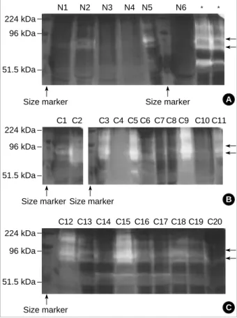
To identify the profile of MMPs in the lung tissue proteins of 26 subjects, we first performed zymographic analysis of the protein extracts. It revealed the presence of a major gelati- nase species having molecular weights of 92-kDa and 82-kDa, which are equivalent to pro- and active- forms of MMP-9 molecules in the cigarette smokers (Fig. 1B). For the non- smokers, gelatinolytic bands were rarely observed (Fig. 1A).
Comparison of MMP-9 and TIMP-1 between Non- smokers and Cigarette Smokers
According to the zymographic analysis, we selected MMP- 9 as one of the most probable candidate enzyme, and then measured the concentrations of MMP-9 and one of its inhi- bitor, TIMP-1. The concentrations of MMP-9 were higher in protein extracts of the cigarette smokers (median: 51.45,
*Results are expressed as median, with ranges in parentheses. �p value by 2test for sex variable. For other variables, by Mann-Whitney U test.
NS, not significant; PYs, pack-years; FEV1, forced expiratory volume in one second; FVC, forced vital capacity.
Characteristics Non-smokers Cigarette Smokers p value� (n=6) (n=20)
Age (yr) 62.5 (31-67) 58 (27-78) NS
Sex, male : female 0:6 19:1 <0.001
Cigarette Smoking (PYs) 0 40 (3-104) <0.001 Spirometry
FEV1(% predicted) 105.5 (89-118) 85.5 (45-118) <0.005 FVC (% predicted) 93.5 (80-106) 88.0 (54-110) NS FEV1/FVC (%) 82.5 (73-87) 73.5 (54-84) <0.03 Table 1.Characteristics of the subjects*
Fig. 1.The profile of matrix metalloproteinases (MMPs) in lung tis- sues by gelatin zymography. The figure shows multiple clear bands in the cigarette smokers (C1-C20) (B), as compared with the non-smokers (N1-N6), in which gelatinolytic bands are rarely seen (A). Arrows indicate the positions of pro- and active- forms of MMP-9 (92 and 82 kDa, respectively). Zones of enzymatic activity are indicated by negative staining, with areas of proteoly- sis appearing as clear bands against a blue background. *Posi- tive control.
N1 N2 N3 N4 N5 N6 * *
Size marker Size marker
224 kDa 96 kDa
51.5 kDa
A
C12 C13 C14 C15 C16 C17 C18 C19 C20
Size marker 224 kDa
96 kDa
51.5 kDa
C C1 C2 C3 C4 C5 C6 C7 C8 C9 C10 C11
Size marker Size marker 224 kDa
96 kDa
51.5 kDa
B
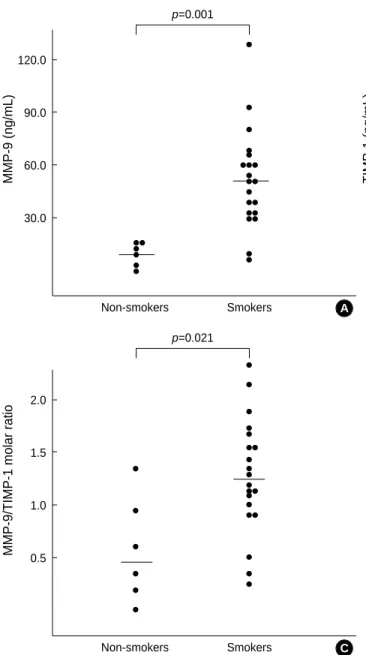
range: 7.20 to 129.90 ng/mL) than the non-smokers (medi- an: 14.70, range: 1.40 to 16.10 ng/mL) (p<0.001, Mann- Whitney U test) (Fig. 2A).
The concentrations of TIMP-1 were higher in the cigarette smokers (median: 11.15, range: 7.90 to 20.80 ng/mL) than the non-smokers (media: 8.10, range: 5.40 to 14.40 ng/mL) (p<0.045, Mann-Whitney U test) (Fig. 2B).
The molar ratio of MMP-9 to TIMP-1 (MMP-9/TIMP- 1) was higher in the cigarette smokers (median: 1.26, range:
0.24 to 2.36) than the non-smokers (median: 0.49, range:
0.01 to 1.35) (p<0.021, Mann-Whitney U test) (Fig. 2C).
Correlation of MMP-9 and MMP-9/TIMP-1 with Cigarette Smoking
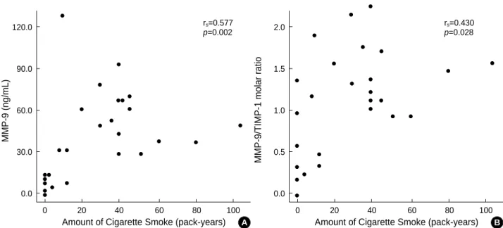
Among the 26 subjects, there was a positive correlation of
MMP-9 concentrations with the amount of cigarette smok- ing (rs=0.577, p<0.002) (Fig. 3A). In addition, there was also a positive correlation of the molar ratio of MMP-9 to TIMP-1 with the amount of cigarette smoking (rs=0.430, p<0.028) (Fig. 3B).
Correlation of MMP-9 and MMP-9/TIMP-1 with FEV1
Among the 26 subjects, there was an inverse correlation of MMP-9 concentrations with the FEV1(% predicted) val- ues (rs=-0.553, p<0.003) (Fig. 4A). However, correlation analysis of the molar ratio of MMP-9 to TIMP-1 with the FEV1(% predicted) values failed to show statistical signifi- cance, although it tended to increase according to the sever- ity of obstruction of airflow (rs=-0.356, p<0.074) (Fig. 4B).
Fig. 2.Concentrations of MMP-9 (A), TIMP-1 (B), and the molar ratios of MMP-9 to TIMP-1 (MMP-9/TIMP-1) (C) in lung tissues from the non-smokers and cigarette smokers. Horizontal lines indicate medians. Statistical analysis by Mann-Whitney U test.
MMP-9 (ng/mL)
120.0
90.0
60.0
30.0
Non-smokers Smokers p=0.001
A
MMP-9/TIMP-1 molar ratio
2.0
1.5
1.0
0.5
Non-smokers Smokers p=0.021
C
TIMP-1 (ng/mL)
20.0
15.0
10.0
5.0
Non-smokers Smokers p=0.045
B
DISCUSSION
This study showed that in human lung parenchyma, MMP- 9 expression and the molar ratio of MMP-9 to TIMP-1 were increased in cigarette smokers when compared with non- smokers, and that both the MMP-9 expression and the molar ratio of MMP-9 to TIMP-1 were correlated with the amount of cigarette smoking. Furthermore, this study also revealed inverse correlation of the concentration of MMP-9 in lung parenchyma with the FEV1(% predicted) value, an index of
the obstruction of airflow of cigarette smokers.
Obviously, cigarette smoking is the most important cause of obstruction of airflow, which is the characteristic of COPD.
In humans, however, there has been no direct evidence con- firming that MMPs expression is increased according to the amount of cigarette smoking or that their expression is asso- ciated with the degree of obstruction of airflow observed among cigarette smokers. To our knowledge, this is the first study showing that MMP-9 expression in human lung parenchyma is associated with the amount of cigarette smok-
Fig. 3.Correlation of MMP-9 (A), and the molar ratio of MMP-9 to TIMP-1 (MMP-9/TIMP-1) (B) with the amount of cigarette smoking in lung tissues from the non-smokers and cigarette smokers. PYs, pack-years. Statistical analysis by Spearman’s rank correlation analysis.
MMP-9 (ng/mL)
120.0
90.0
60.0
30.0
0.0
0 20 40 60 80 100
Amount of Cigarette Smoke (pack-years) rs=0.577 p=0.002
A
MMP-9/TIMP-1 molar ratio
2.0
1.5
1.0
0.5
0.0
0 20 40 60 80 100
Amount of Cigarette Smoke (pack-years) B rs=0.430 p=0.028
Fig. 4.Correlation of MMP-9 (A), and the molar ratio of MMP-9 to TIMP-1 (MMP-9/TIMP-1) (B) with the FEV1(% predicted) value in lung tissues from the non-smokers and cigarette smokers. Statistical analysis by Spearman’s rank correlation analysis.
MMP-9 (ng/mL)
120.0
90.0
60.0
30.0
0.0
0 40 60 80 100 120
FEV1(% predicted)
rs=-0.553 p=0.003
A
2.0
1.5
1.0
0.5
0.0
0 40 60 80 100 120
FEV1(% predicted)
rs=-0.356 p=0.074
B
MMP-9/TIMP-1 molar ratio
ing, and also with the obstruction of airflow in cigarette smokers.
Previous studies have shown that MMP-9 is consistently increased in emphysema patients or cigarette smokers. Fin- lay and coworkers demonstrated that the secretion of MMP- 1 and MMP-9 and the expression of MMP-1 and MMP-9 mRNA were increased in the alveolar macrophages from emphysema patients (8). Ohnishi and coworkers demonstrat- ed that the elastolytic activities of MMP-2 and MMP-9 were increased from emphysematous lung tissues when compared with nonemphysematous lung tissues (9). Betsuyaku and coworkers demonstrated that in bronchoalveolar lavage fluid from 65 community-based elderly volunteers, the levels of MMP-9 and MMP-8 were significantly higher in current smokers with emphysema than in those without emphysema (16). However, none of the above human studies analyzed data in terms of the amount of cigarette smoking; instead they merely considered the presence or the absence of emphy- sematous change. Recently, Lim and coworkers demonstrat- ed that alveolar macrophages obtained from bronchoalveo- lar lavage fluid in current healthy smokers released greater amount of MMP-9 and TIMP-1 compared with non-smok- ers (11). In addition, Russell and coworkers studied the pro- duction and the activities of macrophage-derived MMP-9 and TIMP-1 from alveolar macrophages from smokers with COPD, healthy smokers and non-smokers (12). Their study revealed that alveolar macrophages from smokers with COPD released the greatest amounts of MMP-9, followed by healthy smokers, and with MMP-9 expression from non-smokers being the smallest. There is also a report that MMP-9 was increased in serums from cigarette smokers when compared with those from non-smokers (17). Therefore, we consider that our results are well in agreement to the previously pub- lished experimental data.
There exists a study that failed to verify increased expres- sion of MMP-9 of lung parenchyma from patients with emphysema (18). They examined the lung parenchyma of 23 patients with emphysema and of 8 normal control sam- ples for the expression of MMP-1, MMP-9, and MMP-12 and reported that MMP-1 mRNA, protein, and activities were present in the lung parenchyma of patients with emphy- sema but not in the lungs of the controls. However, they did not investigate further for the expression of MMP-9 protein and its activities because they observed that MMP-9 mRNA was expressed throughout the study subjects, irrespective of the presence or absence of emphysema. In addition, they did not analyze the data from the aspect of cigarette smok- ing status or its amount.
Our study showed that MMP-9 expression and the molar ratio of MMP-9 to TIMP-1 were increased among cigarette smokers when compared with non-smokers, and that both the MMP-9 expression and the molar ratio of MMP-9 to TIMP-1 were correlated with the amount of cigarette smok- ing. There exists a study showing similar results, in which
alveolar macrophages from cigarette smokers released greater amounts of both MMP-9 and TIMP-1 than do alveolar macro- phages from non-smokers, with a trend toward a higher molar ratio of MMP-9 to TIMP-1 (11). However, in anoth- er study that evaluated an imbalance between MMP-9 and TIMP-1 in induced sputum of chronic bronchitic subjects compared with controls, MMP-9 and TIMP-1 concentra- tions were greater in sputum of patients with chronic bron- chitis than in control subjects but the molar ratio of MMP- 9 to TIMP-1 was lower in chronic bronchitics than in con- trol subjects (19). The difference might be explained from that induced sputum is mainly obtained from large airways and are unlikely to reflect pathophysiologic changes of lung parenchyma. The major site of the chronic airflow limita- tion in COPD are the small airways and lung parenchyma.
Therefore, we suggest that our study presents a more close explanation for cigarette smoke-induced obstruction in air- flow, which is characteristic of COPD.
Several studies have shown that cigarette smoking causes an inflammatory process in central airways (20, 21), periph- eral airways (22-24), and lung parenchyma (25). Cigarette smoking leads to the accumulation of various inflammatory cells including macrophages and neutrophils that are well known to produce MMP-9. MMP-9 expression of these inflammatory cells may increase in response to various inflam- matory cytokines and oxidative stress caused by cigarette smoking. It has previously been shown that an increase in oxidant stress may increase MMP-9 activation (26, 27).
MMP-9 release from alveolar macrophages can be stimulat- ed by lipopolysaccharide, interleukin-1 , and tumor necro- sis factor- (11, 12, 28). These findings suggest that proin- flammatory stimuli induced by cigarette smoking might regulate MMP-9 activity in the COPD airways.
In conclusion, this study shows that MMP-9 expression in human lung parenchyma is associated with cigarette smoking and also with the obstruction of airflow. Further study should follow to investigate the role of MMP-9 in the pathogenesis of cigarette smoke induced airflow obstruc- tion, which is the characteristic of COPD.
REFERENCES
1. Pauwels RA, Buist AS, Ma P, Jenkins CR, Hurd SS, GOLD Scien- tific Committee. Global strategy for the diagnosis, management, and prevention of chronic obstructive pulmonary disease: NHLBI/WHO Global Initiative for Chronic Obstructive Lung Disease (GOLD) executive summary. Am J Respir Crit Care Med 2001; 163: 1256-76.
2. Fletcher CM, Peto R, Tinker C, Speizer FE. The natural history of chronic bronchitis and emphysema. Oxford, Oxford University Press 1976.
3. Tager IB, Segal MR, Speizer FE, Weiss ST. The natural history of forced expiratory volumes: effect of cigarette smoking and respira- tory symptoms. Am Rev Respir Dis 1988; 138: 837-49.
4. Xu X, Laird N, Dockery DW, Schouten JP, Rijcken B, Weiss ST.
Age, period, and cohort effects on pulmonary function in a 24-year longitudinal study. Am J Epidemiol 1995; 141: 554-66.
5. Doll R, Peto R, Wheatley K, Gray R, Sutherland I. Mortality in rela- tion to smoking: 40 years’ observations on male British doctors. Br Med J 1994; 309: 901-11.
6. Tetley TD. Protease imbalance: its role in lung disease. Thorax 1993; 48: 560-5.
7. 9th Transatlantic Airway Conference. Proteases and antiproteases.
Am J Respir Crit Care Med 1994; 150: S1-189.
8. Finlay GA, O’Driscoll LR, Russell KJ, D’Arcy EM, Masterson JB, FitzGerald MX, O’Connor CM. Matrix metalloproteinase expres- sion and production by alveolar macrophages in emphysema. Am J Respir Crit Care Med 1997; 156: 240-7.
9. Ohnishi K, Takagi M, Kurokawa Y, Satomi S, Konttinen YT. Matrix metalloproteinase-mediated extracellular matrix protein degrada- tion in human pulmonary emphysema. Lab Invest 1998; 78: 1077-87.
10. Hautamaki RD, Kobayashi DK, Senior RM, Shapiro SD. Require- ment for macrophage elastase for cigarette smoke-induced emphy- sema in mice. Science 1997; 277: 2002-4.
11. Lim S, Roche N, Oliver BG, Mattos W, Barnes PJ, Chung KF. Bal- ance of matrix metalloprotease-9 and tissue inhibitor of metallopro- tease-1 from alveolar macrophages in cigarette smokers. Regulation by interleukin-10. Am J Respir Crit Care Med 2000; 162: 1355-60.
12. Russell REK, Culpitt SV, DeMatos C, Donnelly L, Smith M, Wig- gins J, Barnes PJ. Release and activity of matrix metalloproteinase- 9 and tissue inhibitor of metalloproteinase-1 by alveolar macrophages from patients with chronic obstructive pulmonary disease. Am J Respir Cell Mol Biol 2002; 26: 602-9.
13. Nuorti JP, Butler JC, Farley MM, Harrison LH, McGeer A, Kolczak MS, Breiman RF. Cigarette smoking and invasive pneumococcal disease. N Engl J Med 2000; 342: 681-9.
14. Fukuda Y, Ishizaki M, Okada Y, Seiki M, Yamanaka N. Matrix metalloproteinases and tissue inhibitor of metalloproteinase-2 in fetal rabbit lung. Am J Physiol Lung Cell Mol Physiol 2000; 279:
L555-61.
15. Kleiner DE, Stetler-Stevenson WG. Quantitative zymography: detec- tion of picogram quantities of gelatinases. Anal Biochem 1994; 218:
325-9.
16. Betsuyaku T, Nishimura M, Takeyabu K, Tanino M, Venge P, Xu S, Kawakami Y. Neutrophil granule proteins in bronchoalveolar lavage fluid from subjects with subclinical emphysema. Am J Respir Crit Care Med 1999; 159: 1985-91.
17. Nakamura T, Ebihara I, Shimada N, Koide H. Effect of cigarette
smoking on plasma metalloproteinase-9 concentration. Clin Chim Acta 1998; 276: 173-7.
18. Imai K, Dalal SS, Chen ES, Downey R, Schulman LL, Ginsburg M, D’Armiento J. Human collagenase (matrix metalloproteinase-1) expression in the lungs of patients with emphysema. Am J Respir Crit Care Med 2001; 163: 786-91.
19. Vignola AM, Riccobono L, Mirabella A, Profita M, Chanez P, Bel- lia V, Mautino G, D’accardi P, Bousquet J, Bonsignore G. Sputum metalloproteinase-9/tissue inhibitor of metalloproteinase-1 ratio correlates with airflow obstruction in asthma and chronic bronchi- tis. Am J Respir Crit Care Med 1998; 158: 1945-50.
20. Jeffery PK. Morphology of the airway wall in asthma and in chron- ic obstructive pulmonary disease. Am Rev Respir Dis 1991; 143:
1152-8.
21. Saetta M, Di Stefano A, Maestrelli P, Ferraresso A, Drigo R, Potena A, Ciaccia A, Fabbri LM. Activated T-lymphocytes and macrophages in bronchial mucosa of subjects with chronic bronchitis. Am Rev Respir Dis 1993; 147: 301-6.
22. Niewoehner DE, Kleinerman J, Rice DB. Pathologic changes in the peripheral airways of young cigarette smokers. N Engl J Med 1974;
291: 755-8.
23. Cosio MG, Hale KA, Niewoehner DE. Morphologic and morpho- metric effects of prolonged cigarette smoking on the small airways.
Am Rev Respir Dis 1980; 122: 265-71.
24. Bosken CH, Hards J, Gatter K, Hogg JC. Characterization of the inflammatory reaction in the peripheral airways of cigarette smokers using immunohistochemistry. Am Rev Respir Dis 1992; 145: 911-7.
25. Eidelman D, Saetta M, Ghezzo H, Wang NS, Hoidal JR, King M, Cosio MG. Cellularity of the alveolar walls in smokers and its rela- tion to alveolar destruction: functional implications. Am Rev Respir Dis 1990; 141: 1547-52.
26. Rajagopalan S, Meng XP, Ramasamy S, Harrison DG, Galis ZS.
Reactive oxygen species produced by macrophage-derived foam cells regulate the activity of vascular matrix metalloproteinases in vitro: implications for atherosclerotic plaque stability. J Clin Invest 1996; 98: 2572-9.
27. Frears ER, Zhang Z, Blake DR, O’Connell JP, Winyard PG. Inacti- vation of tissue inhibitor of metalloproteinase-1 by peroxynitrite.
FEBS Lett 1996; 381: 21-4.
28. Bond M, Fabunmi RP, Baker AH, Newby AC. Synergistic upregu- lation of metalloproteinase-9 by growth factors and inflammatory cytokines: an absolute requirement for transcription factor NF-kappa B. FEBS Lett 1998; 435: 29-34.