ISSN 0378-6471 (Print)⋅ISSN 2092-9374 (Online)
http://dx.doi.org/10.3341/jkos.2016.57.7.1056
Original Article
두 샤임플러그 전안부사진기를 이용한 전안부 계측치의 비교
Comparison of Anterior Segment Measurements between Dual and Single Scheimpflug Camera
안영주1⋅김효진2⋅주천기1,3
Youngju An, MPH1, Hyojin Kim, MPH, PhD2, Choun-Ki Joo, MD, PhD1,3
가톨릭대학교 의과대학 시과학연구소1, 백석대학교 보건학부2, 가톨릭대학교 의과대학 안과 및 시과학교실3 Catholic Institute of Visual Science, College of Medicine, The Catholic University of Korea1, Seoul, Korea
Division of Health Science, Baekseok University2, Cheonan, Korea
Department of Ophthalmology and Visual Science, College of Medicine, The Catholic University of Korea3, Seoul, Korea
Purpose: To assess the degree of agreement of two rotating Scheimpflug cameras, Galilei G6 and Pentacam HR, in measuring corneal refractive power (K), anterior chamber depth (ACD), and central corneal thickness (CCT).
Methods: Measurement agreement was assessed in 40 eyes of 40 outpatients at our hospital. Measurements of anterior and posterior corneal refractive power (K), ACD, and CCT were compared between the Galilei G6 and Pentacam HR.
Results: For Galilei G6 (4 mm), Pentacam HR (3 mm) and Pentacam HR (4 mm), the anterior corneal refractive powers (K) were 44.35 ± 1.38 D, 44.09 ± 1.32 D, and 44.12 ± 1.35 D, respectively, and the posterior corneal refractive powers (K) were 6.39 ± 0.23 D, 6.45 ± 0.23 D, 6.45 ±0.22 D. The differences in the results were statistically significant. The average ACD measurements using Galilei G6 and Pentacam HR were 3.26 ± 0.42 mm and 3.17 ± 0.42 mm, respectively, and the average CCT measure- ments were 556.65 ± 30.12 µm and 553.78 ± 29.42 µm. The differences in the measurements were statistically significant. In ad- dition, ACD 95% limits of agreement (LoA) between Galilei G6 and Pentacam HR were in the range of -0.14~0.32 mm, and CCT 95% LoA were in the range of -12.54~18.29 µm.
Conclusions: There were significant differences in measurements of anterior and posterior corneal refractive power (K), ACD, and CCT between the two cameras. Agreement analysis suggests that Galilei G6 and Pentacam HR should not be used interchangeably.
J Korean Ophthalmol Soc 2016;57(7):1056-1062
Keywords: Anterior chamber depth, Central corneal thickness, Corneal refractive power, Scheimpflug camera
■Received: 2015. 12. 3. ■ Revised: 2016. 4. 11.
■Accepted: 2016. 5. 23.
■Address reprint requests to Choun-Ki Joo, MD, PhD Department of Ophthalmology, The Catholic University of Korea Seoul St. Mary’s Hospital, #222 Banpo-daero, Seocho-gu, Seoul 06591, Korea
Tel: 82-2-2258-1188, Fax: 82-2-599-7405 E-mail: [email protected]
ⓒ2016 The Korean Ophthalmological Society
This is an Open Access article distributed under the terms of the Creative Commons Attribution Non-Commercial License (http://creativecommons.org/licenses/by-nc/3.0/) which permits unrestricted non-commercial use, distribution, and reproduction in any medium, provided the original work is properly cited.
각막굴절력, 전방깊이 및 중심각막두께를 포함한 전안부 의 정확한 측정은 백내장과 각막교정굴절수술에서 매우 중 요하다. 특히 각막굴절력과 전방깊이는 인공수정체 삽입을
위한 도수 계산에 필요하며, 중심각막두께는 굴절교정레이 저각막절제술(photorefractive keratectomy)과 레이저각막절 삭가공성형술(laser in situ keratomileusis, LASIK)을 시행 할 때 수술의 종류나 수술 후 합병증을 예측하고 진단함에 있어 중요한 요소가 된다.1
현재 전안부 측정에 사용되고 있는 방법은 주사 세극등 (slit-scanning tomography), 전안부 빛간섭단층촬영계(anterior segment optical coherence tomography), 회전샤임플러그 카 메라(rotating scheimpflug camera) 등이 있으며,2 그중 회전 샤임플러그 카메라의 일종인 Pentacam과 Galilei는 전면각
Table 1. Patient demographics
Parameter Data
No. of eyes (patients) 40 (40)
Age (years) 62.28 ± 10.20
Sex (male:female) 13:27
Laterality (right:left) 17:23
Spherical equivalent (D) -0.12 ± 2.22
Values are presented as mean ± SD.
Table 2. Comparison of corneal refractive power (K) measured by Galilei G6 and Pentacam HR
Anterior corneal surface Posterior corneal surface
Kmean (D) Difference (D) p-value* Kmean (D) Difference (D) p-value*
Galilei G6 (4 mm) 44.35 ± 1.38 6.39 ± 0.23
Pentacam HR (3 mm) 44.09 ± 1.32 0.26 ± 0.27† <0.001 6.45 ± 0.23 -0.06 ± 0.10† <0.001 Pentacam HR (4 mm) 44.12 ± 1.35 0.23 ± 0.23‡ <0.001 6.45 ± 0.22 -0.06 ± 0.13‡ 0.004 Values are presented as mean ± SD.
*Paired t-test; †Galilei G6 (4 mm) vs. Pentacam HR (3 mm); ‡Galilei G6 (4 mm) vs. Pentacam HR (4 mm).
막곡률계뿐만 아니라 각막두께, 후면각막곡률계의 정확한 측정이 가능하다는 장점 때문에 임상에서 그 유용성이 증가 하고 있다.3
Pentacam HR은 360° rotating Scheimpflug camera를 이 용하여 450 nm 파장의 blue light-emitting diode를 방출함으 로써 전안부의 이미지를 단면적으로 얻는다. 그러나 Galilei G6는 rotating dual-Scheimpflug camera와 Placido top- ography가 결합된 광학 진단 장치로 880 µm 파장의 저결합 간섭 원리를 이용하여 각막, 홍채, 동공, 각막윤부, 전방, 렌 즈 등의 생체계측치를 얻을 수 있다.4 따라서 Pentacam HR 은 single rotating camera인 반면 Galilei G6는 placido disk 가 결합된 dual rotating camera로 하드웨어의 구성이 다르 다는 차이점이 있다.5
최근 개발된 Galilei G6는 한 번의 측정으로 인공수정체 도수 계산까지 가능하기 때문에 환자에게 편리성을 제공한 다. 기존 연구에서 Galilei G6와 Pentacam HR의 재현성 및 반복성은 매우 높은 것으로 밝혀져 있지만,5 같은 회전샤임 플러그 카메라의 일종인 두 기기를 서로 대치하여 사용할 수 있는지에 대해서는 이견이 있는 상태이다.5-9 또한 최근 국내에서 Galilei G6와 IOLMaster의 안구생체계측치 비교 를 비롯하여10 Galilei에 대한 몇몇 연구가 진행되었으나, Galilei G6와 Pentacam HR로 측정한 전안부 계측치를 비교 한 논문은 부족한 실정이다. 이에 본 연구에서는 백내장 수 술 예정 환자를 대상으로 Galilei G6와 Pentacam HR로 측 정한 각막굴절력, 전방깊이 및 중심각막두께를 비교하여 두 기기를 임상적으로 대치하여 사용할 수 있는지에 대하 여 알아보고자 하였다.
대상과 방법
2015년 4월부터 6월까지 백내장 수술을 위해 본원을 방 문한 40명 40안을 대상으로 의무기록을 후향적으로 분석하 였으며(IRB 승인 넘버: KC16RESI0555), 백내장과 굴절이 상을 제외한 다른 안과적 질환을 가지고 있는 경우, 안과 수 술의 과거력, 외상, 전신질환이 있는 경우는 제외하였다. 모 든 환자들을 대상으로 술 전 나안 및 교정시력, 안압, 자동 굴절검사, 세극등 검사를 시행하였다.
수술 전 대상 환자들에게 동일한 검사자가 Galilei G6 (Ziemer Ophthalmic Systems AG, Zurich, Switzerland)와 Pentacam HR (Oculus, Wetzlar, Germany)을 이용하여 각 막굴절력, 전방깊이 및 중심각막두께를 측정하였다. Penta- cam HR을 먼저 시행하고 Galilei G6를 나중에 측정하였으 며, 두 검사 간 시간은 15분을 넘지 않도록 하였다. 검사 전 눈을 깜박거리게 하여 눈물층을 균일하게 하였으며, 동일 한 검사자에 의해 3회 측정하여 그 평균값을 분석에 이용 하였다. Galilei G6와 Pentacam HR은 모두 quality에 대한 정보를 제공하며, ‘OK’나 ‘measurement successful’이 아닌 경우는 분석대상에서 제외하였다.
각막굴절력은 Galilei G6의 경우 중심 4 mm 영역(simula- ted keratometry)의 전면과 후면의 각막굴절력을 측정하였 고, Pentacam HR의 경우 중심 3 mm 영역(simulated kera- tometry)과 중심 4 mm 영역(sagittal [axial] power)의 전면 과 후면의 각막굴절력을 측정하여 분석하였다. 전방깊이는 각막내피에서 수정체 전면까지의 거리로 정의하였다.11,12
통계학적인 분석은 SPSS 18.0 version (SPSS Inc., Chica- go, IL, USA)을 이용하였다. Paired t-test를 이용하여 기기 에 따른 측정값 평균의 차이를 비교하였다. 또한 Pearson correlation을 이용하여 측정방식 간 상관계수를 구하였으 며, p<0.05를 유의한 차이가 있다고 판단하였다. 두 기기로 측정한 값의 일치도는 Bland-Altman plots 분석을 시행하여 95% 일치도 한계(일치도 하한, 일치도 상한)로 나타내었다.
결 과
전체 대상자의 평균 나이는 62.28 ± 10.20세(남자 13명, 여자 27명)였고, 현성굴절검사상 평균 구면렌즈 대응치는
A B
C D
E F
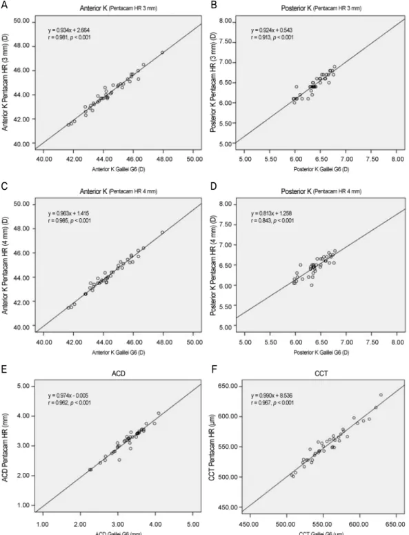
Figure 1. Pearson correlation of Galilei G6 and Pentacam HR in anterior segment parameters. (A) Anterior mean keratometric diop-
ter (Kmean) (Pentacam HR 3 mm), (B) Posterior Kmean (Pentacam HR 3 mm), (C) Anterior Kmean (Pentacam HR 4 mm), (D) Posterior Kmean (Pentacam HR 4 mm), (E) anterior chamber depth (ACD), (F) central corneal thickness (CCT). K = keratometric diopter.-0.12 ± 2.22D였다(Table 1). Galilei G6와 Pentacam HR을 이용하여 측정한 각막굴절력은 다음과 같다(Table 2). Galilei G6로 측정한 전면 각막굴절력은 44.35 ± 1.38D로 Pentacam HR (3 mm)의 44.09 ± 1.32D와 Pentacam HR (4 mm)의 44.12 ± 1.35D보다 통계적으로 유의하게 높았다(Paired
t-test, all p<0.001). Pearson correlation 결과, Galilei G6의 전면 각막굴절력은 Pentacam HR (3 mm)과 Pentacam HR (4 mm)의 측정치와 각각 r=0.981과 r=0.985의 통계학적으로 유의하게 높은 양의 상관관계를 보였고(all p<0.001) (Fig. 1), Bland-Altman plots 일치도 분석 결과 Galilei G6와 Pentacam
A B
C D
E F
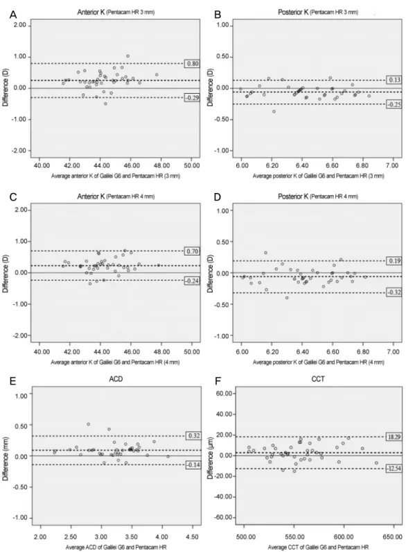
Figure 2. Bland-Altman plots of Galilei G6 and Pentacam HR in anterior segment parameters. (A) Anterior mean keratometric diop-
ter (Kmean) (Pentacam HR 3 mm), (B) Posterior Kmean (Pentacam HR 3 mm), (C) Anterior Kmean (Pentacam HR 4 mm), (D) Posterior Kmean (Pentacam HR 4 mm), (E) anterior chamber depth (ACD), (F) central corneal thickness (CCT). K = keratometric diopter.HR (3 mm)의 95% 일치도 한계는 -0.29~0.80D였으며, Galilei G6와 Pentacam HR (4 mm)의 95% 일치도 한계는 -0.24~
0.70D였다(Fig. 2).
후면 각막굴절력은 Galilei G6, Pentacam HR (3 mm)과 Pentacam HR (4 mm)에서 각각 6.39 ± 0.23D, 6.45 ± 0.23D
와 6.45 ± 0.22D로 통계학적으로 유의한 차이가 있었다 (Paired t-test, p<0.001, p=0.004, respectively). Pearson cor- relation 결과, Galilei G6의 후면 각막굴절력은 Pentacam HR (3 mm)과 Pentacam HR (4 mm)의 측정치와 각각 r=0.913과 r=0.843의 통계학적으로 유의하게 높은 양의 상
Table 3. Comparison of anterior chamber depth (ACD) and central corneal thickness (CCT) measured by Galilei G6 and Pentacam HR
Galilei G6 Pentacam HR Difference p-value*
ACD (mm) 3.26 ± 0.42 3.17 ± 0.42 0.09 ± 0.12 <0.001
CCT (µm) 556.65 ± 30.12 553.78 ± 29.42 -2.88 ± 7.71 0.023
Values are presented as mean ± SD.
*Paired t-test.
관관계를 보였고(Fig. 1), Bland-Altman plots 일치도 분석 결과 Galilei G6와 Pentacam HR (3 mm)의 95% 일치도 한 계는 -0.25~0.13D였으며, Galilei G6와 Pentacam HR (4 mm)의 95% 일치도 한계는 -0.32~0.19D였다(Fig. 2).
전방깊이는 Galilei G6와 Pentacam HR에서 각각 3.26 ± 0.42 mm, 3.17 ± 0.42 mm로 Galilei G6에서 더 깊게 측정 되었고, 통계적으로 유의한 차이를 보였다(Paired t-test, p<0.001) (Table 3). Pearson correlation 결과 통계적으로 유 의한 상관관계를 보였으며(r=0.962, p<0.001) (Fig. 1), Bland-Altman plots 일치도 분석 결과 95% 일치도 한계는 -0.14~0.32 mm였다(Fig. 2).
중심각막두께는 Galilei G6와 Pentacam HR에서 각각 556.65 ± 30.12 µm, 553.78 ± 29.42 µm로 Galilei G6에서 더 두껍게 측정되었고, 통계적으로 유의한 차이를 보였다 (Paired t-test, p=0.023). Pearson correlation 결과 통계적으 로 유의한 상관관계를 보였으며(r=0.967, p<0.001) (Fig. 1), Bland-Altman plots 일치도 분석 결과 95% 일치도 한계는 -12.54~18.29 µm였다(Fig. 2).
고 찰
정확한 전안부의 계측은 노안 교정을 위한 각막굴절교정 수술이나 인공수정체삽입술에 매우 중요하다. 회전샤임플 러그 카메라의 일종인 Galilei G6와 Pentacam HR은 비접촉 식으로 측정시간이 2초 이내이며, 전면각막곡률계뿐만 아 니라 각막두께, 후면각막곡률계의 정확한 측정이 가능하다 는 장점이 있다.3 하지만 두 기기 간 호환성에 대해서는 이 견이 있는 상태이며,5-9 Galilei G6와 Pentacam HR로 측정 한 전안부 계측치를 비교한 연구는 국내에서 보고된 바가 없다. 이에 본 연구에서는 Galilei G6와 Pentacam HR로 측 정한 각막굴절력, 전방깊이 및 중심각막두께를 비교하여 두 기기를 서로 대치하여 사용할 수 있는지 알아보고자 하 였다.
본 연구에서 각막굴절력의 경우 Galilei G6와 Pentacam HR (3 mm)을 비교하였을 때 전면과 후면 각막굴절력에서 모두 유의한 차이를 보였으며, Galilei G6와 Pentacam HR (4 mm)을 비교하였을 때도 동일하였다. Galilei G6는 기본 적으로 중심 4 mm 영역의 각막굴절력을 제공하며, Penta-
cam HR은 중심 3 mm 영역의 각막굴절력을 제공한다. 측 정 영역의 차이는 각막굴절력에 영향을 미치게 되는데,13 본연구에서 Galilei G6와 Pentacam HR은 같은 측정 영역 에서도 각막굴절력의 유의한 차이를 보여 측정 영역 차이 로 인한 영향은 크지 않았던 것으로 생각된다. 각막굴절력 의 경우 기존의 연구들에서 측정기기 간에 차이가 나는 경 우가 많았다. Anayol et al6은Galilei, Pentacam 및 Sirius를 이용하여 측정한 각막굴절력을 비교한 결과 Galilei가 Pen- tacam보다 평균 0.41D 유의하게 높게 측정되었다고 하였으 며, 이는 본 연구의 결과와 유사하였다. Hernández-Camare- na et al9은 평균 31.42세의 정상인을 대상으로 Galilei G2, Pentacam HR 및 Sirius 3D를 이용하여 측정한 평균 각막굴 절력을 비교하였으며, Pentacam HR과 Galilei G2의 평균 각막굴절력은 각각 43.70D, 44.00D로 측정되어, Galilei G2 가 Pentacam HR보다 약 0.30D 높게 측정되었지만 두 기기 간 유의한 차이는 없다고 하였다. 또한 Aramberri et al5은 Pentacam HR과 Galilei G2를 이용하여 평균 34.91세의 정 상인을 대상으로 전면과 후면의 각막굴절력을 비교하였는 데, 전면 각막굴절력은 각각 43.19 ± 1.39D, 43.22 ± 1.43D 로 측정되었고 유의한 차이를 보이지 않아 본 연구결과와 상이하였다. 후면 각막굴절력의 경우는 각각 6.24 ± 0.24D, 6.21 ± 0.24D로 측정되어 Pentacam HR의 각막굴절력이 더 높게 측정되었다는 점에서는 일치하지 않았지만, 두 기기 로 측정한 후면 각막굴절력이 유의한 차이를 보였다는 점 에서는 본 연구결과와 유사하였다. Youn et al2은 Pentacam (3 mm)과 AL-Scan (2.4 mm)을 이용하여 측정한 각막굴절 력은 각각 43.36 ± 1.35D, 43.32 ± 1.40D로 절대적인 차이 는 적으나, 95% 일치도 한계가 -0.38~0.46D로 서로 대치하 여 사용할 수 없으며 다른 각막 측정계들과의 상호 보완이 필요하다고 하였다. 본 연구에서 후면 각막굴절력의 경우 이전 연구가 부족하여 절대적인 비교는 불가능했지만, 전 면 각막굴절력의 경우 Galilei G6와 Pentacam HR (3 mm) 의 95% 일치도 한계는 -0.29~0.80D였으며, Galilei G6와 Pentacam HR (4 mm)의 95% 일치도 한계는 -0.24~0.70D 로 측정되어 두 기기를 대치하여 사용하기는 어려울 것으 로 생각한다.
본 연구에서 전방깊이는 Galilei G6가 Pentacam HR보다 0.09 mm 더 깊게 측정되는 것으로 나타나 이전 연구와 유
사한 경향을 보였다. Aramberri et al5은 정상 성인 35안을 대상으로 Galilei G2와 Pentacam HR을 이용하여 측정한 전 방깊이를 비교하였으며, Galilei G2로 측정한 전방깊이가 Pentacam HR보다 0.10 mm 더 깊게 측정되었고 통계적으 로 유의한 차이가 있었다고 하였다. 또한 Hernández-Cama- rena et al9은 Pentacam HR과 Galilei G2로 측정한 전방두 께는 각각 3.10 mm와 3.60 mm로 경계역 수준에서 유의한 차이(p=0.059)를 보였다고 하였으며, Anayol et al6은Gal- ilei, Pentacam, Sirius에서의 전방깊이를 비교하였고, 세 기 기로 측정한 전방깊이는 통계적으로 유의한 차이를 보여 대치하여 사용할 수 없다고 하였다. Han et al8의 연구에서 는Galilei G2, Pentacam 및 Lenstar를 이용하여 측정한 전 방깊이는 각각 3.23 mm, 3.22 mm, 3.19 mm로 Galilei G2 와 Pentacam은 유의한 차이를 보이지 않았고, Galilei G2와 Lenstar만 유의한 차이를 보였으나 세 기기 간의 일치도 범 위가 넓어 측정치를 대치하여 사용할 수 없다고 하였다. 기 존 연구에서 Galilei G4와 Pentacam HR을 이용하여 측정한 전방깊이의 95% 일치도 한계는 -0.29~0.14 mm로 이러한 차이는 인공수정체 삽입 후 vault와 관련된 문제를 발생시 킬 수 있다고 하였으며,7 본 연구에서도 95% 일치도 한계 가 -0.14~0.32 mm로 유사하여 두 기기로 측정한 전방깊이 는 서로 대치하여 사용할 수 없을 것으로 생각된다.
본 연구 결과에서 중심각막두께는 Galilei G6가 2.88 µm 더 두껍게 측정되었으며 통계학적으로 유의한 차이를 보였 다. 이전 연구에서 Crawford et al14은 평균나이 38.0세의 정 상 성인을 대상으로 Galilei와 Pentacam을 이용하여 측정한 중심각막두께를 비교하였으며, Pentacam이 Galilei보다 2 µm 더 두껍게 측정되어 큰 차이는 없었으나 95% 일치도 한계가 -11~47 µm로 넓어 상호보완적으로 사용할 수 없다 고 하였다. 반면, Aramberri et al5은 Galilei의 경우 2개의 Scheimpflug camera를 이용해 x-y축의 중심이탈을 보정하 기 때문에 Galilei로 측정한 중심각막두께의 정확성이 Pen- tacam보다 더 높았으나, 두 기기로 측정한 중심각막두께의 차이는 2.76 µm로 크지 않아 임상에서 상호보완적으로 사 용할 수 있다고 하였다. Han et al8의 연구에서는 정상 성인 을 대상으로 Galilei, Pentacam 및 Lenstar를 이용하여 측정 한 중심각막두께를 비교하였는데, Galilei와 Pentacam의 일 치도가 가장 높았지만, 95% 일치도 한계는 -7.30~24.65 µm 로 넓어 임상에서 대치하여 사용할 수 없다고 하였다. 본 연구에서는 Galilei G6의 측정치가 Pentacam HR보다 두껍 게 측정되기는 하였지만, 차이가 2.88 µm로 적어 임상적으 로 큰 의미는 없을 것으로 생각한다. 하지만 95% 일치도 한계가 -12.54~18.29 µm로 일치도가 높지는 않아 측정치를 대치하여 사용할 때는 주의가 필요할 것으로 생각된다.
Galilei G6와 Pentacam HR로 측정한 각막굴절력과 전방 깊이 및 중심각막두께에 차이가 생기는 원인은 두 기기의 하 드웨어적인 차이점 때문으로 생각해 볼 수 있다. Pentacam HR은 카메라가 눈의 광축(optical axes) 주위를 360° 회전 하면서15 25,000개의 true elevation data point를 이용하여16 전안부의 이미지를 3차원적으로 재구성한다. 반면, Galilei G6는 Pentacam HR과 달리 placido disc를 통해 얻은 이미 지를 보정하여 각막굴절력을 얻으며,11 122,000개 이상의 data point를 이용하여 전안부(각막, 홍채, 동공, 전방깊이, 수정체)를 직접적으로 측정하고, 각막의 형상과 두께, 동공 크기를 분석하며,17 두 개의 Scheimpflug camera 이미지를 동시에 기록하여 검사 시 생길 수 있는 안구의 중심이탈을 보정한다.18
Galilei, Pentacam과 Sirius를 비교한 이전 연구에서 Gal- ilei로 측정한 중심각막두께는 Pentacam보다 두껍게 측정되 었고 통계적으로 유의한 차이를 보였으나, 실제 값(true value)을 알 수 없기 때문에 어떠한 장비로 측정한 값이 더 정확한지 제시하지 못하였다.6 그러나 Aramberri et al5은 Galilei와 Pentacam으로 측정한 각막곡률, 난시 및 중심각 막두께에 대해 급내상관계수(intraclass correlation coeffi- cient)와 개체 내 표준편차(2.77 × within subject standard deviation [Sw])를 계산하여, simK와 각막난시는 Pentacam 이 측정자 간, 측정자 내에서 더 높은 재현성을 보였고, 중 심각막두께는 Galilei가 높은 재현성으로 더 정확히 측정한 다고 제시하였다. 또 다른 연구19에서는 Galilei가 Pentacam 보다 각막 고위수차의 측정에 있어서 더 높은 재현성을 보 였지만 어느 장비에서 각막 고위수차값이 기준보다 크게 혹은 작게 측정되는 것인지 모르기 때문에 각 기기 고유의 특성을 고려하여 판단하는 것이 좋을 것이라 하였다. 본 연 구 역시 실제 값(true value)을 제시하지 못하였다는 제한점 을 가지고 있으며, 이에 대해서는 추가적인 연구가 필요할 것으로 생각된다.
본 연구는 정상 성인을 대상으로 한 기존의 연구들과 달 리 대상자의 평균나이가 62.28세로 연령에 따른 각막의 morphology에 차이가 있을 수 있기 때문에 직접적인 비교 에는 한계가 있다. 하지만 백내장 수술 시 두 기기의 활용 빈도가 높다는 점을 감안할 때 백내장 환자를 대상으로 기 기 간 측정치의 비교 결과를 제시했다는 점에서 충분히 의 의가 있을 것이라고 생각한다. 결론적으로, 회전샤임플러그 카메라를 이용하는 Galilei G6와 Pentacam HR는 각막굴절 력, 전방깊이 및 중심각막두께의 유의한 차이를 보이며, 일 치도가 낮아 두 기기를 임상에서 대치하여 사용할 수 없을 것으로 생각한다.
= 국문초록 =
두 샤임플러그 전안부사진기를 이용한 전안부 계측치의 비교
목적: Galilei G6와 Pentacam HR로 측정한 각막굴절력과 전방깊이, 중심각막두께의 측정치를 비교하고, 기기 간 일치도를 분석하고 자 하였다.
대상과 방법: 백내장 수술을 목적으로 방문한 환자 40명(40안)을 대상으로 두 기기를 이용하여 측정한 전면과 후면의 각막굴절력, 전방깊이, 그리고 중심각막두께를 비교하였다.
결과: Galilei G6 (4 mm), Pentacam HR (3 mm), Pentacam HR (4 mm)로 측정한 전면 각막굴절력은 각각 44.35 ± 1.38D, 44.09
± 1.32D, 44.12 ± 1.35D였고, 후면 각막굴절력은 각각 6.39 ± 0.23D, 6.45 ± 0.23D, 6.45 ± 0.22D였다. Galilei G6 (4 mm)와 Pen- tacam HR (3 mm), Galilei G6 (4 mm)와 Pentacam HR (4 mm) 사이에서 전면과 후면 각막굴절력 모두 유의한 차이를 보였다. Galilei G6와 Pentacam HR로 측정한 전방깊이는 각각 3.26 ± 0.42 mm, 3.17 ± 0.42 mm, 중심각막두께는 각각 556.65 ± 30.12 μm, 553.78
± 29.42 μm였고, 측정치 간에 유의한 차이를 보였으며, 측정값의 95% 일치도 한계는 각각 -0.14~0.32 mm, -12.54~18.29 μm였다.
결론: Galilei G6와 Pentacam HR로 측정한 전면과 후면의 각막굴절력, 전방깊이, 그리고 중심각막두께는 측정치에서 유의한 차이를 보이고 일치도가 낮아 임상에서 대치하여 사용할 수 없을 것으로 생각한다.
<대한안과학회지 2016;57(7):1056-1062>
REFERENCES
1) Ou RJ, Shaw EL, Glasgow BJ. Keratectasia after laser in situ kera- tomileusis (LASIK): evaluation of the calculated residual stromal bed thickness. Am J Ophthalmol 2002;134:771-3.
2) Youn SM, Lim SH, Lee HY. Measurement comparison of anterior segment parameters between AL-Scan(R) and Pentacam(R). J Korean Ophthalmol Soc 2014;55:801-8.
3) Chen D, Lam AK. Intrasession and intersession repeatability of the Pentacam system on posterior corneal assessment in the normal human eye. J Cataract Refract Surg 2007;33:448-54.
4) Patel RP, Pandit RT. Comparison of anterior chamber depth meas- urements from the Galilei Dual Scheimpflug Analyzer with IOLMaster. J Ophthalmol 2012;2012:430249.
5) Aramberri J, Araiz L, Garcia A, et al. Dual versus single Scheim- pflug camera for anterior segment analysis: precision and agree- ment. J Cataract Refract Surg 2012;38:1934-49.
6) Anayol MA, Güler E, Yağci R, et al. Comparison of central corneal thickness, thinnest corneal thickness, anterior chamber depth, and simulated keratometry using Galilei, Pentacam, and Sirius Devi- ces. Cornea 2014;33:582-6.
7) Domínguez-Vicent A, Monsálvez-Romín D, Águila-Carrasco AJ- D, et al. Measurements of anterior chamber depth, white-to-white distance, anterior chamber angle, and pupil diameter using two Scheimpflug imaging devices. Arq Bras Oftalmol 2014;77:233-7.
8) Han SH, Hwang HS, Shin MC, Han KE. Comparison of central corneal thickness and anterior chamber depth measured using three different devices. J Korean Ophthalmol Soc 2015;56:694-701.
9) Hernández-Camarena JC, Chirinos-Saldaña P, Navas A, et al.
Repeatability, reproducibility, and agreement between three differ- ent Scheimpflug systems in measuring corneal and anterior seg- ment biometry. J Refract Surg 2014;30:616-21.
10) Lee JW, Park SH, Seong MC, et al. Comparison of ocular biometry and postoperative refraction in cataract patients between Galilei-G6(R) and IOL Master(R). J Korean Ophthalmol Soc 2015;56:515-20.
11) Savini G, Carbonelli M, Barboni P, Hoffer KJ. Repeatability of au- tomatic measurements performed by a dual Scheimpflug analyzer in unoperated and post-refractive surgery eyes. J Cataract Refract Surg 2011;37:302-9.
12) Cınar Y, Cingü AK, Sahin M, et al. Comparison of optical versus ul- trasonic biometry in keratoconic eyes. J Ophthalmol 2013;2013:481238.
13) Saad E, Shammas MC, Shammas HJ. Scheimpflug corneal power measurements for intraocular lens power calculation in cataract surgery. Am J Ophthalmol 2013;156:460-7.e2.
14) Crawford AZ, Patel DV, McGhee CN. Comparison and repeat- ability of keratometric and corneal power measurements obtained by Orbscan II, Pentacam, and Galilei corneal tomography systems.
Am J Ophthalmol 2013;156:53-60.
15) Engren AL, Behndig A. Anterior chamber depth, intraocular lens position, and refractive outcomes after cataract surgery. J Cataract Refract Surg 2013;39:572-7.
16) Miyake T, Shimizu K, Kamiya K. Distribution of posterior corneal astigmatism according to axis orientation of anterior corneal astigmatism. PLoS One 2015;10:e0117194.
17) Salouti R, Nowroozzadeh MH, Zamani M, et al. Comparison of an- terior and posterior elevation map measurements between 2 Schei- mpflug imaging systems. J Cataract Refract Surg 2009;35:856-62.
18) Menassa N, Kaufmann C, Goggin M, et al. Comparison and re- producibility of corneal thickness and curvature readings obtained by the Galilei and the Orbscan II analysis systems. J Cataract Refract Surg 2008;34:1742-7.
19) Choi YJ, Kang NH, Jun RM. Comparison of corneal higher-order aberrations measured with two instruments using Scheimpflug Camera System. J Korean Ophthalmol Soc 2015;56:1497-1504.