Characteristics of Dynamic and Kinetic Visual Acuity in Young Myopic Eyes
Young-Cheong Kim
1,2,*, Sang-Woo Park
3, and Hyun-Suk Shim
21
Dept. of Medical science, Graduate School Chonnam National University, Gwangju 61469, Korea
2
Dept. of Ophthalmic Optics, Gwangju Health University, Gwangju 62287, Korea
3
Dept. of Ophthalmology, Chonnam National University Medical School and Hospital, Gwangju 61469, Korea (Received August 9, 2017: Revised September 1, 2017: Accepted December 11, 2017)
···
Purpose: To investigate the characteristics of dynamic visual acuity (DVA) and kinetic visual acuity (KVA) in young myopic eyes and to investigate the clinical or ocular factors affecting DVA and KVA. Methods: Eighty subjects aged between 20-32 years old were recruited. Before measuring the DVA and KVA, visual acuity, spherical equivalent refractive error, intraocular pressure were measured, and axial length, central corneal thickness, anterior chamber depth, lens thickness, pupil diameter were obtained by optical low coherence reflectometry (Haag-Streit, Köniz, Switzerland) and central subfield thickness, cube volume, cube average thickness parameters of macular were measured using a Cirrus high-definition optical coherence tomography (HD-OCT) device (Carl Zeiss Meditec, Dublin, CA, USA). Monocular and binocular DVA were measured using a dynamic visual acuity analyzer (designed by Jangill Moon, Hyunsuk Shim, and Youngcheong Kim). Monocular and binocular KVA were measured using a kinetic visual acuity meter AS-4A (KOWA, Tokyo, Japan). All measurements were repeated at baseline, 1 week, and 4 weeks. Results: In comparison between monocular and binocular viewing, binocular DVA was higher than monocular DVA. However, there was no difference in KVA. Men showed higher DVA and KVA than women at baseline, 1 week, and 4 weeks. The lower degree of myopia, shorter axial length, and longer pupil diameter were the higher DVA. Similarly, the lower degree of myopia and shorter axial length were the higher KVA. However, KVA did not show association with pupil diameter. Conclusions: DVA and KVA may be different by sex, degree of myopia, axial length, pupil diameter. Clinical factors and ocular biometry should be considered when measuring DVA and KVA.
Key words: DVA, Dynamic visual acuity, KVA, Kinetic visual acuity, Clinical factor, Myopia, Ocular biometry, Pupil diameter, Axial length
···
INTRODUCTION
Dynamic visual acuity (DVA) refers to the ability to dis- criminate the fine parts of a horizontally moving object during relative motion between the object and the observer.
Kinetic visual acuity (KVA) refers to the ability to dis- criminate the fine parts of an object moving from a dis- tant point towards the subject.
[1-3]DVA and KVA are suitable for estimating the visual functions of athletes who con- tinue to pursue a moving ball during their play, so these two parameters have been widely studied to measure the ability to visually discern a moving object in athletes of various sports especially using ball such as baseball, volleyball,
and basketball.
[1-5]Winograd reported that college baseball players showed better visual efficiency and better stereoacuity than the general public and students who did not qualify for the team.
[6]Rouse et al. also reported that DVA of college baseball players was better than nonathletic students.
[7]Laby et al showed that visual acuity, contrast sensitivity, and distance stereoacuity of Los Angeles Dodgers base- ball players were better than general public.
[4]But all above mentioned studies were conducted without consider- ing the ocular factors such as degree of myopia, gender, axial length and pupil diameter.
Recent technological advances in optical biometry have
*Corresponding author: Young-Cheong Kim, TEL: +82-62-232-6520, E-mail: [email protected]
been achieved by latest introduction of swept-source opti- cal coherence tomography.
[8,9]Ocular parameters such as axial length, corneal thickness, anterior chamber depth, lens thickness and pupil diameter can be measured by the swept-source optical coherence tomography, and many studies have been reported that above mentioned ocular parameters can be measured with high repeatability and reproducibility.
[10-13]Until now, the most studies
[14,15]have considered correlations of KVA and DVA with refractive errors, corrected visual acuity, static visual acuity(SVA), dominant eye and pupil distance(PD), and none of studies have considered the biometric ocular elements provided by optical low coherence reflectometry when analyzing DVA and KVA.
The purpose of this study was to investigate the charac- teristics of DVA and KVA according to the sex, refractive error, and pupil diameter in young myopic eyes with nor- mal anatomical macular. Also, we investigated if clinical or ocular factors such axial length, affecting DVA and KVA.
MATERIALS AND METHODS
1. Subjects
Healthy volunteers were prospectively and consecutively recruited from September 2014 to July 2015. The subjects were selected from students attending the Chonnam National University Medical School. The participants were informed about the study objectives, and signed informed consent was obtained from all participants. During the enrollment period, 86 subjects were evaluated and 4 sub- jects were excluded because of prior refractive surgery, 2 subjects were excluded due to follow up loss. Total 80 subjects (male 42, female 38) were enrolled in this study.
All subjects underwent complete ophthalmic examination including measurement of best-corrected visual acuity, intraocular pressure (IOP) by Goldmann applanation tonome- try, manifest refraction, slit-lamp examination and stereo- scopic disc photography and red-free disc photography.
1) The following inclusion criteria were used Healthy subjects aged between 20 and 40 years, a spher- ical equivalent (SE) refractive error between −9.0 and −0.5 diopters (D), astigmatism within ± 2D, monocular best-cor- rected visual acuity ≥ 20/25, binocular best-corrected visual
acuity ≥ 20/20, IOP ≤ 21 mmHg, normal anterior chamber angles, nonglaucomatous ONHs (Optic Nerve Heads) on stereoscopic photographs (an intact neuroretinal rim with- out peripapillary hemorrhage, thinning, or localized pal- lor), and absence of any RNFL (Retinal Nerve Fiber Layer) abnormalities on red-free fundus photographs. We excluded subjects with a family history of glaucoma in a first- degree relative, history of intraocular or refractive surgery, pathologic myopia (patch chorioretinal atrophy, lacquer crack lesions, intrachoroidal cavitations, choroidal neovas- cularization), other evidence of retinal pathology, or opaque media.
2. Clinical factors and Ocular biometry
Axial length, central corneal thickness, anterior chamber depth, lens thickness and pupil diameter were measured in a dimly lighted room by optical low-coherence reflectome- try (Lenstar; Haag-Streit AG, Koeniz, Switzerland). The central subfield thickness, cube volume, cube average thick- ness parameters of macular were measured using a Cirrus high-definition optical coherence tomography (HD-OCT) device (Carl Zeiss Meditec). All measurements were done at baseline, 1 week, 4 weeks and performed by the same examiner who was experienced in taking OCT images. A detailed medical history was also recorded for each sub- ject.
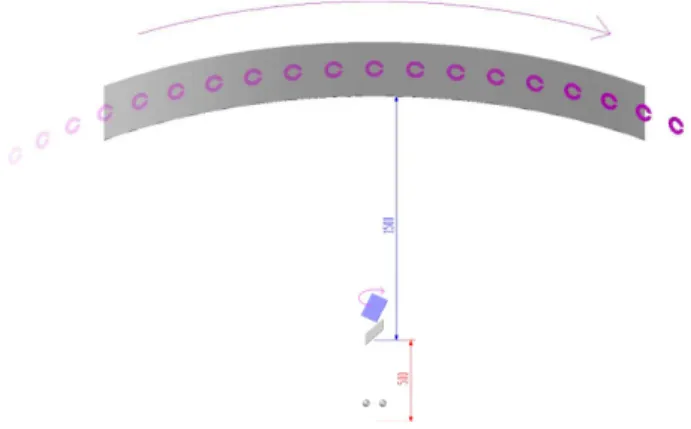
Fig. 1. Schematic diagram of dynamic visual acuity analyzer (designed by Jangill Moon, Hyunsuk Shim, and Youngcheong Kim) Radius of curvature and length of arc of screen was 1.5 m, and Landolt-C ring size was 0.05 visual acuity chart with outer diameter 60 mm.
When measuring DVA, Landolt-C ring moving from left
to right, on the screen 2 m distant from the subject. If
subject identified the Landolt-C ring gap, the examiner
changed the moving velocity of the Landolt-C ring
more faster.
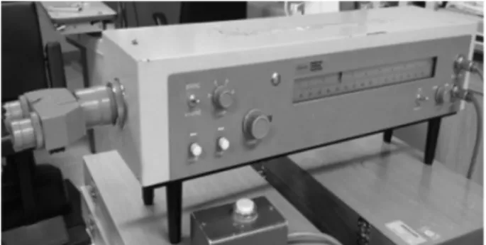
3. Dynamic Visual Acuity and Kinetic Visual Acuity DVA was measured using a dynamic visual acuity ana- lyzer (designed by J.I.M, H.S.S, and Y.C.K, Fig. 1), and measured with monocularly (right to left) and binocularly and then KVA was measured in the same order using a kinetic visual acuity meter AS-4A (KOWA, Tokyo, Japan, Fig. 2).
For the measuring DVA, the radius of curvature screen was 1.5 m and visual acuity chart was 0.05 Landolt-C ring. The subjects were directed to put their chins on the chin rest, and their eyes followed the Landolt-C ring mov- ing from left to right, on the screen at a 2 m distant from the subjects. Subjects were not allowed to move or turn their heads during the measurements. Subjects were asked to tell the identified direction of Landolt-C ring. If subject answered wrong direction, the examiner randomly changed the direction with the same speed, and then the subject had two more chances to tell the right answer. Fastest moving velocity of the Landolt-C ring when the subject could identify the ring gap was used for analysis.
For the measuring KVA, the subjects were directed to look through the eyepiece. Black Landolt-C ring in a bright white circular shape would appear in the eyepiece.
AS-4A showed the Landolt-C ring moving towards the subjects horizontally from a 50 m distance at a rate of 30 km/hour. Subjects were asked to immediately push the but- ton when they could identify the direction of the ring gap.
Landolt-C ring size according to the identification dis-
tance was converted to decimal visual acuity and used for analysis (Table 1). The identification of the smaller the Landolt-C ring size, indicates the better KVA.
DVA and KVA were measured at base line, 1 week, and 4 weeks by the same examiner. Subjects were asked to respond consecutive three times for each measurement until they get the right answer. The mean value of the three measurements was used in analysis.
4. Statistical Analysis
SPSS version 18.0 (SPSS Inc., Chicago, IL) was used for all statistical analyses. Baseline characteristics were reported in mean ± standard deviation values. Groups were compared using student t test and pearson’s correlation analysis. P-values < 0.05 were considered statistically sig- nificant.
RESULTS AND DISCUSSION
Subject's characteristics are presented in Table 2. The mean age were 26.11±2.61 for male, and 25.75±1.58 for female. There were no significant difference between male and female in age, SE refractive error, IOP, axial length, central corneal thickness, anterior chamber depth, lens thick- ness, pupil diameter, central subfield thickness, cube vol- ume, and cube average thickness.
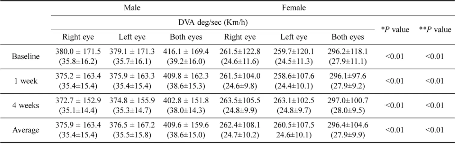
Table 3 shows the mean value of DVA at baseline, 1 week, and 4 weeks. In both gender, there were significant differences between monocular DVA and binocular DVA.
Binocular DVA was significantly higher than monocular DVA (P < 0.01). But there was no significant differences between right DVA and left DVA. In comparison by gen- der, average right, left, and binocular DVA of men were significantly higher than those of women (P < 0.01).
Table 4 shows the mean value of KVA at baseline, 1 week, and 4 weeks. Similar to DVA, average right, left, and binocular KVA of men were higher than those of women (P < 0.01). However, no significant difference was noted between the monocular KVA and binocular KVA.
Fig. 2. Kinetic visual acuity meter AS-4A (KOWA, Tokyo, Japan).
Table 1. The conversion chart of kinetic visual acuity according to the identification distance of the Landolt-C ring in the kinetic visual acuity meter
Distance (m) 3 9 15 21 27 30 33 37 48
KVA 0.1 0.3 0.5 0.7 0.9 1.0 1.1 1.3 1.6
KVA = kinetic visual acuity converted to decimal visual acuity
Table 2. Demographic characteristics of subjects
Variables Male ( n=42) Female ( n=38) P value
Age (years) 26.11±2.610 25.75±2.610 0.24
SE refractive error (D) −4.06±2.28 0 −3.53±2.28 0 0.32
IOP (mmHg) 15.90±3.170 16.12±3.410 0.24
Axial length (mm) 23.56±1.410 23.20±1.650 0.33
Central corneal thickness (µm) 544.46±54.37 540.6±45.20 0.29
Anterior chamber depth (mm) 3.11±0.270 3.07±0.430 0.14
Lens thickness (mm) 3.58±0.240 3.60±0.260 0.16
Pupil diameter (mm) 5.10±1.030 5.13±0.840 0.21
Central subfield thickness (µm) 258.07±23.06 251.43±20.08 0.31
Cube volume (mm
3) 10.09±0.610 9.98±0.430 0.30
Cube average thickness (µm) 282.34±17.47 279.87±12.06 0.16
Data are mean ± standard deviation.
D = diopters; IOP = intraocular pressure; SE = spherical equivalent.
Table 3. Mean value of dynamic visual acuity at baseline, 1 week, and 4 weeks
Male Female DVA deg/sec (Km/h)
*P value **P value Right eye Left eye Both eyes Right eye Left eye Both eyes
Baseline 380.0 ± 171.5 (35.8±16.2)
379.1 ± 171.3 (35.7±16.1)
416.1 ± 169.4 (39.2±16.0)
261.5±122.8 (24.6±11.6)
259.7±120.1 (24.5±11.3)
296.2±118.1
(27.9±11.1) <0.01 <0.01 1 week 375.2 ± 163.4
(35.4±15.4)
375.9 ± 163.3 (35.4±15.4)
409.8 ± 162.3 (38.6±15.3)
261.5±104.0 (24.6±9.8)
258.6±107.6 (24.4±10.1)
296.1±97.6
(27.9±9.2) <0.01 <0.01 4 weeks 372.7 ± 152.9
(35.1±14.4)
374.8 ± 155.9 (35.3±14.7)
402.8 ± 151.8 (38.0±14.3)
263.5±105.5 (24.8±9.9)
263.1±102.5 (24.8±9.7)
297.0±100.7
(28.0±9.5) <0.01 <0.01 Average 375.9 ± 163.4
(35.4±15.4)
376.5 ± 167.2 (35.5±15.8)
409.6 ± 159.6 (38.6±15.0)
262.4±108.1 (24.7±10.2)
260.5±107.5 24.6±10.1)
296.4±104.6
(27.9±9.9) <0.01 <0.01 Data are mean ± standard deviation.
DVA; dynamic visual acuity
* Student t test for monocular DVA between male and female.
** Student t test for binocular DVA between male and female.
Table 4. Mean value of kinetic visual acuity at baseline, 1 week, and 4 weeks
Male Female
KVA (Decimal visual acuity)
*P value **P value Right eye Left eye Both eyes Right eye Left eye Both eyes
Baseline 0.41 ± 0.15 0.39 ± 0.14 0.40 ± 0.13 0.29 ± 0.17 0.28 ± 0.16 0.29 ± 0.15 <0.01 <0.01 1 week 0.39 ± 0.13 0.40 ± 0.12 0.39 ± 0.10 0.29 ± 0.16 0.28 ± 0.17 0.29 ± 0.14 <0.01 <0.01 4 weeks 0.38 ± 0.15 0.39 ± 0.14 0.39 ± 0.12 0.31 ± 0.14 0.30 ± 0.15 0.30 ± 0.13 <0.01 <0.01 Average 0.38 ± 0.13 0.39 ± 0.15 0.39 ± 0.11 0.30 ± 0.13 0.29 ± 0.18 0.29 ± 0.13 <0.01 <0.01 Data are mean ± standard deviation.
KVA = kinetic visual acuity
* Student t test for monocular KVA between male and female.
** Student t test for binocular KVA between male and female.
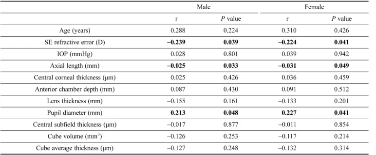
Table 5 and 6 show the pearson’s correlation between clinical or ocular factors and DVA and KVA, respectively.
In men, DVA was associated with SE refractive error (r =
−0.239, P = 0.039), axial length (r = −0.025, P = 0.033) and pupil diameter (r = 0.213, P = 0.048) and KVA was associ- ated with SE refractive error (r = −0.227, P = 0.034), axial length (r = −0.016, P = 0.018). In women, DVA was asso- ciated with SE refractive error (r = −0.224, P = 0.041), axial length (r = −0.031, P = 0.049) and pupil diameter (r = 0.227, P = 0.041) and KVA was associated with SE refractive error (r = −0.204, P = 0.024), axial length (r = −0.029, P = 0.039).
This study shows that DVA and KVA are related to the several clinical and ocular factors, and men have better DVA and KVA than women. Compared to women, men are likely to spend more time for sports activity, this seems to be a reason that men have better DVA and KVA. To our knowl- edge, this is the first study investigating in domestic about DVA and KVA with ocular factors measured by optical low coherence reflectometry. Results of this study showed that SE refractive error and axial length could affect DVA and KVA, and pupil diameter could affect the DVA. Many studies
[1-5]investigating DVA and KVA of various athletes have been Table 5. Pearson’s correlation analysis to dynamic visual acuity
Male Female
r P value r P value
Age (years) 0.288 0.224 0.310 0.426
SE refractive error (D) −0.239 0.039 −0.224 0.041
IOP (mmHg) 0.028 0.801 0.039 0.942
Axial length (mm) −0.025 0.033 −0.031 0.049
Central corneal thickness (µm) 0.025 0.426 0.036 0.459
Anterior chamber depth (mm) 0.087 0.430 0.091 0.512
Lens thickness (mm) −0.155 0.161 −0.133 0.201
Pupil diameter (mm) 0.213 0.048 0.227 0.041
Central subfield thickness (µm) −0.017 0.877 −0.011 0.854
Cube volume (mm
3) −0.126 0.253 −0.117 0.214
Cube average thickness (µm) −0.127 0.248 −0.132 0.314
D = diopters; IOP = intraocular pressure; SE = spherical equivalent.
Table 6. Pearson’s correlation analysis to kinetic visual acuity
Male Female
r P value r P value
Age (years) 0.215 0.194 0.288 0.329
SE refractive error (D) −0.227 0.034 −0.204 0.024
IOP (mmHg) −0.072 0.516 −0.061 0.484
Axial length (mm) −0.016 0.018 −0.029 0.039
Central corneal thickness (µm) 0.027 0.808 0.034 0.744
Anterior chamber depth (mm) 0.097 0.378 0.102 0.413
Lens thickness (mm) −0.029 0.793 −0.038 0.647
Pupil diameter (mm) −0.159 0.148 −0.146 0.201
Central subfield thickness (µm) −0.011 0.920 −0.004 0.841
Cube volume (mm
3) −0.177 0.107 −0.138 0.264
Cube average thickness (µm) −0.169 0.124 −0.147 0.134
D = diopters; IOP = intraocular pressure; SE = spherical equivalent.
reported but there never been studies considered ocular factors to the DVA and KVA.
Myopia is common ocular condition and continue to increases in prevalence, particularly in Asian populations.
[16-19]As subjects had higher myopia and longer axial length, they showed worse DVA and KVA. Because more-myopic eyes tend to have lower SE refractive error and longer axial length, our results suggest that myopia might have nega- tive correlation with DVA and KVA.
[19]DVA is typically tested by asking subjects to judge or identify dynamic objects while their head is fixed. This involves reporting the location of a small opening in a moving Landolt-C ring. This examines the ability of mini- mum resolvable capacity, which means the ability to dis- criminate two small shapes in space. Combination of different types of eye and head movements enables to stabilize the image of subject close to the fovea, the small area on ret- ina where photoreceptor density and visual acuity are max- imal.
[20,21]Smooth pursuit eye movements aim to match the speed of gaze with that of small, moving visual tar- gets, and can be used to track objects travelling at speeds of up to 50 degrees of visual angle per second. At higher target speeds, when gaze lags behind the target, the eyes use fast catch-up saccades to compensate for position and velocity errors.
[22]In this study, pupil diameter had posi- tive correlation with DVA in men and women, and it was statistically significant. However, it is unclear which mech- anism lead pupil diameter to such DVA advantages. Fur- ther studies of relationship among the pupil size, smooth pursuit eye movements, and DVA would be needed.
There are several limitations of this study. First, we didn’t include athletic ability parameters of subjects. Some studies reported differences in DVA or KVA among the groups of different performance levels.
[5,23]But Hoshina K et al reported there were no significant differences in DVA or KVA among the player groups.
[24]Cumulated perfor- mance data were available for professional athletes but there was no data and no standard method to evaluate ath- letic performance of normal subjects. Further study will be needed to investigate standardized method evaluating ath- letic performance when measuring DVA or KVA. Another limitation of this study was that we measured DVA with the target moving from only right to left. Because effec- tive eye movements are related to DVA, measuring DVA with moving targets of various directions such as left to
right, superior to inferior, inferior to superior could be helpful for further studies.
[25,26]CONCLUSIONS
We investigated the characteristics of DVA and KVA in young myopic eyes and found that both DVA and KVA were significantly different between men and women, and showed that men had better DVA and KVA than women.
Also, DVA and KVA can be measured significantly differ- ent by degree of myopia, axial length, pupil diameter. The lower degree of myopia, shorter axial length, and longer pupil diameter were associated with the higher DVA. Simi- larly, the lower degree of myopia and shorter axial length were associated with high KVA. However, KVA did not show association with pupil diameter. When measuring DVA and KVA, clinical factors, such as myopia and ocu- lar biometry should be considered.
REFERENCES
[1] Ludvigh E. The visibility of moving objects. Science.
1948;108(2794):63-64.
[2] Miller JW, Ludvigh E. The effect of relative motion on visual acuity. Surv Ophthalmol. 1962;7:83-116.
[3] Suzumura A. Studies on the kinetic visual acuity espe- cially on accommodation to the moving object: on the trial manufactured kinetic vision tester, model A.S. 3.
Nippon Ganka Gakkai Zasshi. 1964;68(10):1277-1288.
[4] Laby DM, Rosenbaum AL, Kirschen DG, Davidson JL, Rosenbaum LJ, Strasser C et al. The visual function of professional baseball players. Am J Ophthalmol. 1996;
122(4):476–485.
[5] Morris GS, Kreighbaum E. Dynamic visual acuity of var- sity women volleyball and basketball players. Res Q.
1977;48(2):480-483.
[6] Winograd S. The relationship of timing and vision to baseball performance. Res Q. 1942;13(4):481–483.
[7] Rouse MW, DeLand P, Christian R, Hawley J. A compar- ison study of dynamic visual acuity between athletes and nonathletes. J Am Optom Assoc. 1988;59(12):946–950.
[8] Akman A, Asena L, Güngör SG. Evaluation and compari- son of the new swept source OCT-based IOLMaster 700 with the IOLMaster 500. Br J Ophthalmol. 2016;100(9):
1201-1205.
[9] McAlinden C, Wang Q, Gao R et al. Axial length mea-
surement failure rates with biometers using swept-source
optical coherence tomography compared to partial-coher-
ence interferometry and optical low-coherence interfer-
ometry. Am J Ophthalmol. 2017;173:64-69.
[10] Buckhurst PJ, Wolffsohn JS, Shah S, Naroo SA, Davies LN, Berrow EJ. A new optical low coherence reflectome- try device for ocular biometry in cataract patients. Br J Ophthalmol. 2009;93(7):949-953.
[11] Hoffer KJ, Shammas HJ, Savini G. Comparison of 2 laser instruments for measuring axial length. J Cataract Refract Surg. 2010;36(4):644-648.
[12] Bjeloš Rončević M, Bušić M, Cima I, Kuzmanović Elab- jer B, Bosnar D, Miletić D. Intraobserver and interobserver repeatability of ocular components measurement in cataract eyes using a new optical low coherence reflectometer. Graefes Arch Clin Exp Ophthalmol. 2011;249(1):83-87.
[13] Shammas HJ, Hoffer KJ. Repeatability and reproducibil- ity of biometry and keratometry measurements using a noncontact optical low-coherence reflectometer and ker- atometer. Am J Ophthalmol. 2012;153(1):55-61.
[14] Shim HS, Kim SH, Kang HS. Changes in KVA resulting from correction condition of refractive error. J Korean Ophthalmic Opt Soc. 2013;18(2):165-171.
[15] Shim MS, Kim SH, Kang HS, Shim HS. The study of kinetic visual acuity in college students. J Korean Oph- thalmic Opt Soc. 2012;17(4):419-424.
[16] Sperduto RD, Seigel D, Roberts J, Rowland M. Preva- lence of myopia in the United States. Arch Ophthalmol.
1983;101(3):405-407.
[17] Wong TY, Foster PJ, Hee J, Ng TP, Tielsch JM, Chew SJ.
Prevalence and risk factors for refractive errors in adult
Chinese in Singapore. Invest Ophthalmol Vis Sci. 2000;
41(9):2486-2494.
[18] Sawada A, Tomidokoro A, Araie M, Iwase A, Yamamoto T. Refractive errors in an elderly Japanese population: the Tajimi study. Ophthalmology. 2008;115(2):363–370.
[19] Mc Brien NA, Gentle A. Role of the sclera in the devel- opment and pathological complications of myopia. Prog Retin Eye Res. 2003;22(3):307-338.
[20] Jacobs RJ. Visual resolution and contour interaction in the fovea and periphery. Vision Res. 1979;19(11):1187-1195.
[21] Hirsch J, Curcio CA. The spatial resolution capacity of human foveal retina. Vision Res. 1989;29(9):1095-1101.
[22] de Brouwer S, Yuksel D, Blohm G, Missal M, Lefèvre P.
What triggers catch-up saccades during visual tracking?. J Neurophysiol. 2002;87(3):1646-1650.
[23] Stine CD, Arterburn MR, Stern NS. Vision and sports: a review of the literature. J Am Optom Assoc. 1982;53(8): 627- 633.
[24] Hoshina K, Tagami Y, Mimura O, Edagawa H, Matsubara M, Nakayama T. A study of static, kinetic, and dynamic visual acuity in 102 Japanese professional baseball play- ers. Clin Ophthalmol. 2013;7:627-632.
[25] Barmack NH. Dynamic visual acuity as an index of eye movement control. Vision Res. 1970;10(12):1377-1391.
[26] Brown B. Dynamic visual acuity, eye movements and peripheral acuity for moving targets. Vision Res. 1972;
12(2):305-321.
젊은 근시안에서 동체시력과 동적시력의 특징
김영청
1,2,
*, 박상우3
, 심현석2
1
전남대학교 일반대학원 의학과, 광주 61469
2
광주보건대학교 안경광학과, 광주 62287
3