ISSN 0378-6471 (Print)⋅ISSN 2092-9374 (Online)
https://doi.org/10.3341/jkos.2019.60.10.959
Original Article
유리체절제술과 백내장병합수술 시 동시 시행한 후낭절개술 후 앞방 깊이와 굴절력 변화
The Changes of Anterior Chamber Depth and Refractive Errors after Phacovitrectomy with Posterior Capsulotomy
유웅선1,2,3⋅서진석1,2⋅정지성1,2⋅신민호4⋅김성재1,2,3⋅정인영1,2,3
Woong-Sun Yoo, MD1,2,3, Jin-Seok Seo, MD1,2, Ji-Sung Jeong, MD1,2, Min-Ho Shin, MD4, Seong-Jae Kim, MD, PhD1,2,3, In Young Chung, MD, PhD1,2,3
경상대학교 의과대학 안과학교실1, 경상대학교병원 안과2, 경상대학교 건강과학연구원3, 조선대학교 의과대학 안과학교실4 Department of Ophthalmology, Gyeongsang National University College of Medicine1, Jinju, Korea
Department of Ophthalmology, Gyeongsang National University Hospital2, Jinju, Korea Health Science Institute, Gyeongsang National University3, Jinju, Korea Department of Ophthalmology, Chosun University College of Medicine4, Gwangju, Korea
Purpose: To evaluate the changes in anterior chamber depth (ACD) and refractive error after combined phacovitrectomy with posterior capsulotomy using a vitrectomy probe.
Methods: In 20 eyes of 20 patients who underwent combined phacovitrectomy with posterior capsulotomy using a vitrectomy probe, the ACD was measured with Scheimpflug imaging (Pentacam®, OCULUS Optikgeräte GmbH, Wetzlar, Germany) pre- operatively and postoperatively. We compared the preoperative desired refraction and postoperative refraction using an autokeratorefractometor.
Results: The preoperative ACD was 2.58 ± 0.248 mm; the ACD significantly increased in 1 month postoperatively to 3.65 ± 0.475 mm (p < 0.001), and it was maintained as 3.70 ± 0.452 mm (p = 0.213) at 3 months postoperatively. The preoperative target spherical equivalent was -0.60 ± 0.809 diopters (D). Myopic shifting was noticed at 1 month postoperatively as -1.45 ± 1.252 D, and it changed between 1 month and 3 months postoperatively (-1.48 ± 1.235 D at 3 months postoperatively was not statistically significant). There was no increased intraocular pressure or intraocular lens-related complication.
Conclusions: Phacovitrectomy with posterior capsulotomy using a vitrectomy probe might be a useful way to stabilize the axial position of an intraocular lens without constriction of the capsular bag. However, using this procedure, the surgeon must consid- er the possibility of myopic shifting in the postoperative refractive error.
J Korean Ophthalmol Soc 2019;60(10):959-965
Keywords: Anterior chamber, Posterior capsulotomy, Refractive error, Vitrectomy
■Received: 2019. 1. 3. ■ Revised: 2019. 2. 24.
■Accepted: 2019. 9. 24.
■Address reprint requests to In Young Chung, MD, PhD Department of Ophthalmology, Gyeongsang National University Hospital, #79 Gangnam-ro, Jinju 52727, Korea Tel: 82-55-750-8171, Fax: 82-55-758-4158
E-mail: [email protected]
*Conflicts of Interest: The authors have no conflicts to disclose.
ⓒ2019 The Korean Ophthalmological Society
This is an Open Access article distributed under the terms of the Creative Commons Attribution Non-Commercial License (http://creativecommons.org/licenses/by-nc/3.0/) which permits unrestricted non-commercial use, distribution, and reproduction in any medium, provided the original work is properly cited.
유수정체안에서 망막 질환을 치료하는 데 있어서 유리체 절제술과 백내장병합수술이 최근 많이 사용되고 있으며 유 리체절제술 후에 백내장수술을 따로 한 경우에 비해 경제 적, 시간적 이점을 가지고 있으며 한 번의 수술로 빠른 시 력회복을 가져오는 이점도 있다.1,2 두 단계로 나누어 유리 체절제술과 백내장수술을 따로 하는 경우에 비해 병합수술 이 이점을 가지고 있지만, 병합수술 시행 시 술 중 혹은 술
Characteristic Baseline demographic (n = 20)
Age (years) 61.0 ± 7.49
Sex
Male 6 (30)
Female 14 (70)
Laterality
Right 7 (35)
Left 13 (65)
Disease
Epiretinal membrane 6 (30)
PDR 5 (25)
Macular hole 2 (10)
Vitreous opacity 5 (25)
Other 2 (10)
Values are presented as mean ± standard deviation or number (%).
PDR = proliferative diabetic retinopathy.
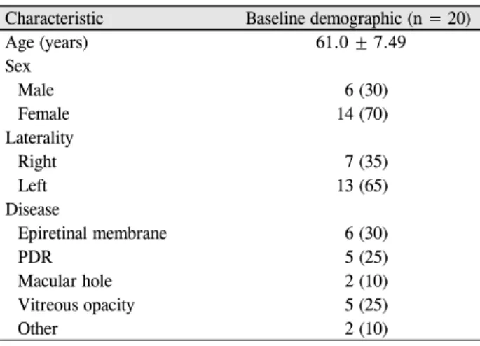
Table 1. Baseline characteristic of patients
후 합병증 발생 위험도가 증가함을 보고하기도 한다.1 수술시간이 길어짐에 따른 합병증뿐만 아니라 홍채후유착, 근 시 이행, 후낭혼탁 등이 잘 생긴다고 보고되고 있다.3-5 특히 야그후낭절개술이 필요한 후낭혼탁은 병합수술환자에서 10%에서 51%까지 발생한다고 보고되고 있으며 유리체절 제술이 후낭혼탁의 위험인자라고 알려져 있다.6-11 또한 후 낭혼탁의 진행은 수정체낭의 수축을 유발시키고 이로 인한 인공수정체의 위치 변화를 일으켜 예측한 굴절력과 다른 결과를 낳기도 하며 인공수정체의 기울어짐이나 위치 이상 을 일으키기도 한다.12,13 이러한 합병증을 예방하기 위해 백 내장수술과 동시에 후낭절개술을 시행하기도 하며, 후낭절 개술 유무에 따른 비교 연구에서 후낭절개술을 동시에 시 행한 경우, 하지 않은 경우에 비해 인공수정체의 앞방 이동 이 적음을 보고하기도 하였다.14 이에 본 연구에서는 유리 체절제술과 백내장병합수술 시 유리체절단침을 이용하여 후낭절개술을 동시에 시행한 경우 수술 후 앞방 깊이 및 굴 절력 변화를 확인하고자 한다.
대상과 방법
2018년 1월에서 2018년 7월까지 본원에서 유리체절제술 과 백내장병합수술을 시행받은 환자 중 3개월 이상 추적 관찰이 가능했던 환자를 대상으로 의무기록을 통한 후향적 연구를 진행하였다. 본 연구는 헬싱키선언(Declaration of Helsinki)을 준수하였으며, 경상대학교병원 임상연구윤리위 원회(institutional review board, IRB)의 승인하에 진행되었 다(GNUH 2018-11-016). 유리체절제술 및 백내장병합수술 을 시행하면서 유리체절단침을 이용한 후낭절개술을 받은 20명 20안을 대상으로 하였다. 이전에 안내수술을 받은 경 험이 있는 경우, 과거에 유리체절제술을 시행받은 경우, 녹 내장의 병력이 있는 경우, 과거에 포도막염이나 안내염증 을 앓았던 경우, 외상의 병력이 있는 경우는 연구 대상에서 제외하였고, 술 중 유리체강 내 실리콘 기름 주입을 시행한 경우, 술 중 후낭파열이 일어난 경우, 수정체소대의 이상이 발견된 경우 및 인공수정체를 섬모체고랑에 삽입한 경우 또한 연구에 포함시키지 않았다. 모든 수술은 한 명의 술자 (I.Y.C.)에 의해 표준화된 방법으로 시행하였다. 유리체절 제술은 전신마취 혹은 구후부마취하에 3개의 공막 천자를 통한 섬모체 평면부를 통해 시행하였고, 백내장적출술은 투명각막절개를 통해 수정체유화술을 시행하였다. 모든 환 자에서 인공수정체는 동일한 1-piece 비구면 아크릴 인공수 정체를 사용하였으며(ZCB00 TECNIS®, Abbott Medical Optics Inc., Santa Ana, CA, USA), 인공수정체 삽입 후 공 막 천자 부위를 통해 유리체절단침을 이용하여 후낭에 약
4.0 mm 크기의 원형의 후낭절개술을 시행하였다. 모든 환자 들에게 시력, 세극등현미경, 안압, 자동각막굴절계, 안저검 사와 함께 샤임플러그 사진기(Pentacam®, OCULUS Optikgeräte GmbH, Wetzlar, Germany)를 이용하여 앞방 깊이 술 전, 술 후 1개월, 3개월째에 측정하였으며 스펙트럼영역 빛간섭단 층촬영기(HRA Spectralis+OCT, Heidelberg Engineering, Heidelberg, Germany)를 이용하여 술 전, 술 후 1개월, 3개 월의 황반부 단층촬영을 시행하였다. 또한 IOL master® (Carl Zeiss Meditec AG, Jena, Germany)를 이용하여 안구 길이 측정을 술 전 및 술 후 3개월째에 시행하였다. 검사의 조건은 산동 상태로 동일하게 하였으며, 검사 및 측정 모두 동일한 검사자가 시행하였다. 인공수정체의 도수 결정은 SRK-T 공식을 이용하였고, 굴절력은 spherical equivalent 로 계산하였다. 질환군별 측정치 비교는 analysis of variance test를 이용하였으며, 수술 전 후의 측정치 비교는 paired samples t-test를 이용하였다. 모든 통계 분석은 SPSS (version 21.0, software for windows; IBM Corp., Armonk, NY, USA)를 이용하였으며, p값이 0.05 미만인 경우에 통계적으 로 유의하다고 판단하였다.
결 과
남자가 6안, 여자가 14안이었으며, 평균 나이는 61.0 ± 7.49세였다. 유리체절제술 및 백내장병합수술의 원인 질환 으로 망막전막 6안, 증식성 당뇨망막병증에 의한 합병증 5안, 황반원공 2안, 유리체혼탁 5안, 기타 2안이었다(Table 1).
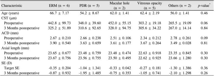
원인 질환에 따라 나누어 비교하였을 때 연령은 질환에 따 른 차이가 없었으며 술 전, 술 후 3개월째 중심황반부두께 와 앞방 깊이, 안축장길이 및 굴절력은 질환에 따른 유의한 차
Characteristic ERM (n = 6) PDR (n = 5) Macular hole (n = 2)
Vitreous opacity
(n = 5) Others (n = 2) p-value*
Age (years) 66.7 ± 7.17 54.2 ± 8.67 62.0 ± 1.41 62.4 ± 2.19 56.0 ± 1.41 0.46
CST (μm)
Preoperative 442.8 ± 99.73 348.0 ± 39.60 452.0 ± 55.15 303.2 ± 19.18 265.5 ± 19.09 0.06 3 Months postoperative 325.2 ± 51.89 310.6 ± 92.65 328.0 ± 94.75 305.6 ± 34.22 267.0 ± 14.14 0.84 ACD (mm)
Preoperative 2.67 ± 0.210 2.66 ± 0.238 2.51 ± 0.106 2.34 ± 0.212 2.78 ± 0.261 0.09
3 Months postoperative 3.90 ± 0.540 3.63 ± 0.659 3.61 ± 0.177 3.67 ± 0.264 3.49 ± 0.028 0.81 Axial length (mm)
Preoperative 23.65 ± 0.677 23.48 ± 0.759 23.48 ± 0.474 22.63 ± 0.918 23.35 ± 0.845 0.30 3 Months postoperative 23.67 ± 0.756 23.56 ± 0.755 23.50 ± 0.495 22.62 ± 0.925 23.66 ± 1.280 0.30 SE (D)
Target SE -0.35 ± 0.204 -1.04 ± 1.341 -0.33 ± 0.042 -0.27 ± 0.181 -1.30 ± 1.386 0.36
3 Months postoperative -0.87 ± 0.932 -1.95 ± 1.485 -0.75 ± 0.353 -1.05 ± 0.741 -2.10 ± 1.298 0.26 Values are presented as mean ± standard deviation unless otherwise indicated.
CST = central subfield thickness; ACD = anterior chamber depth; SE = spherical equivalent; ERM = epiretinal membrane; PDR = pro- liferative diabetic retinopathy; D = diopter.
*p-value was calculated by analysis of variance.
Table 2. Comparison of CST, ACD, axial length and SE in preoperative and postoperative 3 months between diagnoses
Characteristic Preoperative 1 Month postoperative 3 Months postoperative
BCVA (logMAR) 0.67 ± 0.619 0.24 ± 0.217 0.20 ± 0.223
IOP (mmHg) 14.6 ± 2.06 15.0 ± 1.96 14.6 ± 1.82
ACD (mm) 2.58 ± 0.248 3.65 ± 0.475 3.70 ± 0.452
Axial length (mm) 23.35 ± 0.189 N/A 23.36 ± 0.193
SE (D) -0.60 ± 0.809* -1.45 ± 1.252 -1.48 ± 1.235
Values are presented as mean ± standard deviation.
BCVA = best corrected visual acuity; IOP = intraocular pressure; ACD = anterior chamber depth; logMAR = logarithm of minimal angle of resolution; SE = spherical equivalent; D = diopter; N/A = not associated.
*Target spherical equivalent.
Table 3. Changes of BCVA, IOP, ACD, axial length and SE in postoperative 1 month and 3 months
Figure 1. Changes of best corrected visual acuity (BCVA) log-
arithm of minimal angle of resolution in postoperative 1 month and 3 months. BCVA increased statistically significant in post- operative 1 month, and it maintained in postoperative 3 months.LogMAR = logarithm of minimal angle of resolution.
이를 보이지 않았다(Table 2). 술 전 최대교정시력은 logMAR 0.67 ± 0.619였으며 술 후 1개월에 logMAR 0.24 ± 0.217로 유의하게 증가하였으며(p=0.003), 술 후 3개월에 logMAR 0.20 ± 0.223으로 술 후 1개월에 비해 통계적으로 유의한 증가를 보였다(p=0.01) (Table 3, Fig. 1).
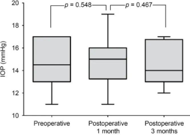
술 전 안압은 14.6 ± 2.06 mmHg였으며 술 후 1개월, 3개 월째 각각 15.0 ± 1.96, 14.6 ± 1.82 mmHg였으며 유의한 안압의 상승이 나타나지 않았다(p=0.548, p=0.467) (Table 3, Fig. 2). 수술 전후의 앞방 깊이의 변화를 살펴보면 수술 전 앞방 깊이는 2.58 ± 0.248 mm였으며 술 후 1개월에 3.65
± 0.475 mm로 유의한 증가가 나타났으며(p<0.001), 술 후 3개월에 3.70 ± 0.452 mm로 앞방 깊이의 감소 없이 유지되 는 양상이었다(p=0.213) (Table 3, Fig. 3). 술 후 안구 길이의 변화를 비교해보면 술 전 안구 길이는 23.35 ± 0.189 mm였 으며 수술 후 3개월째 안구 길이는 23.36 ± 0.193 mm로 유 의한 변화를 보이지 않았다(p=0.953) (Table 3, Fig. 4). 술
Figure 2. Changes of intraocular pressure (IOP) in postoperative
1 month and 3 months. IOP has no significant differences be- tween preoperative and postoperative 1 month and 3 months.Figure 3. Changes of anterior chamber depth (ACD) in post-
operative 1 month and 3 months. ACD showed statistically in- creased in postoperative 1 month and it maintained until 3 months.Figure 4. Changes of axial length in postoperative 3 months.
Axial length showed no changes in postoperative 3 months compared with preoperative.
Figure 5. Changes of refractory error in postoperative 1 month
and 3 months. Postoperative 1 month and 3 months revealed statistically significant myopic shifting compared with pre- operative target spherical equivalent.후 굴절력의 변화 양상을 보면, 술 전 목표 굴절력은 -0.60
± 0.809D였으며 술 후 1개월째 -1.45 ± 1.252D로 통계적 으로 유의한 근시성 이동이 관찰되었으며(p<0.001), 술 후 3개월째는 -1.48 ± 1.235D로 수술 1개월과 비교하였을 때 변화 없이 유지됨을 알 수 있었다(p=0.781) (Table 3, Fig. 5).
술 중 앞방출혈, 섬모체소대 손상, 인공수정체 관련 이상, 맥락막상강출혈 등의 합병증은 없었으며, 술 후 3개월까지 인공수정체의 위치 이상, 고안압증 및 신생혈관녹내장, 유 리체출혈 등의 합병증은 관찰되지 않았다.
고 찰
유리체절제술 및 백내장병합수술은 황반원공 및 망막전 막 등의 유리체망막 질환에서 치료로 많은 술자들이 선호
하고 있다. 백내장이 경도인 경우에도 유리체절제술 단독 수술 후 백내장 진행이 빠름을 감안하여 빠른 시력회복을 위해 단계적 수술보다는 병합수술이 최근 증가하고 있다.15 Rogers et al16은 황반원공과 망막전막에서 유리체절제술 후 백내장수술과 비교하여 백내장병합수술이 안전하고 효 과적이라 보고하였다. 본 연구에서도 단계적 수술과 직접 적 비교는 어려우나 병합수술 전후 의미 있는 시력 상승을 확인하였다.
유리체절제술 및 백내장병합수술의 가장 흔한 술 후 합 병증으로 야그후낭절개술이 필요한 후낭혼탁으로 10-51%
에서 보고되고 있으며6-11 최근 망막전막과 황반원공에서 시행한 병합수술의 대규모 연구에서는 10.6%의 후낭혼탁 이 발생함을 보고하였다.15 후낭혼탁의 진행은 수정체낭의 수축을 유발 및 인공수정체의 위치 변화를 일으키며 인공
수정체의 기울어짐 등을 유발하기도 한다.12,13 또한 후낭혼 탁이 발생된 경우 시행하게 되는 야그후낭절개술은 환자의 비문증을 일으켜 주관적 불편감을 흔히 나타내며, 기계적 인 인공수정체의 이동이나 손상을 입히며, 낭포황반부종을 일으키거나 망막 박리와 같은 심각한 합병증을 유발하기도 한다.17 따라서 Sato et al18은 유리체절제술 및 백내장병합 수술에서 인공수정체를 삽입한 후 유리체절제침을 이용한 후낭절개술을 처음으로 보고하였고 효과적이며 안전한 방 법으로 소개하였다. 본 연구에서도 유리체절제침을 이용한 후낭절개술을 병합수술을 시행하였을 때 술 중, 술 후 합병 증을 보이지 않았으며 술 후에도 의미 있는 시력 상승을 확 인할 수 있었다. 또한 야그후낭절개술 후 발생할 수 있는 안압상승 역시 본 연구에서는 술 후 3개월까지 나타나지 않았다. 다만 유리체절제술 및 백내장수술 후 후낭절개술 을 시행하는 경우 술 중에 액체공기치환술을 하는 도중 인 공수정체의 후면에 결로가 생겨 안저 관찰이 용이하지 않 은 경우가 있으나 이런 경우 백내장수술 후 남은 점탄 물질 을 이용하여 인공수정체의 후면에 도포하면 쉽게 해결할 수 있었다.
Stifter et al14은 백내장수술 중 후낭절개술을 시행한 환 자에서 앞방의 깊이를 측정한 경우 하지 않은 대조군과 비 교하였을 때 수술 후 1개월까지 안정적으로 유지하며 대조 군에서 수정체낭 수축으로 발생하는 인공수정체의 앞방 이 동이 술 중 후낭절개술을 시행한 군에서 의미 있게 감소함 을 보고하였다. 저자들의 연구에서 대조군 설정이 되어있 지 않아 직접적인 비교를 할 수는 없으나 이전 연구와 유사 하게 환자군에서 병합수술 시 시행한 후낭절개술 후 샤임 플러그를 이용한 앞방 깊이 측정 시 술 전에 비해 술 후 1개 월에 의미 있게 증가하였으며 특히 증가한 앞방의 깊이가 3개월까지 변화 없이 잘 유지됨을 알 수 있었다. 이는 인공 수정체의 앞방 이동 등으로 인한 앞방 깊이의 변화가 없음 을 보여주며 인공수정체의 굴절력에 안정성을 나타내는 지 표로 생각된다. 대조군이 설정되어 있지 않은 제한점은 있 으나 이전 연구와는 다르게 유리체절제술을 시행한 후 후 낭절개술을 시행하였으며 유리체에 의한 후낭지지 역할과 유리체 탈출 등으로 인한 앞방 깊이의 변화에 영향을 주지 않는 점이 있어 이전 연구와 차별점이 있다.
이전에 보고된 백내장수술 중 후낭절개술을 시행한 연구 에서 술 후 1개월에 통계적으로 의미는 없지만 술 전에 비 해 술 후 경한 근시성 굴절 이동을 나타냈으며,14 본 연구에 서는 술 전에 예측한 굴절력에 비해 의미 있는 근시성 굴절 이동이 나타났음을 알 수 있었다. 유리체절제술 및 백내장 수술 후 발생하는 근시성 굴절 이동은 유리체와 앞방수의 굴절계수의 차이, 안구 길이의 변화 등의 가설이 제시되었
으나 정확한 원인을 이전 연구에서 밝히지는 못하였으며, 병합수술 중 인공수정체를 삽입 시 0.5디옵터의 원시화를 하는 것이 제시되었다.19 저자들도 근시성 변화를 확인하기 위해 술 전과 술 후 3개월째 안구 길이를 측정하였으나 의 미 있는 차이를 보이지 않아 병합수술 후 발생한 근시성 변 화의 원인에 대한 추가적 연구가 필요할 것으로 생각되며 대조군 설정을 통한 후낭절개술에 의한 근시성 이동 유무 를 비교해보아야 할 것이다. 하지만 이전에 후낭절개술을 시행하지 않은 병합수술 후 굴절력 근시화의 보고에서 근 시화의 정도가 -0.76디옵터로19 직접적인 비교는 어려우나 저자들의 연구에서 보인 굴절력 근시화 이동과 큰 차이를 보이지는 않아 후낭절개술이 굴절력 근시화에 큰 영향을 미치지 않을 것으로 보인다. 다만 유리체절제술 및 백내장 병합수술 시 인공수정체 도수를 예측 인공수정체 도수보다 0.5디옵터 원시화하는 것이 좋을 것으로 생각된다.
본 연구의 가장 큰 제한점으로 대조군 설정되어 있지 않 은 다수 환자군 분석으로 직접적인 유리체절제침을 이용한 후낭절개술 후 앞방 깊이 변화 비교가 어려운 점이 있으며, 이는 추후 전향적 환자-대조군 연구가 필요할 것으로 생각 된다. 또한 환자군에서 앞방 깊이와 굴절력의 변화가 술 후 3개월까지 잘 유지되었으나 관찰 기간이 짧아 장기적 결과 를 관찰하지 않은 제한점이 있어 추후 장기간의 경과 관찰 을 통한 추가 연구가 필요할 것이다.
결론적으로 본 연구는 유리체절제술 및 백내장병합수술 후 유리체절제침을 이용하여 후낭절개술을 시행한 후 샤임 플러그를 이용한 앞방의 깊이의 변화와 굴절력 변화를 측 정한 첫 연구로 유리체절제술 및 백내장병합수술은 빠른 시력 회복을 도모하며 안전한 수술이며, 병합수술의 흔한 합병증으로 알려진 후낭혼탁은 병합수술 중 유리체절제침 을 이용한 후낭절개술을 통해 방지할 수 있고 이를 통해 수 정체낭의 수축으로 인한 인공수정체의 위치 변화 및 굴절 력 변화를 방지할 수 있는 하나의 방법으로 생각된다.
REFERENCES
1) Steel DH. Phacovitrectomy: expanding indications. J Cataract Refract Surg 2007;33:933-6.
2) Muselier A, Dugas B, Burelle X, et al. Macular hole surgery and cataract extraction: combined vs consecutive surgery. Am J Ophthalmol 2010;150:387-91.
3) Manvikar SR, Allen D, Steel DH. Optical biometry in combined phacovitrectomy. J Cataract Refract Surg 2009;35:64-9.
4) Kim YK, Woo SJ, Hyon JY, et al. Refractive outcomes of com- bined phacovitrectomy and delayed cataract surgery in retinal detachment. Can J Ophthalmol 2015;50:360-6.
5) Pinarci EY, Bayar SA, Sizmaz S, et al. Anterior segment complica-
tions after phacovitrectomy in diabetic and nondiabetic patients.
Eur J Ophthalmol 2013;23:223-9.
6) Chang MA, Parides MK, Chang S, Braunstein RE. Outcome of phacoemulsification after pars plana vitrectomy. Ophthalmology 2002;109:948-54.
7) Pinter SM, Sugar A. Phacoemulsification in eyes with past pars plana vitrectomy: case-control study. J Cataract Refract Surg 1999;
25:556-61.
8) Ling R, Simcock P, McCoombes J, Shaw S. Presbyopic phacovitrectomy.
Br J Ophthalmol 2003;87:1333-5.
9) Mochizuki Y, Kubota T, Hata Y, et al. Surgical results of combined pars plana vitrectomy, phacoemulsification, and intraocular lens implantation. Eur J Ophthalmol 2006;16:279-86.
10) Toda J, Kato S, Oshika T, Sugita G. Posterior capsule opacification after combined cataract surgery and vitrectomy. J Cataract Refract Surg 2007;33:104-7.
11) Roh JH, Sohn HJ, Lee DY, et al. Comparison of posterior capsular opacification between a combined procedure and a sequential pro- cedure of pars plana vitrectomy and cataract surgery. Ophthalmologica 2010;224:42-6.
12) Gimbel HV. Posterior capsulorhexis with optic capture in pediatric cataract and intraocular lens surgery. Ophthalmology 1996;103:
1871-5.
13) Gimbel HV. Posterior continuous curvilinear capsulorhexis and
optic capture of the intraocular lens to prevent secondary opacifica- tion in pediatric cataract surgery. J Cataract Refract Surg 1997;23 Suppl 1:652-6.
14) Stifter E, Menapace R, Luksch A, et al. Anterior chamber depth and change in axial intraocular lens position after cataract surgery with primary posterior capsulorhexis and posterior optic buttonholing.
J Cataract Refract Surg 2008;34:749-54.
15) Fajgenbaum MAP, Neffendorf JE, Wong RS, et al. Intraoperative and postoperative complications in phacovitrectomy for epiretinal membrane and macular hole: a clinical audit of 1,000 consecutive eyes. Retina 2018;38:1865-72.
16) Rogers S, Madhusudhana KC, Kang HK, et al. Combined phacovi- trectomy for macular hole: long-term results. Ophthalmic Surg Lasers Imaging 2007;38:452-6.
17) Karahan E, Er D, Kaynak S. An overview of Nd:YAG laser capsulotomy. Med Hypothesis Discov Innov Ophthalmol 2014;3:
45-50.
18) Sato S, Inoue M, Kobayashi S, et al. Primary posterior capsu- lotomy using a 25-gauge vitreous cutter in vitrectomy combined with cataract surgery. J Cataract Refract Surg 2010;36:2-5.
19) Ehmann D, García R. Investigating a possible cause of the myopic shift after combined cataract extraction, intraocular lens im- plantation, and vitrectomy for treatment of a macular hole. Can J Ophthalmol 2009;44:594-7.
= 국문초록 =
유리체절제술과 백내장병합수술 시 동시 시행한 후낭절개술 후 앞방 깊이와 굴절력 변화
목적: 유리체절제술과 백내장병합수술 시 유리체절단침을 이용하여 후낭절개술을 동시에 시행한 경우 수술 후 앞방 깊이 및 굴절력 변화를 확인하고자 하였다.
대상과 방법: 유리체절제술과 백내장병합수술 및 유리체절단침을 이용한 후낭절개술을 받은 20명 20안을 대상으로 하였다. 술 전후의 앞방의 깊이는 샤임플러그 사진기(Pentacam®, OCULUS Optikger te GmbH, Wetzlar, Germany)를 이용하여 측정했으며, 술 후 굴절 력은 자동각막굴절측정기를 이용하여 예측한 굴절력과 차이를 비교하였다.
결과: 수술 전 앞방 깊이는 2.58 ± 0.248 mm였으며, 수술 후 1개월째 3.65 ± 0.475 mm로 유의하게 깊어졌으며(p<0.001), 수술 후 3개월째 3.70 ± 0.452 mm로 1개월째와 유의한 차이를 보이지 않았다(p=0.213). 술 전 예측 굴절력은 -0.60 ± 0.809D였고, 수술 후 1개월째 -1.45 ± 1.252D로 술 전 예측 굴절력에 비해 의미 있는 근시성 이동이 관찰되었으며, 이는 3개월째 -1.48 ± 1.235D로 술 후 1개월에 비해 유의한 변화를 보이지는 않았다. 안압상승이나 인공수정체 관련 합병증은 관찰되지 않았다.
결론: 유리체절제술과 백내장병합수술 시 유리체절단침을 이용한 후낭절개술은 술 후 3개월까지 수정체 낭 수축 등으로 인한 인공수 정체의 앞방 이동을 방지할 수 있는 유용한 방법이 될 수 있으나 근시화되는 점을 고려하여 인공수정체 도수 계산을 해야할 것으로 생각된다.
<대한안과학회지 2019;60(10):959-965>
유웅선 / Woong-Sun Yoo
경상대학교 의과대학 안과학교실 Department of Ophthalmology, Gyeongsang
National University College of Medicine