www.krspine.org
Lumbar Transforaminal Epidural Steroid Injection Using a Nerve Stimulator: The Effect of Needle Tip
Position on Short-Term Effectiveness
Shi-Uk Lee, M.D., Ph.D., Chang Han Lee, M.D.
J Korean Soc Spine Surg 2021 Mar;28(1):1-12.
Originally published online March 31, 2021;
https://doi.org/10.4184/jkss.2021.28.1.1
Korean Society of Spine Surgery
SMG-SNU Boramae Medical Center, 20, Boramae-ro 5-gil, Dongjak-gu, Seoul 07061, Korea Tel: +82-2-831-3413 Fax: +82-2-831-3414
©Copyright 2017 Korean Society of Spine Surgery pISSN 2093-4378 eISSN 2093-4386
The online version of this article, along with updated information and services, is located on the World Wide Web at:
http://www.krspine.org/DOIx.php?id=10.4184/jkss.2021.28.1.1
This is an Open Access article distributed under the terms of the Creative Commons Attribution Non-Commercial License (http://
creativecommons.org/licenses/by-nc/4.0) which permits unrestricted non-commercial use, distribution, and reproduction in any medium, provided the original work is properly cited.
Spine Surgery
Lumbar Transforaminal Epidural Steroid Injection Using a Nerve Stimulator: The Effect of Needle Tip Position on Short-Term Effectiveness
Shi-Uk Lee, M.D., Ph.D., Chang Han Lee, M.D.
*Department of Rehabilitation Medicine, Seoul National University Boramae Medical Center, Seoul, Korea
*Department of Rehabilitation Medicine, Gyeongsang National University School of Medicine and Gyeongsang National University Hospital, Jinju, Korea
Study Design: Retrospective study.
Objectives: We aimed to determine the effect of needle tip position on the short-term effectiveness of transforaminal epidural steroid injection (TFESI) using a nerve stimulator (NS).
Summary of Literature Review: NS enables the needle to be located as close to the nerve root as possible; however, it is not known whether needle proximity affects the performance of TFESI.
Materials and Methods: Forty-five patients were grouped according to number of injected roots. Group I included patients administered TFESI in a single spinal nerve root, and the remainder belonged to group II. Needle tip positions were assessed by dividing the intervertebral foramen into 4 quadrants. Group A referred to all needle positions in quadrant 1 of the injected spinal nerve roots. Location of the needle tip in quadrants 2-4 in at least one spinal nerve root was seen in group B. The Oswestry Disability Index (ODI), Brief Pain Inventory (BPI), and residual pain (RP) were evaluated.
Results: After 2 weeks, a significant decrease in the ODI, BPI, and RP score was noted in all patients, as well as in group I and group II separately. In group I, changes in ODI and BPI scores were statistically significantly greater and RP scores improved to a significantly greater extent in patients who had their needle position in quadrant 1. In all patients, group A had a significant therapeutic effect than group B. In group II, there was no statistically significant difference between group A and group B.
Conclusions: Our study showed a better effect in quadrant 1 than in quadrant 2. NS guidance for TFESI is useful.
Key words: Injections, Needle, Stimulator, Radicular pain
Received: September 21, 2020 Revised: October 12, 2020 Accepted: February 2, 2021 Published Online: March 31, 2021 Corresponding author: Chang Han Lee, M.D.
ORCID ID: Shi-Uk Lee: https//orcid.org/0000-0003-0850-5217 Chang Han Lee: https//orcid.org/0000-0001-8351-5226 Department of Rehabilitation Medicine, Gyeongsang National University School of Medicine and Gyeongsang National University Hospital, Jinju, Korea 79 Gangnam-ro, Jinju, 52727 Korea
TEL: +82-55-750-9639 E-mail: [email protected]
Introduction
Epidural steroid injections have been used to treat lumbar radicular pain syndrome since 1952.1) In theory, transforaminal epidural steroid injection (TFESI) has been preferred in many cases because it can deliver the injectate closer to the dorsal root ganglion (DRG) and facilitates ventral epidural flow to the involved nerve root complex.2,3)
In conventional TFESI, for the safety standpoint, the target needle position should be in the so called “safe triangle” (ST), which has a base tangential to the pedicle, one leg in line with the outer margin of the intervertebral foramen, and a hypotenuse coincident with the upper margin of the spinal nerve and DRG.4) It is mandatory to locate the needle tip
as close as possible to the compromised neural structure.2) However, injections administered through the ST can trigger complications, such as dural tears and subarachnoid drug
infusion.5) In the anatomical relations, the DRGs are located below the pedicle in 90% of the cases, in the medial portion of the pedicle in 2%, and below the lateral aspect of the pedicle in 8%.6) Therefore, even without deformation of the anatomic structures in the intervertebral foramen, the injections via ST cannot ensure safety. In addition, usual method to locate the needle close to the nerve root depend on reproduction of pain by mechanical stimulation with the needle tip, as well manifestation of pain relief after local anesthetic infusion.7,8) Provocation of the nerve root by mechanical stimulation with the needle tip can damage to the nerve root.9,10)
Using a nerve stimulator (NS), it is possible to locate the needle tip in the ST close enough to the target without iatrogenic damage to the nerve root by the needle. Roberet al11) reported a new technique that used electrical stimulation during selective nerve root blocks to avoid problems inherent with TFESI in 2003. Locating needle with the NS has resulted in successful epidural spread during fluoroscopy-assisted procedures.12)
Although the NS enables needle location as close as possible to the nerve root, it is not known whether the proximity of the needle to the DRG affects the performance of TFESI.
During the fluoroscopic guidance, there is a tendency among practitioners to locate the needle tip as medial as possible in the ST because the nerve root runs from superior medial to inferior lateral direction. To the best of our knowledge and based on a comprehensive literature review, no published study has yet reported whether the needle tip position affects the performance of TFESI using a NS. In this study, we retrospectively evaluated the effect of needle tip position on the short-term benefit of TFESI using the NS in patients with lumbar radiculopathy and lumbar spinal stenosis.
Materials and Methods
1. Subjects
We retrospectively reviewed the medical records of patients who received TFESI using the NS at our department from August 2011 to April 2013. Ninety-eight patients were treated with TFESI using the NS for lumbar radicular pain. Inclusion criteria were: patients suspected of back pain and lower extremity radiating pain due to lumbar nerve root compromise.
The exclusion criteria included patients with a history of
previous lumbar surgery or spinal intervention in the prior month, radiculitis without disc herniation, uncontrolled medical illnesses, patients who require long-term oral steroid treatment, and those who fell into one of the following categories:
pregnancy, cognitive impairment, use of anti-coagulant, a history of or potential for any type of adverse reactions to steroids or local anesthetics. The degree and location of lumbar root compromise was confirmed with magnetic resonance imaging (MRI) of the lumbar spine in all the patients. A total of 45 patients were consecutively enrolled. This study was approved by the Institutional Review Board (26-2013-48) of our hospital.
2. Outcome measurement
Outcome measurement was performed before and 2 weeks after the injection. The degree of physical disability, status of pain, and degree of pain relief after the injection were measured.
The degree of physical disability were measured using the Oswestry Disability Index (ODI).13) The patients were asked to complete a questionnaire containing six statements (denoted levels 0 to 5) in each of the 10 sections related to impairments:
pain, personal care, lifting, walking, sitting, standing, sleeping, sex life, social life, and traveling. In each section, the patient was asked to select the statement that best described his/
her status. The total scores can range from 0 (highest level of function) to 50 (lowest level of function). To accommodate patients who failed to respond to every section, the percentage of disability was calculated depending on the total number of possible points.
The severity of pain was evaluated using the Brief Pain Inventory (BPI) questionnaire.14) The BPI is composed of pain severity score and pain interference score. The BPI pain severity score includes 4 items that are scored with numeric rating scales that ask the patient to rate their pain intensity on a 0 to 10 point scale. Each scale is presented as a row of equidistant numbers where 0 = no pain and 10 = worst possible pain.
The patient’s functional status was measured according to the 7 items using the BPI pain interference score. The BPI pain interference score is measured on a scale of 0 to 10, with 0 denoting “no interference” and 10 suggesting “interference completely” with regard to the extent of pain interferes with enjoyment of each item.
Lastly, the residual pain (RP) after injection compared with the baseline was determined by asking the patient for improvement felt after the injection considering the maximum intensity of pain before the injection as 100%. For example, if the patient stated that his/her pain after the injection was half of the pain before the injection, the residual pain was recorded as 50%.
3. Injection technique
The conventional TFESI using the NS was conducted under fluoroscopic guidance. Each patient was placed in the prone position, and a Scotty dog shadow was used to identify the desired lumbar region. The C-arm (Flexiview 8800, GE medical system, USA) was adjusted to align with the inferior endplate of the spine and rotated by 15-30 degrees to an oblique position, so that the Scotty dog shadow became visible.
After disinfecting the skin, local anesthetic (2% lidocaine) was administered using a 25-gauge needle. Under fluoroscopic guidance, 22-gauge spinal needle with two way extension tubes (ContiplexR Tuohy set, BRAUN, Melsungen, Germany) was advanced into the ST.
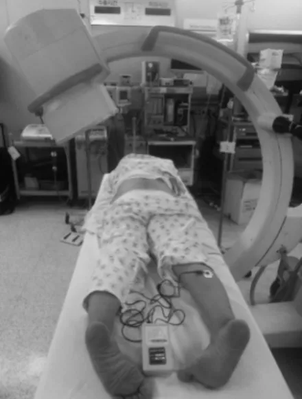
Using the NS (Stimuplex DIG., BRAUN, FL, USA), the needle tip was stimulated with a current output set to 2.0 mV, at a frequency of 2 Hz. The reference electrode was attached to the patient’s calf muscle (Fig. 1). The needle was advanced slowly to the ST. When the patients reported an electrical
shock-like sensation in the corresponding dermatome, the stimulation intensity was slowly decreased by 0.2 mV at a time. When the stimulation was no longer felt by the patient, the intensity was increased by 0.2 mV and the needle position was slightly adjusted to obtain the sensation of the patient.
When the stimulation intensity corresponded to the minimal level of NS, which is 0.2 mV and the patient still reported the tingling sensation, the needle position was defined as optimal.
The optimal position of the needle was the position in the ST with the patient reporting stimulation sensation with minimal stimulation intensity of NS which is 0.2 mV.
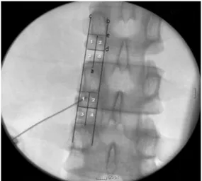
The repositioned needle tip in the anterior-posterior view was saved as the digital imaging and communication in medicine (DICOM) files of the picture archiving communication system (PACS) (M-View 5.4, Marotech, Korea). Next, approximately 1 mL of contrast material (IobrixR, Accuzen, Korea) was injected to confirm epidural flow and to avoid intravascular, intradural, or soft tissue infiltration. Soon after the procedure, anterior-posterior and lateral fluoroscopic view of lumbosacral spine was performed to confirm the presence of epidurogram within the canal (Fig. 2). A 2 cc drug solution (0.5 mL of 1%
Fig. 1. The reference electrode of the nerve stimulator was attached at the calf muscle of the patient.
Fig. 2. C-arm images during a lumbar transforaminal epidural steroid injection of left L4 and L5. (A) Anterior-posterior view. The needle tip remained lateral to the midpedicular line. (B) Lateral fluoroscopic view, showing the spread of the contrast medium into the epidural space.
A
B
lidocaine+0.5 mL of bupivacaine hydrochloride+40 mg (1 mL) of triamcinolone) were slowly injected.
In patients with a unilateral radiating pain of lower extremity and corresponding root compromise on MRI at only a single spinal level, the TFESI was administered at the corresponding level. However, in patients who complained of unilateral or bilateral radiating pain with intermittent claudication and with root compromise involving more than 2 levels on MRI, all the corresponding roots that may trigger symptom were blocked with a 2 cc drug solution (0.5 mL of 1% lidocaine+0.5 mL of bupivacaine hydrochloride+40 mg (1 mL) of triamcinolone) in each corresponding root. However, the MRI findings that were not associated with the patients’ symptoms were considered as irrelevant.
4. Classification of needle tip position
The optimal needle tip position using the NS was assessed retrospectively using the stored image during the procedure.
The classification of needle tip position was based on the method previously reported by Wolff et al.12) The intervertebral foramen was divided into four quadrants in the anterior- posterior view (Fig. 3). Medio- and laterocranial and medio- and laterocaudal quadrants were designated as quadrant 1 to 4, respectively.
5. Grading nerve root compression with MRI
Grading of nerve root compression was performed with the axial view of the corresponding intervertebral discs on the lumbar spine MRI. The grading system used was a 3-tier system described previously by Choi et al15), where grade I is abutment of the disc to the nerve root, grade II is displacement of the nerve root by the herniated disc, and grade III is entrapment of the nerve root between the herniated disc and lamina or facet joint at that level (Fig. 4).
6. Statistical analysis
The patients were grouped according to the number of roots that were injected. Group I included patients who were administered TFESI in only one spinal nerve root (i.e., patients with radiculopathy involving single nerve root). Group II included patients who were administered TFESI in more two spinal nerve roots such as left L4 and L5 (i.e., patients with lumbar spinal stenosis or multiple herniated disc). The number of roots, which was injected, was determined according to the patient’s symptoms and abnormal findings on MRI. Excluding the effects of other variables such as needle tip position or nerve root compression, changes in ODI, BPI, and RP before and after the injection were compared with the Wilcoxon signed rank test and the paired T-test for groups I, II, and all patients.
To evaluate the effect of needle position in group I, changes of ODI, BPI, and RP were compared with the Mann-Whitney test. The Kruskal-Wallis test was used to evaluate the mean scores of changes in the outcome measurements according to
Fig. 3. Anterior-posterior view of the lumbar spine, with a superimposed line (a) bisecting the pedicle. This line was drawn halfway between the farthest medial (b) and farthest lateral (c) points on the pedicle. Two lines (d and e, respectively) perpendicular to lines b and c were drawn at the superior endplate margin of the vertebra and the lower margin of the pedicle. This divided the foramen into four quadrants. Anterior-posterior view showing laterocranial (1), mediocranial (2), laterocaudal (3), and me- diocaudal (4) quadrants.
Fig. 4. Grading nerve root compression with magnetic resonance imag- ing. (A) Grade I: Abutment of the disk to the nerve root. (B) Grade II: The herniated disk displaces the nerve root. (C) Grade III: Entrapment of the nerve root between the herniated disk and the lamina or facet joint at that level. (From Choi SJ, Song JS, Kim C, et al. The use of magnetic res- onance imaging to predict the clinical outcome of non-surgical treatment for lumbar intervertebral disc herniation. Korean J Radiol 2007;8:156-163, with permission).
Grade I Grade II Grade III
A B C
MRI grade in group I.
In group II where multiple injections were performed, the outcome measure was not evaluated for each spinal nerve root.
For the statistical analysis, grouping based on the distribution was administered. When all the needle positions during the procedure were located in quadrant 1 for all the injected spinal nerve roots, the position was considered as group A. When the needle position was located in other quadrant (i.e., quadrant 2-4) in at least one spinal nerve root, the position was classified as group B. For the nerve root compression, when all nerve root compression was grade I for all the injected spinal nerve roots, the compression was considered as Compression group 1. When grade II or III nerve root compression was found in at least one spinal nerve root, the compression was considered as Compression group 2. A linear regression analysis was performed to reveal the correlation between the short-term therapeutic effect and possible outcome predictors: needle tip position, nerve root compression, duration of symptoms, and frequency of injections in group II and all patients. The SAS statistical software (SAS system for Windows, version 9.2; SAS institute, Cary, NC) was used throughout the analysis. p values
<0.05 were considered statistically significant.
Results
1. General characteristics of the subjects
Forty-five subjects (31 females and 14 males) with a mean age of 56.4 years were analyzed. The mean duration of lumbar radicular pain, frequency of injections, MRI findings, and target root are presented in Table 1.
2. Needle tip position and number of spinal levels during injections
Ninety-one TFESIs were conducted in the total of 45 patients. In 13 patients (28.9%), TFESI was performed on one spinal nerve root. A total of 2 nerve roots were blocked in 25 patients (55.6%), and a total of 4 nerve roots were blocked in 7 patients (15.6%). The needle tip was located in quadrant 1 in 68 TFESIs, in quadrant 2 in 19 TFESIs, and in quadrant 3 in 4 TFESIs. Of the 13 patients who were injected in only one spinal nerve root (group I), the needle tips were localized in the quadrant 1 in 10 patients. In the other 3 patients, the needle tip positions were located all in the quadrant 2. In group II who
were injected in more than two spinal roots, 25 patients were injected in 2 spinal nerve roots and 7 in 4 nerve roots. The needle tips were located in the quadrant 1 during 37 TFESIs and in the quadrant 2 during 13 TFESIs in the 25 patients who were injected in 2 spinal nerve roots. The needle tips of the other 7 patients who were injected in 4 spinal nerve roots were localized in the quadrant 1, 2, and 3 (Table 2).
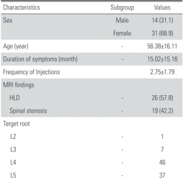
3. Additional analysis of needle tip position and nerve root compression In the total of 45 patients, the TFESI was performed on one spinal nerve root in 13 patients. The needle tips were localized Table 1. Demographic characteristics of the patients (n=45).
Characteristics Subgroup Values
Sex Male 14 (31.1)
Female 31 (68.9)
Age (year) - 56.38±16.11
Duration of symptoms (month) - 15.02±15.16
Frequency of Injections 2.75±1.79
MRI findings
HLD - 26 (57.8)
Spinal stenosis - 19 (42.2)
Target root
L2 - 1
L3 - 7
L4 - 46
L5 - 37
MRI: magnetic resonance imaging, HLD: herniated lumbar disc.
Table 2. Number of spinal levels during injections and classification of needle tip position.
All one level 2 level 4 level Number of Patients 45 13 (28.9) 25 (55.6) 7 (15.6) Needle tip positions
Quadrant 1 68 (74.7) 10 37 21
Quadrant 2 19 (20.9) 3 13 3
Quadrant 3 4 (4.5) 0 0 4
Quadrant 4 0 0 0 0
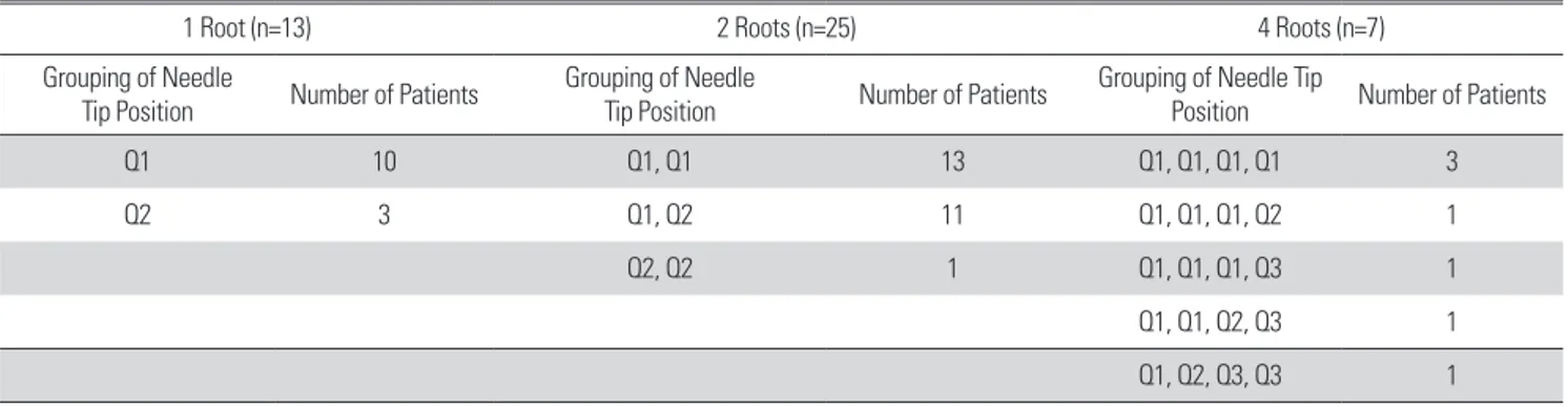
in the quadrant 1 of 10 patients and in the quadrant 2 of 3 patients. In 32 patients, multi-level TFESIs were performed in 16 patients belonging to group A, and in 16 patients classified under group B. Analysis of 16 patients in group B showed that 15 of them had the needle tip at least in the quadrant 1 (Table 3). For the nerve root compression, 29 patients belonged to Compression group 1 and the remainder of 16 patients were in Compression group 2. It was not showed in a table form.
4. The short-term effectiveness of TFESI using a NS regardless of other variables
Two weeks after the injection, significant decrease in ODI,
BPI, and RP score were noted in the all patients, group I, and group II without regard to needle tip position (p<0.05) (Fig.
5). In all the patients, before the injection, mean scores of outcome measurement obtained in total ODI, severity of BPI, interference of BPI, total BPI, and RP were 18.22±7.985, 18.53
±6.986, 34.82±14.562, 53.36±20.357, and 100 respectively.
After the injection, the mean scores were 12.62±7.958, 12.64
±8.488, 22.33±15.693, 34.98±23.468, and 46.22±25.342 respectively. In group I (13 patients), before the injection, mean scores of outcome measurement obtained in total ODI, severity of BPI, interference of BPI, total BPI, and RP were 16.85±
5.031, 18.69±4.090, 38.46±12.745, 57.15±16.041, and 100 respectively. After the injection, mean scores were 7.46±
4.095, 9.00±7.360, 15.54±13.605, 24.54±20.177, and 35.77
±29.144 respectively. In group II (32 patients), before the injection, mean scores of outcome measurement obtained in total ODI, severity of BPI, interference of BPI, total BPI, and RP were 18.78±8.922, 18.47±7.923, 33.34±15.176, 51.81±
21.911, and 100 respectively. After the injection, mean scores were14.72±8.228, 14.13±8.571, 25.09±15.833, 39.22±
23.661, and 50.47±22.767 respectively. The Wilcoxon signed rank test and the paired T-test indicated a significant decrease in ODI, BPI, and RP scores (all p<0.05)
5. In consideration of other variables, treatment effects in the one level TFESI (group I)
In 13 patients, only one spinal level was injected during the procedure. When compared with the baseline, outcome measurement was improved in both needle tip positions. The changes in ODI and BPI were statistically greater and RP was statistically more improved in patients who had their Table 3. Additional analysis of needle tip position
1 Root (n=13) 2 Roots (n=25) 4 Roots (n=7)
Grouping of Needle
Tip Position Number of Patients Grouping of Needle
Tip Position Number of Patients Grouping of Needle Tip
Position Number of Patients
Q1 10 Q1, Q1 13 Q1, Q1, Q1, Q1 3
Q2 3 Q1, Q2 11 Q1, Q1, Q1, Q2 1
Q2, Q2 1 Q1, Q1, Q1, Q3 1
Q1, Q1, Q2, Q3 1
Q1, Q2, Q3, Q3 1
Q: quadrant.
Fig. 5. Mean scores of outcome measurements before and 2 weeks after injection in all patients, group I, and group II. Values are mean±SD. *p
<0.05.
*Comparisons between before and 2 weeks after injection were per- formed using the Wilcoxon signed-rank test for group I and the paired t-test for group II and all patients.
Before 2 weeks Before 2 weeks Before 2 weeks Before 2 weeks Before 2 weeks ODI (total) BPI (severity) BPI (interference) BPI (total) RP
* *
*
* all patients *
Group I Group II 100
80 60 40 20 0
needle position in quadrant 1 (Table 4). The grade of nerve root compression did not affect the results of TFESI (Table 5). However, the change in each outcome measurement varied according to the grade of nerve root compression. The change in ODI and BPI were greater in the grade III and II, respectively, and the RP was greater in the grade I of nerve root compression.
6. In consideration of other variables, treatment effects in the all patients and multi-level TFESI (group II) By using simple linear regression, we analyzed the possible predictors of outcome for ODI, BPI, and RP to determine the effectiveness of TFESI using NS in the all patients and group II. In the all patients, based on the needle tip position, group A showed a significantly better therapeutic effect than group B (p<0.05) (Table 6). Multi-level TFESIs (group II) was conducted in the 32 patients by injecting two or four spinal levels during the procedure. No significant differences were seen in terms of ODI, BPI and RP between group A (i.e., with needle position in quadrant 1 at all the injected levels) and group B (Table 7). In all the patients and group II, we found
a significant correlation between nerve root compression on MRI and ODI scores. However, no significant correlation was found between other outcome measures such as BPI and RP.
No correlations were found between outcome measurement and other investigated factors, such as symptom duration and injection frequency. Our results for the nerve root compression showed an interesting trend suggesting results similar to those involving only one spinal level TFESI (group I). The changes in ODI and BPI were greater in the compression group 2 and the RP was greater in the compression group 1 in the all patients and group II (Table 6 and 7).
7. Safety
No patients experienced severe side effects. One patient (group II) developed a slight headache after injection. This symptom soon got better.
Discussion
We analyzed the effectiveness of TFESI using a NS in 45 patients. Our results suggest a significant decrease in ODI, Table 4. Clinical assessment after 2 weeks in the group I
Quadrant 1 (n=10) Quadrant 2 (n=3) p
Change of ODI (total) 9.40±5.948 2.67±1.528 <0.042
Change of BPI (severity) 11.90±5.820 2.33±3.215 <0.027
Change of BPI (interference) 29.10±14.768 2.33±2.082 <0.028
Change of BPI (total) 41.00±19.247 4.67±4.726 <0.018
RP 24.50±19.501 73.33±25.166 <0.021
*p-value <0.05.
ODI: Oswestry Disability Index, BPI: Brief Pain Inventory, RP: residual pain.
Table 5. Difference of treatment efficacy according to magnetic resonance imaging (MRI) grade in the group I
NRC Gr I NRC Gr II NRC Gr III p
Change of ODI (total) 7.29±5.648 8.40±7.603 9.00±0.000 0.895
Change of BPI (severity) 8.29±6.237 13.00±6.892 3.00±0.000 0.306
Change of BPI (interference) 16.43±14.965 30.80±20.241 29.00±0.000 0.349
Change of BPI (total) 24.71±20.164 43.80±27.031 32.00±0.000 0.460
RP 42.86±32.514 25.00±26.926 40.00±0.000 0.401
*p-value <0.05.
NRC: nerve root compression, Gr: grade, ODI: Oswestry Disability Index, BPI: Brief Pain Inventory, RP: residual pain.
Table 6. Simple linear regression analysis for the possible predictors of outcome for Oswestry Disability Index, Brief Pain Inventory, and residual pain as the effectiveness of transforaminal epidural steroid injection using a nerve stimulator in the all patients
Factor OM Estimate SE 95% CI p
Needle tip position
Group A versus ODI -3.225 1.419 -6.006 - -0.443 <0.028
Group B BPI -11.091 5.234 -21.351 - -0.832 <0.040
RP -16.559 7.313 -22.224 - 3.893 <0.029
Nerve root compression
Compression group 1 versus ODI 3.724 1.442 0.898 - 6.550 <0.013
Compression group 2 BPI 8.856 5.513 -1.950 - 19.661 0.116
RP -13.534 7.712 -28.650 - 1.581 0.086
Duration of Symptoms ODI -0.022 0.020 -0.061 - 0.018 0.293
BPI -0.088 0.074 -0.233 - 0.056 0.238
RP 0.074 0.105 -0.132 - 0.280 0.483
Frequency of injections ODI -2.089 0.709 -3.479 - -0.699 0.105
BPI -4.377 2.768 -9.802 - 1.048 0.121
RP 2.899 3.980 -4.902 - 10.700 0.470
*p-value <0.05.
OM: outcome measurement, SE: standard error, CI: confidence interval, ODI: Oswestry Disability Index, BPI: Brief Pain Inventory, RP: residual pain.
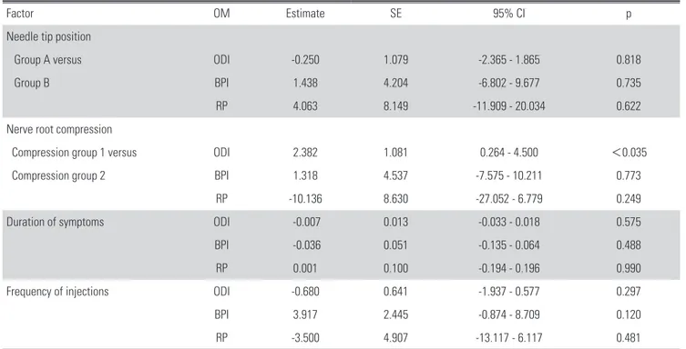
Table 7. Simple linear regression analysis for the possible predictors of outcome for Oswestry Disability Index, Brief Pain Inventory, and residual pain as the effectiveness of transforaminal epidural steroid injection using a nerve stimulator in the group II
Factor OM Estimate SE 95% CI p
Needle tip position
Group A versus ODI -0.250 1.079 -2.365 - 1.865 0.818
Group B BPI 1.438 4.204 -6.802 - 9.677 0.735
RP 4.063 8.149 -11.909 - 20.034 0.622
Nerve root compression
Compression group 1 versus ODI 2.382 1.081 0.264 - 4.500 <0.035
Compression group 2 BPI 1.318 4.537 -7.575 - 10.211 0.773
RP -10.136 8.630 -27.052 - 6.779 0.249
Duration of symptoms ODI -0.007 0.013 -0.033 - 0.018 0.575
BPI -0.036 0.051 -0.135 - 0.064 0.488
RP 0.001 0.100 -0.194 - 0.196 0.990
Frequency of injections ODI -0.680 0.641 -1.937 - 0.577 0.297
BPI 3.917 2.445 -0.874 - 8.709 0.120
RP -3.500 4.907 -13.117 - 6.117 0.481
*p-value <0.05.
OM: outcome measurement, SE: standard error, CI: confidence interval, ODI: Oswestry Disability Index, BPI: Brief Pain Inventory, RP: residual pain.
BPI, and RP scores were noted in the all patients, group I, and group II regardless of other variables such as needle tip position or nerve root compression after the injection.
When considering needle tip position, in 13 patients with only one spinal level, significant decrease in ODI, BPI, and RP scores were observed after 2 weeks. Especially there was a statistically better therapeutic effect in quadrant 1 compared with quadrant 2. In all the patients, group A had a significantly better therapeutic outcome compared with group B. However, in 32 patients with multi-level TFESIs, no significant difference was found in the outcomes measured between group A and group B. Twenty of the 40 needle tip positions in group B were located in quadrant 1 and only a single patient showed both the needle positions located in quadrant 2. Therefore, except for the single patient, 15 patients who were treated with multi- level TFESIs, had their needle located in quadrant 1 in at least one to three TFESIs during the procedures. The absence of statistical significance of the needle position in group B may have been caused by the abundance of needle tip locations in quadrant 1.
In the present study, we analyzed the possible predictors of outcome for ODI, BPI, and RP scores as a measure of effectiveness of TFESI using NS in the all patients and group II, via multiple linear regression analysis. It was not described in the results due to the lack of statistical significance. However, the analysis showed that group A had a lower physical disability (decrease in ODI score by 2.107; 95% confidence interval [CI], -4.932-0.717), lower status of pain (decrease in BPI score by 9.728; 95% CI, -21.059-1.604), and more pain relief (decrease in RP score by 12.995; 95% CI, -23.425- 29.415) versus group B in the all patients. Group A showed a higher physical disability (increase in ODI score by 0.265;
95% confidence interval [CI], -1.960-2.490), higher status of pain (increase in BPI score by 3.015; 95% CI, -6.067- 12.097), and higher pain relief (decrease in RP score by 1.899;
95% CI, -20.194-16.397) compared with group B in the multi-level TEFSIs (group II). Consequentially, group A had a better therapeutic effect than group B although the multiple linear regression analysis showed no significant difference in the all patients probably due to a small sample size. In the group II, no statistically significant difference was found in the outcome measurement between group A and group B in the simple and multiple regression analysis. This results may
be attributed to the abundance of needle tip positions located in the quadrant 1 of group B. Considering the dosage-based analysis of triamcinolone, multi-level TFESIs (group II) was performed in the 32 patients by injecting two or four spinal levels. The triamcinolone of total 1520 and 1600 mg was used in the group A (16 patients) and the group B (16 patients) respectively. The average usage per patient of each group was 95 and 100 mg, so there was little difference between the two groups.
From the perspective of whether multi-level TFESIs is needed, there may be a gain from performing a 2-level TFESI because a single spinal segment may not be identified as the lone pain generator. Especially symptoms may not match the dermatomal maps exactly, targeting both the cephalad and caudad nerves of the pathologic disk may better address the etiology of pain. Our study is based on the inclusion criteria of patients who complained of unilateral or bilateral radiating pain and with root compromise involving more than 2 levels on MRI. So multi-level TFESIs was inevitable.
The ST described by Bogduk16) corresponds to the quadrant 1 and 2. Our results suggest that quadrant 1 is more effective for the pain relief. Wolff et al12) used an electrical current generated by a radiofrequency pulse and lesion generator system to optimize the position of the needle tip with regard to the DRG. The majority (27) of all needle tips (39) were localized in the lateral upper half (quadrant 1) of the intervertebral foramen. Pfirrmann et al17) also suggested that the lateral part of the ST, which corresponds to quadrant 1 in our study was the best site for the needle tip location because of less treatment-induced pain. Therefore, our study found that lateral position of needle in the ST yields better results with less pain consistent with results of Pfirrmann et al17).
The degree of nerve root compression can affect the effectiveness of the TFESI. Choi et al15) reported that grade II or III of nerve root compression showed more unsatisfactory results than the grade I of nerve root compression. However, in our study, the grade of nerve root compression had no effect on the performance of TFESI in the group I, II, and all patients.
The higher the grade of nerve root compression, the greater the change in ODI and BPI, and the RP was greater in the grade I of nerve root compression. Choi et al15) used a visual numeric pain scale (similar to RP) to see the effectiveness of the TFESI.
There was a similar result in case of RP score. The ODI and
BPI scores were mostly used to evaluate not only the patient’
s pain but also the patient’s disability or functional status of patients.13,14) These outcome measurements is not specific to the patient with radicular symptoms. Sirvanci et al18) showed the absence of correlation between ODI score and nerve root compression on MRI. Further studies concerning the correlation between grade of nerve root compression and a few outcome measurements are needed.
We chose the follow-up of 2 weeks to evaluate the effect of needle tip position on the short-term effectiveness of TFESI using the NS in light of published studies related to the duration of therapeutic effect of corticosteroids. Most investigations involving the duration of pain relief after spinal steroid injections had short- or mid-term follow-up of as long as 3 months.19) Considering the chronic and fluctuating nature of radicular pain, the short-term benefit of epidural corticosteroid injections should be weighed against the latent risks including the systemic effects due to corticosteroids, and radiation exposure associated fluoroscopic guidance.
In our experience, therapeutic TFESI of the lumbar spine can be carried out most conveniently with fluoroscopic guidance.
These fluoroscopy-guided injections have been proven to be safe and rapid in the axial skeleton.20-22) However, even in the hands of a skilled and experienced specialist, the needle is not targeted in the precise direction during the procedure through the ST region. In a few cases, intraneural injection or nerve root penetration by a needle have been reported. In this study, according to the injection approach proposed by Wolff et al12), the pedicle was bisected on a posterior-anterior view and the needle was initially localized in the lateral segment by advancing into the ST. We used the NS to optimize the position of the needle tip with regard to the DRG. The needle tip position was verified by the induction of tingling sense in patients’ corresponding dermatome. Thus, we were able to locate the needle tip as close as possible to the DRG without injuring the nerve root.
The study has several limitations although our findings are likely valid. First, the number of patients was not sufficient.
Power analysis was acquired with Power Analysis and Sample Size Software (PASS, version 12). A sample size of 45 achieves 60% power to detect changes in slope from 0.00 under the null hypothesis to 3.22 under the alternative hypothesis when the standard deviation of the X is 0.49, the standard deviation of Y
is 4.92, and the two-sided significance level is 0.05. Second it is not a controlled, randomized prospective study. Thus, it has the limitations of a retrospective study. Third, other factors such as age, sex, target root, MRI findings, and electromyography findings that probably influenced the short-term effectiveness were not included. This study focused to the effect of needle tip positions on the short-term effectiveness of TFESI using a NS.
Further studies need to be conducted as long-term attempts to overcome such limitations.
Conclusions
Despite these limitations, our study demonstrates the effect of needle tip position on lumbar TFESI using NS. There was a better effect in the quadrant 1 than quadrant 2. No specific short-term side effects were reported in any of the patients.
Electrical stimulation to guide TFESI in addition to fluoroscopy can be a useful and safe therapeutic tool for patients with radicular symptoms.
REFERENCES
1. Botwin KP, Gruber RD, Bouchlas CG, Torres-Ramos FM, Sanelli JT, Freeman ED, et al. Fluoroscopically guided lum- bar transformational epidural steroid injections in degenera- tive lumbar stenosis: an outcome study. American journal of physical medicine & rehabilitation 2002;81(12):898- 905. DOI: 10.1097/00002060-200212000-00003.
2. Slipman CW, Chow DW. Therapeutic spinal corticosteroid injections for the management of radiculopathies. Physi- cal medicine and rehabilitation clinics of North America 2002;13(3):697. DOI: 10.1016/s1047-9651(02)00004-9.
3. Vad VB, Bhat AL, Lutz GE, Cammisa F. Transforaminal epidural steroid injections in lumbosacral radiculopathy:
a prospective randomized study. Spine 2002;27(1):11-5.
DOI: 10.1097/00007632-200201010-00005.
4. Husemeyer R, White D. Topography of the lumbar epi- dural space. Anaesthesia 1980;35(1):7-11. DOI: 10.1111/
j.1365-2044.1980.tb03712.x.
5. Goodman BS, Bayazitoglu M, Mallempati S, Noble B, Gef- fen J. Dural puncture and subdural injection: a complication of lumbar transforaminal epidural injections. Pain Physician 2007;10(5):697.
6. Hamanishi C, Tanaka S. Dorsal root ganglia in the lum- bosacral region observed from the axial views of MRI.
Spine 1993;18(13):1753-6. DOI: 10.1097/00007632- 199310000-00006.
7. Derby R, Kine G, Saal JA, Reynolds J, Goldthwaite N, White AH, et al. Response to steroid and dura- tion of radicular pain as predictors of surgical outcome.
Spine 1992;17(6):S176-S83. DOI: 10.1097/00007632- 199206001-00020.
8. TAJIMA T, FURUKAWA K, KURAMOCHI E. Se- lective lumbosacral radiculography and block. Spine 1980;5(1):68-77. DOI: 10.1097/00007632-198001000- 00013.
9. Murphey F. Sources and patterns of pain in disc disease.
Clinical neurosurgery 1968;15:343. DOI: 10.1093/neuro- surgery/15.cn_suppl_1.343.
10. Steindler A, Luck J. Differential diagnosis of pain low in the back. Journal of the American Medi- cal Association 1938;110(2):106-13. DOI: 10.1001/
jama.1938.02790020020007.
11. Haynsworth R. Selective nerve root blocks: a new technique using electrical stimulation. Pain Physician 2003;6(4):517- 20.
12. Wolff AP, Groen GJ, Wilder-Smith OH. Influence of needle position on lumbar segmental nerve root block selectivity.
Regional anesthesia and pain medicine 2006;31(6):523-30.
DOI: 10.1016/j.rapm.2006.07.008.
13. Fairbank JC, Pynsent PB. The Oswestry disability index.
Spine 2000;25(22):2940-53. DOI: 10.1097/00007632- 200011150-00017.
14. Yun YH, TR RM, Heo DS, Yoo T, Heo BY, Park H-A, et al. Development of a cancer pain assessment tool in Korea: a validation study of a Korean version of the brief pain inventory. Oncology 2004;66(6):439-44. DOI:
10.1159/000079497.
15. Choi S-J, Song JS, Kim C, Shin MJ, Ryu DS, Ahn JH, et al. The use of magnetic resonance imaging to predict the clinical outcome of non-surgical treatment for lumbar in- terverterbal disc herniation. Korean Journal of Radiology 2007;8(2):156-63. DOI: 10.3348/kjr.2007.8.2.156.
16. Bogduk N. Clinical anatomy of the lumbar spine and sa- crum: Elsevier Health Sciences; 2005.
17. Pfirrmann CW, Oberholzer PA, Zanetti M, Boos N, Trudell DJ, Resnick D, et al. Selective Nerve Root Blocks for the Treatment of Sciatica: Evaluation of Injection Site and Ef- fectiveness—A Study with Patients and Cadavers1. Radiolo- gy 2001;221(3):704-11. DOI: 10.1148/radiol.2213001635.
18. Sirvanci M, Bhatia M, Ganiyusufoglu KA, Duran C, Tezer M, Ozturk C, et al. Degenerative lumbar spinal stenosis:
correlation with Oswestry Disability Index and MR imag- ing. European spine journal 2008;17(5):679-85. DOI:
10.1007/s00586-008-0646-5.
19. Jamison RN, VadeBoncouer T, Ferrante FM. Low back pain patients unresponsive to an epidural steroid injec- tion: identifying predictive factors. LWW; 1991. DOI:
10.1097/00002508-199112000-00010.
20. Link SC, El-Khoury GY, Guilford WB. Percutane- ous epidural and nerve root block and percutane- ous lumbar sympatholysis. Radiologic Clinics of North America 1998;36(3):509-21. DOI: 10.1016/s0033- 8389(05)70040-3.
21. El-Khoury G, Ehara S, Weinstein JN, Montgom- ery W, Kathol M. Epidural steroid injection: a pro- cedure ideally performed with fluoroscopic control.
Radiology 1988;168(2):554-7. DOI: 10.1148/radiol- ogy.168.2.2969118.
22. Dussault RG, Kaplan PA, Anderson MW. Fluoroscopy- guided sacroiliac joint injections. Radiology 2000;214(1):273- 7. DOI: 10.1148/radiology.214.1.r00ja28273.
신경 자극기를 사용하여 요추 선택적 추간공 경막외 스테로이드 주입술: 단기간의 유효성에서 바늘 끝 위치 의 효과
이시욱 • 이창한*
서울대학교 의과대학 서울보라매병원 재활의학과교실, *경상대학교병원 재활의학과교실 연구 계획: 후향적 연구
목적: 우리는 신경 자극기를 이용한 선택적 추간공 경막외 스테로이드 주입술의 단기간의 유효성에서 바늘 끝 위치의 영향을 결정하는 것이었다.
선행 연구문헌의 요약: 신경 자극기는 신경근에 가까운 바늘 위치를 가능하게 하며, 바늘의 근접성이 추간공 경막외 스테로이드 주입술의 성능에 영향을 미치는지 여부는 알려져 있지 않다.
대상 및 방법: 45명의 환자들은 주사된 신경근의 수에 따라 분류되었다. 그룹 I는 단일 척수 신경근에서 주사를 시행한 환자를 포함시켰고 나머지는 그 룹 II에 속했다. 바늘 끝 위치는 추간공을 4 사분명으로 나누어 평가하였다. A 그룹은 주사된 척수 신경근의 모든 바늘 끝이 사분면 1에 있을 때를 말하며, 최소한 하나의 척수 신경근에서 사분면 2에서 사분면 4에 바늘 끝의 위치가 있을 때를 B 그룹으로 하였다. Oswestry Disability Index (ODI)와 Brief Pain Inventory (BPI) 및 residual pain (RP)를 평가하였다.
결과: 2주 후에, ODI와 BPI 및 RP 점수의 현저한 감소가 모든 환자와 그룹 I 및 그룹 II에서 나타났다. 다른 변수를 고려했을 때, 그룹 I의 경우, 사분면 1에 바늘 위치를 가졌던 환자에서 ODI와 BPI 점수의 변화가 현저하게 크게 나타났으며, RP 점수는 통계적으로 더욱 향상되었다. 모든 환자에서, A 그룹은 B 그룹보다 현저한 치료적인 효과를 가졌다. 그룹 II에서는, A 그룹과 B 그룹 사이에서 통계적인 차이가 없었다.
결론: 우리는 연구는 사분면 2보다 사분면 1에서 보다 나은 효과를 보여 주었으며, 전기 자극기를 이용한 추간공 경막회 스테로이드 주입술이 유용한 도 구이다.
색인 단어: 주사, 바늘, 자극기, 방사통
약칭 제목: 경막외 주사에서 바늘 끝 위치의 효과
접수일: 2020년 9월 21일 수정일: 2020년 10월 12일 게재확정일: 2021년 2월 2일 교신저자: 이창한
경상남도 진주시 강남로 79 경상대학교병원 재활의학과교실 TEL: 055-750-9639 E-mail: [email protected]