A Comparative Study of Korean mistletoe lectin and bee venom on mechanism in inducing apoptosis of Hep G2,
a liver cancer cell
Seong-Woo Lim
Department of Korean Internal Medicine, Ilsan Oriental Hospital of Dongguk University.
Original Article
⋅Received:20 December 2018 ⋅Revised:26 December 2018 ⋅Accepted:26 December 2018
⋅Correspondence to: Seong-Woo Lim
Dept of Korean Internal Medicine, Ilsan Oriental Hospital of Dongguk University Sik sa-dong Dog-gu Il san Go-yang, Republic of Korea
Tel: +82-31-961-9043, E-mail: omdlsw@naver.com
Objectives: The objective of this study is Korean mistletoe lectin (Viscum album coloratum agglutinin, VCA) and bee venom (BV) to experimental prove comparative study of VCA and BV on the anti-cancer effect and mechanisms of action.
Methods: In this study, it was examined in a human hepatocellular carcinoma cell line, Hep G2 cells. Cytotoxic effects of VCA and BV on Hep G2 cells were determined by 3- (4, 5-dimethylthiazol-2-yl) -2, 5-diphenyltetrazolium bromide (MTT) assay in vitro. VCA and BV killed Hep G2 cells in a time- and dose-dependent manner.
Results: The apoptotic cell death was then confirmed by propidium iodide staining and DNA fragmentation analysis.
The mechanisms of action was examined by the expression of anti-apoptotic proteins and activation of mitogen-activated protein kinases. Treatment of Hep G2 cells with VCA activated poly (ADP-ribose) polymerase-1 (PARP-1) known as a marker of apoptosis, and mitogen-activated protein kinases signaling pathways including SAPK/JNK, MAPK and p38. BV also activated PARP-1, MAPK, p38 but not JNK. The expression level of anti-apoptotic molecule, Bcl-X, was decreased by VCA treatment but not BV. Finally, the phosphorylation level of ERM proteins involved in the cytoskeleton homeostasis was decreased by both stimuli.
Conclusion: We examined the involvement of kinase in VCA or BV - induced apoptosis by using kinase inhibitors. VCA-induced apoptosis was partially inhibited by in the presence
Key Words : Mistletoe, Bee venom, Apoptosis, Liver cancer
Introduction
The rate of liver cancer in Korea is estimated at 30.5 per 100,000 men and 7.6 per 100,000 women, and the third annual report on the Korea Central Cancer Registration Project showed that it was the seventh for men and the seventh for women, which was higher than for other OECD countries
1),2).
The mistletoe is a plant parasitic on shrubs that
has long been used as a medicinal plant, and attempts to use it in malignant tumors have recently been made using the extract
3).
Mistleto extracts are known to cause changes in
the human immune system, and because of these
immune-control effects, malignant tumors such as
malignant lymphoma, multiple myeloma, leukemia are
treated, malignant tumors are intended to prevent
recurrence after primary treatment, or pre-cervative
tumors such as chronic hepatitis B, and primary bone treatment of patients
4).
The anti-cancer effects on Be Venom were actively implemented both at home and abroad, and the effects of cancer were reported due to the ingredients of Bong-dock, immunotherapy, pain, and so on. Be Venom is peptide components (melithin, adolapin, MCD sensitive, etc.), enzymes (PLA2, Hyaluronidase, Glucosidase, etc.), amines (histamin, epinephrine, etc.), non-ptitious components Beef poison is 1.3 percent, ph 5.2 percent bitter, weak-tasting, and 75 percent protein
5).
In this study, we report that mistletoe and Be Venom were carefully studied to induce apoptosis of Hep G2, the liver cancer cell owner.
Material and Methods
1. Preparations of bee venom
1g of the poison Apis Melipera ligustica (West honey bee, Sigma, USA) was dissolved in the poison distilled water.
The mistletoe was purchased from Viscum album colorectum agglutin, VCA (Related per Cheongun, South Gyeongsang and South Korea) and used as the extraction method below.
2) Preparation of a mistleto extract solution After grinding the leaves and stems of mistletoe into water at room temperature for 48 hours, the supernatant obtained by filtering them to freeze and dry them. After 96 hours, 2.5g was filtered and stored at minus 80°C.
2. Method
1) Cellular stock and cell culture
The hepatoma cell line and the Hep G2 cell used in the experiment were purchased from the American
Type Culture Collection (Rockville, MD). Cells were incubated using Dulbecco's modified Eagle's Medium (DMEM, Invitrogen, USA) containing 10% Fetal Bovine Serum (FBS; Invitrogen, USA) at 37°C and 5% CO2 incubator. To prevent contamination, 100ut/ml phenicillin, 100ug/ml streptomin (Gibco/BRL, USA) was added as an antibiotic. Trypsin-EDTA (Gibco/BRL, USA) was treated and stratified. The badge was exchanged every two or three days.
2) Cellular activity measurement (MTT allay) MTT ((4,5-dimethylthiazol-2-yl)-2,5-diphenyltrazolium bromide, Sigma) alay was conducted using Sladowski's method. MTT assay is a test method that utilizes the ability of cells to reduce MTT titrazol, a yellow soluble substrate, to a green-colored MTT FORMazan by dehydrogen enzyme. The absorption of MTT formazan is greatest at a wavelength of 580nm, where the absorbance measured is reflected in the oxidative power of a cell that is alive and has a strong metabolism
6).
MTT assembly was first incubated for 24 hours after 16 hours of Hep G2 cells (5x10³ cells/well) in a 12 cell plate. And 3-(4-Dimethylthiazol-2-yl)-2,5-diphenyl- tetra-zolium bromide (MTT). Sigma) 2 mg/ml was added, incubated in the incubator for 4 hours, then dissolved with dimethyllsulfoxide (DMSO; Sigma) and measured absorbency with a microplate reader (Molecular devices, USA) at a wavelength of 580nm to calculate cell survival.
Cell viability (%) is defined as: If the value of the normal range is controlled and the O.D. value is defined as 100 percent survival of the cell, and the value of the O.D. measured by the rest of the group is calculated as follows.
Cell Viability = (experiment / control group)
3) Medication of inhibitors
PH3K inhibitor (LY2942), p38 MAPK inhibitor
(SB203580), MAPK inhibitor (PD98059), and JNK inhibitor (SP600125) (Calbiochem, USA) treated the extraction and sterilization rates before the end of 30 minutes and then treated them with the MTT method.
4) Western blot analysis method
All cell solvents were made by boiling cells within a sample buffer (66.5 mmol/1 Tris-HCl, pH 6.8, 2%
SDS, 20% glycerol, 10% 2-mercaptoethanol). Protein doses were used in the bicinchonic acid (BCA, Pierce) method. 50 ug of the quantified protein sample was separated by 4-12% sodium diecylsulfate-polyacrylamide gradient gel (Invitrogen) and transferred to the nitrocellulose paper (Amersham). It was confirmed that the protein was completely transferred by dyeing the membrane of protein with Ponceau-S. After washing it with a Tris-buffered saline (TBS-T) containing 0.1%
Tween 20, the protein was blocked with 5% de-fat milk solution for more than 30 minutes. p38 (p-p38), p38, MAPK (p-MAPK), MAPK, ERM (p-EMM), moesin, JNK (p-JNK), JNK antibodies, PARP-1, Bcl-x, and activated (Cell signing) are in the USAch After the second antibody reaction, the curtain was washed and the protein that they wanted was visualized through the advanced chemical protection system (ECL, Pierce).
Visualization and quantitative analysis of proteins was done using image equipment (LAS-3000, Fujitsu
5) DNA fragmentation
10 ug/ml of BV and 50 ug/ml of VCA beef were processed in Hep G2 cells (5x10⁵ cells), then cells were recovered for 16 hours and 24 hours respectively, centrifugation was performed for 5 minutes at 800 rpm, 10 mM Tris (pH7.4), 10 m EDTA (pH 8.0), and the floating solution was filled with a buffer of 10 mM (X00-1). Add 20 mg/l of RNase A and 20 mg/l of Proteinase K to react for another hour at 37 °C and
centrifugation to obtain genomic DNA. Add TE buffer (10mM Tris (pH7.4) and 10mM EDTA (pH 8.0) to dissolve DNA and electro-activate the 1% agarose gel to check the band.
6) FACS Analysis
After processing 10 ug/ml of BV and 50 ug/ml of VCA milk fat in the Hep G2 cell (2x10⁶ cells), recover the cells each 24 hours, wash them with cold PBS, and secure them at room temperature for 30 minutes.
Then, add 0.05 mg/ml of RNase A to remove RNA and react for one hour at 37°C. Then, add 50g/ml of Propium iodide (PI) to the specimen and react in the dark for 30 minutes. DNA analysis used ELITE ESP flow rateometer (Bekman-Coulter Inc., USA).
7) Data analysis and statistical processing All measurements were expressed as the mean value standard deviation (mean ± S.D.) and statistical analysis among each group of experiments performed one-way ANOVA of the SPSS program for windows, and post-verification was verified by Tukey test. The statistical significance of the whole experiment was recognized as significant if the p value was less than 0.05.
Result
1. Effect of Mistleto (VCA) on the survival rate of liver cancer cells
The cell survival rate was observed 24 hours and
48 hours after mistletoe was treated by concentration
to investigate the effect of mistletoe on the survival
rate of liver cancer cells. Concentration-dependent
degradation from 10 ug/ml to 200 ug/ml resulted in
increased apoptosis after 48 hours (Figure 1).
Fig. 1. Dose dependent cytotoxicity effect of VCA in Hep G2 cells. Hep G2 cells were exposed to different concentration of 10-200 ug/ml of VCA. Cell viability was measured by MTT assay at 24 h and 48 h after VCA addition.
Data, expressed as percentage of control (Con), are the mean ±SEM of three separate experiments. *p<0.05 vs control, **p<0.01 vs control
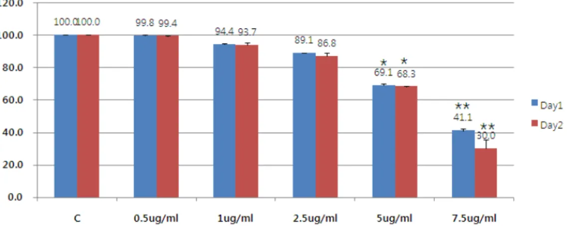
Fig. 2. Dose dependent cytotoxicity effect of BV in Hep G2 cells. Hep G2 cells were exposed to different concentration of 0.5-7.5 ug/ml of BV. Cell viability was measured by MTT assay at 24 h and 48 h after BV addition. Data, expressed as percentage of control (Con), are the mean ±SEM of three separate experiments. *p<0.05 vs control, **p<0.01 vs control
2. The Effect of Bovine (BV) on the Survival Rate of Liver Cancer Cells
Mistletoe used the previously well-known anti-cancer drug to compare and study the anti-cancer effect in liver cancer cells. The MTT method observed the effect on the survival rate of cells 24 hours and
48 hours after the poison was treated with liver cancer
cells by concentration. Bong-dock significantly killed
cells from a concentration of 5 ug/ml and at a
concentration of 7.5 u/ml, more than 50% of the cells
were killed (Figure 2).
Fig. 3. Electrophoretic analysis of genomic DNA of Hep G2 cells treated with VCA (50 ug/ml) and BV (10 ug/ml). Cells were incubated for 16 and 24 h with or without VCA and BV. The genomic DNA was analysed by electrophoresis on a 1 % agarose gel containing SYBR green.
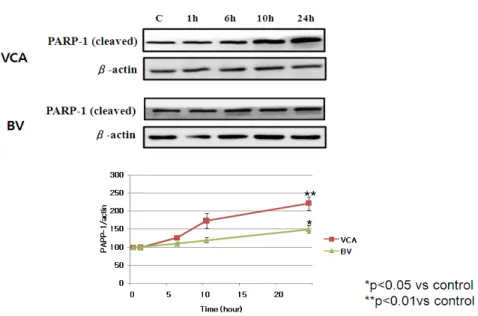
Fig. 4. Effect of VCA and BV on the expression of cleaved PARP-1 in Hep G2 cells. Hep G2 cells treated with VCA (50 ug/ml) and BV (10 ug/ml) for the indicated time. After that, whole cell lysates were electrophoresed in SDS-PAGE and analyzed by immunoblotting with anti-cleaved PARP-1 or actin antibody. The intensity of cleaved PARP-1 and and actin bands were quantitated by densitometric analysis, and the amounts of cleaved PARP-1 were normalized versus actin. The data represent the means ± S.E. of three independent experiments. C:
Unstimulated cells.
3. Mystery mistletoe and Bongtoe are the two most successful liver cancer cells in Korea.
1) DNA fragmentation
The electroactivity was used to observe if chromosome DNA in the nucleus was cut to see if the apoptosis of the liver cancer cells in mistletoe and Bongto was achieved. We were able to see chromeoseome DNA cut into a ladder in 16 hours and 24 hours by Bong-dok and Mistleto (Figure 3).
2) PARP-1 expression
Electrostatic activity was used to observe that the nucleus was cut off when mistletoe and poison induces apoptosis, and it was confirmed that the PARP-1 was activated. PARP-1 increased at activation by a small band of 29 kDa and observed that the cut band of
PARP-1 by mistletoe and mutilation increased over
time (Figure 4).
*p<0.05 vs control **p<0.01 vs control
Fig. 5. Effect of VCA on the expression of Bcl-x in Hep G2 cells. Hep G2 cells treated with VCA (50 ug/ml) for the indicated time. After that, whole cell lysates were electrophoresed in SDS-PAGE and analyzed by immunoblotting with anti-Bcl-x or actin antibody. The intensity of Bcl-xand and actin bands were quantitated by densitometric analysis, and the amounts of Bcl-x were normalized versus actin.
The data represent the means ± S.E. of three independent experiments. C: Unstimulated cells.
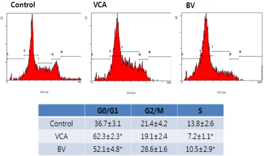
Fig. 6. Effect of VCA and BV on the cell cycle of Hep G2 cells. Hep G2 cells treated with VCA (50 ug/ml) and BV (10 ug/ml) for 9 h and then fixed and stained with PI. *p<0.05 vs control.
3) Bcl-x expression
Observing the accents of Bcl-x on a time-by-hour basis after mistreatment, mistletoe showed that only Mistletoe significantly decreased the amount of Bcl-x from six hours to 24 hours. These results suggest a different system of cell death signal transmission between Bong-dok and Mislotto (Figure 5).
4) FACS analysis
After the treatment, cells were fixed at 9 hours
and the nucleus was dyed to observe the distribution
by cell cycle using flow chromometer. The number
of cells in the G0/G1 phase has increased statistically
significantly and the number of newly synthesized
Sphases has also decreased by treatment of mistletoe
and poison. These results suggest that mistletoe and
bovine may also be partially involved in arresting the
cell cycle of G0/G1 (Figure 6).
*p<0.01 vs control.
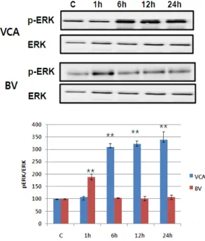
Fig. 7. VCA and BV induces ERK phosphorylation in Hep G2 cells. Hep G2 cells were seeded in 6 well plates and were stimulated with 50 ug/ml of VCA and 10 ug/ml of BV for 1, 6, 12, or 24 h. After that, whole cell lysates were electrophoresed in SDS-PAGE and analyzed by immunoblotting with anti-p-ERK or ERK antibody. The intensity of phosphorylation and total ERK bands were quantitated by densitometric analysis, and the amounts of phosphorylated ERK were normalized versus total ERK. The data represent the means
± S.E. of three independent experiments. C:
Unstimulated cells.
*p<0.05 vs control. **p<0.01 vs control.
Fig. 8. VCA and BV induces p38 phosphorylation in Hep G2 cells. Hep G2 cells were seeded in 6 well plates and were stimulated with 50 ug/ml of VCA and 10 ug/ml of BV for 1, 6, 12, or 24 h. After that, whole cell lysates were electrophoresed in SDS-PAGE and analyzed by immunoblotting with anti-p-p38 or p38 antibody. The intensity of phosphorylation and total p38 bands were quantitated by densitometric analysis, and the amounts of phosphorylated p38 were normalized versus total p38. The data represent the means
± S.E. of three independent experiments. C:
Unstimulated cells.
5) Activating MAPK family
The activity of MAPK/ERK, p38 MAPK, and JNK of the proteins reported to increase in activity in the event of mistletoe and bovine killing the liver cancer cells was observed using phosphorylation-specific antibodies
6),7),8). Mistletoe continued for 24 hours from 6 hours after drug treatment, as phosphorylation of
MAPK/ERK, p38 MAPK, and JNK began to increase
three times more than that of controls. On the other
hand, Bong-dok returned to the control level in six
hours after the drug was processed, with the
phosphorylation of MAPK/ERK and p38 MAPK
doubling in just one hour. No activity of JNK was
observed (Figure 7-9).
*p<0.05 vs control. **p<0.01 vs control.
Fig. 9. VCA induces JNK phosphorylation in Hep G2 cells.
Hep G2 cells were seeded in 6 well plates and were stimulated with 50 ug/ml of VCA 10 ug/ml of BV for 1, 6, 12, or 24 h. After that, whole cell lysates were electrophoresed in SDS-PAGE and analyzed by immunoblotting with anti-p-JNK or JNK antibody. The intensity of phosphorylation and total JNK bands were quantitated by densitometric analysis, and the amounts of phosphorylated JNK were normalized versus total JNK. The data represent the means ± S.E. of three independent experiments. C: Unstimulated cells.
*p<0.05 vs control
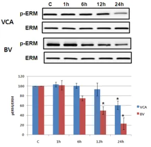
Fig. 10. VCA and BV induces ERM proteins phosphorylation in Hep G2 cells. Hep G2 cells were seeded in 6 well plates and were stimulated with 50 ug/ml of VCA and 10 ug/ml of BV for 1, 6, 12, or 24 h. After that, whole cell lysates were electrophoresed in SDS-PAGE and analyzed by immunoblotting with anti-p-ERM or ERM antibody. The intensity of phosphorylation and total ERM bands were quantitated by densitometric analysis, and the amounts of phosphorylated ERM were normalized versus total ERM. The data represent the means ± S.E. of three independent experiments. C: Unstimulated cells.
6) Activating ERM proteins family
The degree of activity of ERM protein, known to affect the cellular skeleton with another protein related to apoptosis, was performed as immunosuppression
9). As shown in Figure 10, phosphorylation of ERM protein was reduced by mistletoe not more than 24 hours later. However, the number of bong poisons continued to decrease up to 24 hours, beginning with a six-hour decline.
However, the amount of ERM protein has not changed, suggesting that only the phosphorylation of ERM proteins has decreased (Figure 10).
7) Identification of the signal transmission system for apoptosis of cells
We used active inhibitors of JNK, p38, and MAPK
among the proteins that are known to be stressed or
activated in apoptosis to identify the signal path of
apoptosis by mistletoe and poison
6),7),8),9). These
inhibitors were given to the cells in advance, and
mistletoe and poison were treated to perform MTT assay
24 hours after they were killed. As shown in Figure
11, the apoptosis of cells by mistleto was partially
inhibited by JNK and p38 inhibitors. The cell killing
effect of the poison was partially inhibited by the
treatment of p38 inhibitors (Figure 11, 12).
Fig. 11. MAPK family activation involved in the VCA and BV-induced cytotoxicity effect in Hep G2 cells. Hep G2 cells were pre-treated with 15 uM JNK, p38 or MAPK inhibitor, SP600125, SB203580 or PD98059, respectively for 30 min before the addition of 50 ug/ml of VCA and 10 ug/ml of BV. Cell viability was measured by MTT assay at 24 h after addition. Data, expressed as percentage of control (Con), are the mean ±SEM of three separate experiments
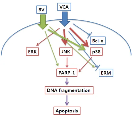
Fig. 12. Signaling pathway activated by VCA and BV treatment in Hep G2 cells.