인터페론 베타로 치료한 소아의 재발-이장 다발성 경화증 1례
건국대학교병원 소아청소년과1, 청담이든 소아청소년과2
조명현1・송민경2・곽병옥1・이란1
Submitted: 6 August, 2015 Revised: 20 September, 2015 Accepted: 24 September, 2015 Correspondence to Ran Lee, MD
Department of Pediatrics, Konkuk University Medical Center, Konkuk University School of Medicine, Seoul, Korea, 120-1 Neungdong-ro, Gwangjin-gu, Seoul 05030, Korea
Tel : +82.2-2030-7557 Fax : +82.2-2030-7748 E-mail : [email protected]
A Case of Childhood Relapsing–remitting Multiple Sclerosis Treated with Interferon Beta
Multiple sclerosis (MS) is an inflammatory demyelinating disease of the central ner- vous system that is primarily a disease of young adulthood. Onset before 10 years of age is considered unusual. We report a boy with MS with onset at 34 months of age who was effectively treated with interferon beta 1b. He visited the emergency room with seizure, left-side limb weakness, and intentional tremor on the left hand. He had received influenza (H1N1) vaccination 1 week prior to that event. Brain mag- netic resonance imaging (MRI) disclosed multifocal edematous T2-weighted hyper- intensity lesions in the left thalamus, bilateral midbrain, pons, medulla, cerebral cortices, and subcortical white matter. This presentation suggested the diagnosis of ADEM. Intravenous methylprednisolone was administered and the symptoms re covered completely. Fifteen months later, he experienced gait disturbance, inten- tional tremor, and dysarthria and a new T2-weighted hyperintensity lesion was found on brain MRI. The diagnosis of early-onset MS was made and the patient was administered interferon beta-1b every other day. He was relapse free after the treat ment. However, he experienced flu-like symptoms and intermittent fever and temporary withdrawal of interferon beta-1b led to relapse of MS. Intravenous me- thylprednisolone was administered again and his symptoms improved gradually.
Currently, at 5 years of age, he has been relapse free from 3 months after interferon beta-1b was administered consistently. This case showed that interferon beta had positive effects to reduce relapses in pediatric MS patients, and pediatric MS relapsed during withdrawal of interferon beta for a short period.
Key Words: Multiple sclerosis, Interferon-beta, Child
Myung Hyun Cho, MD1, Min Kyoung Song, MD2, Byung Ok Kwak, MD1, Ran Lee, MD1
1Department of Pediatrics, Konkuk University Medical Center, Konkuk University School of Medicine, Seoul, Korea, 2Chung Dam Idun Pedia
tric Clinic, Seoul, Korea
Copyright © 2015 by The Korean Child Neurology Society
http://www.cns.or.kr Introduction
Multiple sclerosis (MS), frequently diagnosed in adulthood, is currently also recognized in childhood (4-5% of MS cases)1,2). Juvenile-onset cases presenting between 10-15 years of age are more common (80% of childhood cases) com- pared with true childhood-onset disease presenting before 10 years of age1).
The diagnosis of pediatric MS requires multiple episodes of central nervous system demyelination separated in space and time; at present, the criteria used in adults are also applied to children3). Although acute disseminated encephalo- myelitis-like phenotypes are described currently in 16% of pediatric MS4), the
international panel points out that, after an episode consistent with the features of acute disseminated encephalomyelitis (ADEM), at least two demyelinating events without encephalo- pathy (nonacute disseminated encephalomyelitis events) or one nonacute disseminated encephalomyelitis event plus MRI criteria are needed for the diagnosis of pediatric MS3-8).
We report a boy who was admitted with features mimicking ADEM, but was ultimately diagnosed as a case of MS based on the course of the disease and investigation findings that corro- borated the criteria mentioned above. He had a relapsing-remit- ting disease course of MS after his first attack at the age of 34 months and had another relapse episode and new T2-weighted high-intensity lesions on brain MRI after temporary withdrawal of interferon beta therapy.
Case Report
A 34-month-old boy visited the emergency room with seizure,
left-side limb weakness, and intentional tremor on the left hand.
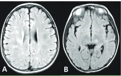
He had received influenza (H1N1) vaccination 1 week prior to that event. Knee jerk and biceps reflex were hypereflexic. Brain MRI disclosed multifocal edematous T2-weighted hyperintense lesions in the left thalamus, bilateral midbrain, pons, medulla, cerebral cortices, and subcortical white matter (Fig.1A, B). This presentation suggested the diagnosis of ADEM. Intravenous me- thylprednisolone was administered and the symptoms recovered completely. The lesions on MRI also completely disappeared 2 weeks later (Fig. 1C). However, 15 months later, he developed a sudden onset of gait disturbance, intentional tremor, and dysar- thria. Cerebrospinal fluid examination revealed pleocytosis (95 cells/μL), negative oligoclonal band, slightly increased IgG (3.73 mg/dL), and IgG index (1). MRI revealed multifocal T2-weighted high-signal-intensity lesions in both subcortical white matter of the cerebral hemisphere, basal ganglia, thalamus, posterior limb of the internal capsule, left midbrain, and deep cerebellum (Fig.
2A, B). He was diagnosed with MS. Intravenous methylpredniso- lone was administered and his symptoms improved. To prevent
A B C
Fig. 1. Brain MRI in first episode. Multifocal edematous T2-weighted hyperintense lesions in the left thalamus, bilateral midbrain, pons, medulla, cerebral cortices, and subcortical white matter (A, B). After intravenous methylprednisolone was administered the lesions on MRI completely disappeared 2 weeks later (C).
A B C
Fig. 2. Brain MRI at first relapse. Multifocal T2-weighted high-signal-intensity lesions in both subcortical white matter of the cerebral hemisphere, basal ganglia, thalamus, posterior limb of the internal capsule, left midbrain, and deep cerebellum (A, B). Complete improvement of the demyelinating foci (C).
relapse of MS, he began to receive subcutaneous injections of interferon beta-1b (Betaferon). Interferon beta-1b at 0.03 mg (0.002 mg/kg) was injected subcutaneously every other day and dose increasing with 2 weeks interval. Maintenance dose (0.12 mg) with gradual escalation was 50% of the adult dose of Inter- feron-beta. Six months later, follow-up MRI showed complete im provement of the demyelinating foci and the patient did not show any abnormal neurological signs (Fig. 2C). Even though he was relapse free after the treatment, he suffered from flu-like symp toms and intermittent fever. Considering these symptoms as side effects of interferon beta-1b therapy, his mother stopped injecting the interferon beta-1b, based on her own decision. One week later, he experienced another relapse characterized by ataxia, dysarthria, and intentional tremor. This occurred 7 months after the first relapse. MRI at that time revealed newly developed fluid attenuated inversion recovery (FLAIR) hyperintense lesions in the bilateral subcortical white matter, periventricular white matter, left thalamus, brain stem, middle cerebellar peduncle, and cere bellar vermis (Fig. 3). Intravenous methylprednisolone was ad ministered again and his symptoms improved gradually. Currently, at 5 years of age, he has been relapse free from 3 months after interferon beta-1b was administered consistently.
Discussion
Pediatric MS is being increasingly recognized worldwide. It has
been estimated that approximately 4-5% of all individuals with MS experience the onset of their disease in childhood1,2). The large majority of patients with pediatric-onset MS have a re- lapsing-remitting course initially. The relapse frequency is high, with 1.9 relapses per year in the first two years of disease, which is higher than that observed in adult-onset MS patients matched for disease duration9).
The most important alternative diagnosis to MS is ADEM. ADEM is an inflammatory demyelinating disease of the central nervous system that often follows a viral illness or vaccination. It is usually monophasic, but relapses have been reported in 5.5-21% of pa- tients2). Depending on whether the new event has or does not have novel clinical manifestations and/or MRI lesions, a diagnosis of multiphasic or recurrent ADEM, respectively, is established5). In the reported patient, the differentiating points favoring the diagnosis of relapsing-remitting MS rather than ADEM were; ob- jective central nerve system (CNS) involvement by examination in each episode, clinical pattern consisting of three episodes of worsening neurodeficits involving two different CNS sites, occur- ring at intervals of 7-15 months, predominant involvement of white matter lesions, and absence of other possible explanations for the disease1-4). Therefore, the patient was diagnosed as having definite MS.
Corticosteroids have been demonstrated to have Class I and Class II evidence on the speed of functional recovery after acute attacks in adults with MS10). Based on these data and on general consensus at the level of expert opinion, intravenous methyl- prednisolone at 10-30 mg/kg for 3-5 days is recommended for
A B
Fig. 3. Brain MRI at second relapse. Newly developed FLAIR hyperintense lesions in the bilateral subcor- tical white matter, periventricular white matter, left thalamus, brain stem, middle cerebellar peduncle, and cerebellar vermis.
relapses of sufficient severity to limit function7,8,11). Intravenous immunoglobulin (IV-Ig) does not have proved efficacy in treating acute relapses of MS7,12). In children with an acute relapse who do not respond to, or do not tolerate corticosteroids, IV-Ig the- rapy can be used at 2 g/kg divided over 2-5 days, depending on tolerance of the infusion rate (level IV evidence: case series and expert opinion)7,10). In our patient, intravenous methylpredniso- lone was administered at all attacks and complete recovery was observed. Seven disease-modifying therapies have been ap- proved for the treatment of relapsing-remitting MS in the adult population, including four first-line (glatiramer acetate, intramu- scular and subcutaneous interferon beta-1a, and subcutaneous interferon beta-1b), and two second-line (mitoxantrone and na- talizumab) therapies. In addition, therapies such as rituximab, daclizumab, and cyclophosphamide have been evaluated in phase 2 trials in adults with breakthrough disease, as have add- on therapies, such as monthly steroids and IV-Ig13).
Several retrospective case series have described the use of interferon beta-1a in the pediatric population. Follow-up in these series has ranged from 12 to 48 months. Interferon beta-1a and -1b appear to be safe and well tolerated in this population, al- though discontinuation rates are in the range of 30-50%2,7,8,14). Many children administered interferon (35-65%) report flu-like symptoms. Other side effects observed relatively frequently include leukopenia (8-27%), thrombocytopenia (16%), anemia (12%), and a transient elevation in the levels of transaminases (10-62%)14). Abnormal results in liver function tests (LFTs) may be more pronounced in younger children taking interferon11). Children with elevated results in LFTs are predominantly younger than 10 years7,11). Temporary interruption of interferon treatment seems to lead to normalization of LFTs in children and is most often accompanied by safe reintroduction of therapy after a temporary withdrawal of medication7,11).
Considering the time of onset and the relapse frequency, our patient began receiving subcutaneous injections of interferon beta-1b at a dose of 0.12 mg every other day. The follow-up MRI performed 6 months later showed complete improvement of the demyelinating foci and the patient did not show any abnormal neurological signs or relapse of MS. Because of flu-like symp- toms and intermittent fever, which are known common side effects of interferon, the medication was temporarily withdrawn, which led to relapse of MS symptoms and newly developed MRI lesions sooner than expected.
Regarding efficacy, there are no randomized controlled trials evaluating the efficacy of interferon beta in the pediatric popula- tion. However, in a prospective, open-label study, Ghezzi and colleagues followed-up 52 patients with pediatric MS who were
treated with interferon beta-1a intramuscularly and found a re- duction in the annualized relapse rate15). In our case, early inter- feron beta treatment at the time of confirmation of diagnosis reduced the MS relapse rate and disease progression. Our ex- perience suggests that short-term withdrawal of interferon beta might cause rapid relapse of MS.
요약
다발성 경화증은 젊은 성인에 주로 발생하는 면역 매개성 탈수초 중추신경계 질환으로 10세 이하에서 발병하는 경우는 드물다. 본 증 례는 34개월 남아로 H1N1 독감 예방 접종하고 일주일 후 발생한 발 작, 좌측 상하지의 근력 저하, 좌측 상지의 의도 진전을 주소로 내원 하였다. 뇌 자기공명영상 T2 강조영상에서 좌측 시상, 양측 중간뇌, 교 뇌, 숨뇌, 대뇌겉질, 피질하 백질에서 고신호강도를 보였다. 처음에는 급성파종뇌척수염으로 생각하여 메틸프레드니솔론을 투여하여 호전 되었으나 15개월 후 보행 장애, 의도 진전, 구어장애를 보여서 다시 시 행한 뇌 자기공명영상 T2 강조영상에서 새로운 병변이 보였다. 이에 다발성 경화증으로 진단하고 인터페론 베타를 격일로 투여하여 증상 이 호전되었다. 인터페론 베타 치료 중 감기 같은 증상 보여서 중단하 자 곧바로 다발성 경화증이 재발하였다. 메틸프레드니솔론으로 치료 하였고 현재 인터페론 베타를 투여 받으며 재발 없이 지내고 있다. 본 증례를 통하여 인터페론 베타는 소아 다발성 경화증의 재발을 억제 하며, 인터페론 베타를 짧은 기간이라도 중단할 경우 다발성 경화증 이 재발할 수 있음을 경험하였다.
References
1) Dale RC, de Sousa C, Chong WK, Cox TC, Harding B, Neville BG.
Acute disseminated encephalomyelitis, multiphasic dissemi
nated encephalomyelitis and multiple sclerosis in children.
Brain 2000;123:240722.
2) Tenembaum S, Chamoles N, Fejerman N. Acute disseminated encephalomyelitis: a longterm followup study of 84 pediatric patients. Neurology 2002;59:122431.
3) Polman CH, Reingold SC, Edan G, Filippi M, Hartung HP, Kappos L, et al. Diagnostic criteria for multiple sclerosis: 2005 revisions to the "McDonald criteria". Ann Neurol 2005;58:8406.
4) Mikaeloff Y, Suissa S, Vallee L, Lubetzki C, Ponsot G, Confavreux C, et al. First episode of acute CNS inflammatory demyelination in childhood: prognostic factors for multiple sclerosis and disa
bility. J Pediatr 2004;144:24652.
5) Krupp LB, Banwell B, Tenembaum S, International Pediatric MS Study Group. Consensus definitions proposed for pediatric mul
tiple sclerosis and related disorders. Neurology 2007;68:S712.
6) Hahn CD, Shroff MM, Blaser SI, Banwell BL. MRI criteria for mul
tiple sclerosis: evaluation in a pediatric cohort. Neurology 2004;
62:8068.
7) Banwell B, Shroff M, Ness JM, Jeffery D, Schwid S, Weinstock
Guttman B ; International Pediatric MS Study Group. MRI fea
tures of pediatric multiple sclerosis. Neurology 2007;68:S4653.
8) Mikaeloff Y, Adamsbaum C, Husson B, Vallee L, Ponsot G, Con
favreux C, et al. MRI prognostic factors for relapse after acute CNS inflammatory demyelination in childhood. Brain 2004;127:1942
7.
9) Renoux C, Vukusic S, Mikaeloff Y, Edan G, Clanet M, Dubois B, et al. Natural history of multiple sclerosis with childhood onset. N Engl J Med 2007;356:260313.
10) Goodin DS, Frohman EM, Garmany GP,Jr, Halper J, Likosky WH, Lublin FD, et al. Disease modifying therapies in multiple sclerosis:
report of the therapeutics and technology assessment subcom
mittee of the american academy of neurology and the MS council for clinical practice guidelines. Neurology 2002;58:16978.
11) Pohl D, Waubant E, Banwell B, Chabas D, Chitnis T, Weinstock
Guttman B, et al. Treatment of pediatric multiple sclerosis and variants. Neurology 2007;68:S5465.
12) Elovaara I, Apostolski S, van Doorn P, Gilhus NE, Hietaharju A, Honkaniemi J, et al. EFNS guidelines for the use of intravenous immunoglobulin in treatment of neurological diseases: EFNS task force on the use of intravenous immunoglobulin in treat
ment of neurological diseases. Eur J Neurol 2008;15:893908.
13) Chitnis T, Krupp L, Yeh A, Rubin J, Kuntz N, Strober JB, et al.
Pediatric multiple sclerosis. Neurol Clin 2011;29:481505.
14) Thannhauser JE, Mah JK, Metz LM. Adherence of adolescents to multiple sclerosis diseasemodifying therapy. Pediatr Neurol 2009;41:11923.
15) Ghezzi A, Amato MP, Capobianco M, Gallo P, Marrosu MG, Mar
tinelli V, et al. Treatment of earlyonset multiple sclerosis with intramuscular interferonbeta1a: longterm results. Neurol Sci 2007;28:12732.