REVIEW ARTICLE
식도암의 내시경 치료
민양원
성균관대학교 의과대학 삼성서울병원 내과
Endoscopic Treatment for Esophageal Cancer
Yang Won Min
Department of Internal Medicine, Samsung Medical Center, Sungkyunkwan University School of Medicine, Seoul, Korea
Esophageal cancer incidence rate per 100,000 is 4.7 in 2013, which accounts for 1.1% of the total cancer incidence in Korea.
Superficial esophageal squamous cell carcinoma is frequently detected in persons undergoing upper endoscopy for gastrointestinal symptoms or for gastric cancer screening. Esophagectomy with lymph node dissection is the standard treatment for esophageal cancer. However, given the considerable morbidity and mortality of esophagectomy, endoscopic resection has become the standard of care for most cases of superficial esophageal squamous cell carcinoma without metastasis. In addition, endoscopic submucosal dissection has increased the cure rate, even when the tumor is large, compared to endoscopic mucosal resection. Thus, endoscopic submucosal dissection is the treatment of choice for superficial esophageal squamous cell carcinoma with a negligible risk of lymph node metastasis. Endoscopic resection is usually associated with a low risk of morbidity and no mortality, and has also shown favor- able long-term outcomes. However, the long-term risk of metastasis remains after endoscopic resection, which varies according to the characteristics of tumor. This review describes the indication and outcomes of endoscopic resection, complications of endoscopic resection, and management after treatment. (Korean J Gastroenterol 2018;71:116-123)
Key Words: Endoscopic mucosal resection; Endoscopic submucosal dissection; Esophageal squamous cell carcinoma
Received February 19, 2018. Revised March 14, 2018. Accepted March 15, 2018.
CC This is an open access article distributed under the terms of the Creative Commons Attribution Non-Commercial License (http://creativecommons.org/licenses/
by-nc/4.0) which permits unrestricted non-commercial use, distribution, and reproduction in any medium, provided the original work is properly cited.
Copyright © 2018. Korean Society of Gastroenterology.
교신저자: 민양원, 06351, 서울시 강남구 일원로 81, 성균관대학교 의과대학 삼성서울병원 내과
Correspondence to: Yang Won Min, Department of Internal Medicine, Samsung Medical Center, Sungkyunkwan University School of Medicine, 81 Irwon-ro, Gangnam-gu, Seoul 06351, Korea. Tel: +82-2-3410-3409, Fax: +82-2-3410-6983, E-mail: [email protected]
Financial support: This paper was supported by the following grant(s): The National R & D Program for Cancer Control, Ministry of Health & Welfare, Korea (1720180).
Conflict of interest: None.
서 론
식도암은 국내에서 2013년 기준 인구 10만 명당 조발생률 4.7건으로, 전체 암 발생의 1.1%를 차지한다. 하지만 식도암 은 진행되어 발견되기 쉽고 림프절 전이가 흔하게 발생하여 예후가 불량하다. 다행히 내시경 검사가 보편화되고 내시경 성능이 향상되면서 식도암의 조기 발견이 늘어나고 있다.1-5 종양을 포함한 식도 절제술 및 주변 림프절 곽청술이 식도암 의 표준 치료법이지만 수술 후 이환율 및 사망률이 낮지 않고, 림프절 전이의 위험이 없는 일부 표재성 식도암의 경우 내시 경적 절제술로도 식도를 보존하면서 근치를 기대해 볼 수 있
기 때문에 조기 식도암 환자에게 내시경 치료가 적용되고 있 다.6-9 조직학적 분류상 국내 식도암의 95% 정도가 편평세포 암이므로 본고에서는 표재성 식도 편평세포암의 내시경 치료 에 대한 최신 경향을 기술하고자 한다.
본 론
1. 시술 전 검사
점막층이나 점막하층에 국한된 식도암을 표재성 식도암이라 고 하는데, 그중 원격 전이가 없고, 림프절 전이 가능성이 거의 없는 조기 식도암의 경우 내시경적 절제술의 좋은 대상이 된다.
시술 전 내시경, 초음파 내시경, 컴퓨터 단층촬영(computed tomography), 양전자 방출 단층촬영(positron emission to- mography) 등으로 치료 대상을 선정하게 된다.
표재성 식도암의 림프절 전이 위험성은 종양의 심달도와 밀접한 관계가 있어서 심달도 진단이 매우 중요하다.10-12 식도 암으로 식도 절제술 및 주변 림프절 곽청술을 받은 환자들의 병 리 조직을 분석한 결과에 따르면 식도암이 점막고유층(M2)까지 만 침윤이 있을 경우 림프절 전이가 거의 없었다(0-8.7%).10-12 따라서, 점막고유층 이상 침윤이 없는 식도암이 내시경적 절제 술의 절대 적응증에 해당된다. 하지만 점막근층(M3)에 침윤하 면 18-27%의 림프절 전이 위험성이 있고, 점막하층(SM)으로 더 깊이 침윤하면 림프절 전이 위험성은 급격히 상승하여 50%까지 올라가게 된다. 치료 전 심달도 진단 방법으로는 내 시경 검사를 통한 육안적 관찰, 초음파 내시경 검사가 주로 시행되며, 최근에는 강조 화상에 의한 확대관찰도 활용된다.
육안적 관찰에 의한 Paris 분류 Ⅰs (융기형 병변)나 Ⅲ (함 요형 병변)에서는 점막하 침윤이 많고, Ⅱb (평탄형 병변)에서 는 적다. Ⅱa (경도융기형)는 Ⅱc (경도함요형)보다는 적지만
Ⅱb보다는 점막하 침윤이 더 많이 동반된다.13 종양의 표면이 불규칙하거나 과립상이 있을 때, 주변부 융기가 동반된 경우 점막하 침윤 가능성이 높고, 종양의 크기가 큰 경우 깊이 침윤 하는 일부분이 섞여 있을 수 있어서 점막하 침윤 가능성을 고려 해야 하겠다. 하지만 이러한 여러 요소의 정량화가 쉽지 않아 전체적으로 판단할 수 있는 경험이 중요하며, 가장 심한 곳을 기준으로 침윤 가능성을 평가한다. 하지만 식도의 굴곡과 점막 고유층의 림프여포 등에 의해 융기가 강조된 경우 실제보다 심 달도를 깊게 판단할 수 있으며, 미소침윤이 있는 경우 실제보다 심달도를 얕게 판단하게 되는 등의 제한점이 있다. 표재성 식도 암의 육안 특징으로 점막근층으로부터 200 μm 이상의 점막 하층 침윤(SM2)을 예측하는 모델을 개발하고, 임상에서 유용 하게 활용되도록 노모그램(normogram)을 보고하였다.14
초음파 내시경 검사는 육안적 관찰을 통한 심달도 진단을 검증하고 보강할 수 있다. 초음파 내시경으로 병리조직상에 대응하는 식도 단면 화상을 얻을 수 있어서 객관적인 심달도 진단이 가능하기 때문이다.15 특히 고주파수(20-30 MHz)의 미세탐촉자(mini-probe)로 관찰 시 상세한 식도암의 침윤도 를 볼 수 있어서 표재성 식도암의 심달도 진단에 유용하다. 하 지만 식도벽의 내시경 초음파 층구조가 환자에 따라서 다르게 보여질 수 있고, 같은 화상을 보고도 다른 해석을 할 수도 있어 서 시술자에 따른 진단율의 차이가 크게 발생할 수 있으며, 위식 도접합부와 같은 위치에서 진단의 정확도가 떨어질 수 있다.15-17 협대혁 화상 강화(narrow band imaging) 확대 내시경으 로 종양 표면의 자세한 혈관 구조 관찰이 가능한데, 식도암 침윤 정도에 따라 미세혈관 구조의 특징적인 변화가 동반되어
표재성 식도암 심달도 진단에 활용될 수 있다. 최근 보고된 일본 식도암 연구회 확대 내시경 분류법은 90% 이상의 훌륭 한 정확도로 표재성 식도암 심달도를 예측할 수 있었다.18 하 지만 종양 표면의 미세혈관 구조 변화만으로 심달도를 예측하 기 때문에 점막하 침윤폭이 작거나 침윤부의 표층을 정상 상 피 또는 상피내암이 덮고 있을 때 실제보다 심달도를 얕게 판단할 수 있다. 실제로, 협대혁 화상 강화 확대 내시경이 초 음파 내시경보다 표재성 식도암 심달도 예측에 더 유용하다는 연구 결과가 있는 반면, 통상적 내시경 관찰만 하였을 때와 비교하여 협대혁 화상 강화 확대 내시경을 추가로 시행한 경 우 심달도 진단의 정확도를 올리지 못하였다는 보고도 있
다.19,20 육안적 관찰, 초음파 내시경, 협대혁 화상 강화 확대
내시경 모두 각기 심달도 예측의 방법에 따른 장애 요소들이 있기 때문에 서로 간의 진단에 차이가 있을 수 있다. 따라서 점막하 침윤 가능성이 낮은 표면이 균일한 Ⅱb 표재성 식도 암에서 일괄적으로 초음파 내시경이나 협대혁 화상 강화 확대 내시경을 추가로 시행하면, 검사 간 예측의 불일치로 내시경 적 절제술이 가능한 식도암을 수술하게 되는 경우도 발생할 수 있으니 주의해야 하겠다. 하지만 표면이 불규칙하거나 Ⅱa 또는 Ⅱc 형태의 표재성 식도암과 같이 점막하 침윤이 의심될 때에는 초음파 내시경이나 협대혁 화상 강화 확대 내시경을 추가로 시행하여 심달도 진단을 검증함으로써 불필요한 수술 및 내시경 치료를 피할 수 있도록 해야겠다.
식도는 림프관이 점막고유층까지 발달해 있기 때문에 표재 성 식도암임에도 불구하고 림프절 전이가 가능하다.21 따라서 내시경 치료 전 식도암 림프절 전이 유무 확인이 매우 중요하
다.22,23 암 진단과정에서 컴퓨터 단층촬영은 원격 전이를 확인
할 수 있는 좋은 도구이지만 식도암, 특히 표재성 식도암에서 림프절 전이 진단 민감도가 매우 낮다. 컴퓨터 단층촬영의 민 감도는 초음파 내시경, 양전자 방출 단층촬영과 비교하였을 때 더 낮았다.24-26 하지만 초음파 내시경의 경우 정확한 진단 을 위해 고도의 숙련도를 필요로 하고, 광범위한 식도암의 림 프절 전이의 범위를 모두 검사할 수 없는 제한점이 있다.27 이에 반해 양전자 방출 단층촬영은 초음파 내시경에 비해 검 사 범위의 제한이 적고, 초음파 내시경과 컴퓨터 단층촬영보 다 림프절 전이 진단 민감도가 높다.25,26,28,29
따라서 식도암 내시경 절제술 전 림프절 전이 유무 평가에 양전자 방출 단층 촬영과 컴퓨터 단층촬영 화상을 겹쳐서 맞춘 양전자 컴퓨터 단층촬영이 최근에 널리 활용되고 있다.
2. 내시경 치료 방법
1980년대 후반에 식도 내시경 점막 절제술(endoscopic mucosal resection)이 식도암 치료법으로 일본에서 시작되 고, 이후 세계적으로 보급되었다. 하지만 내시경 점막 절제술
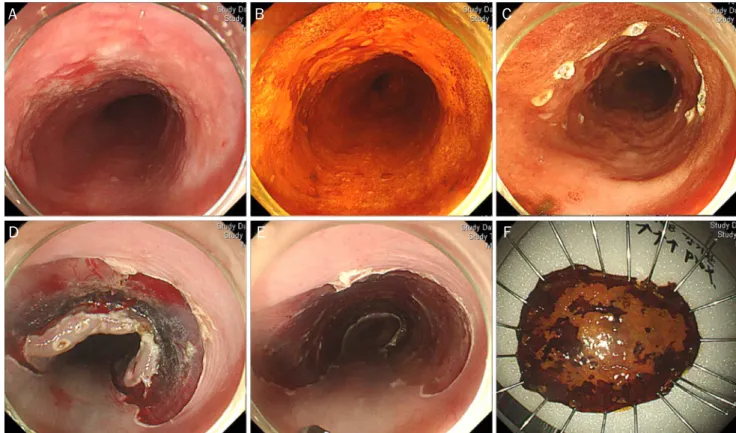
A B C
D E F
Fig. 1. Endoscopic submucosal dissection procedure. (A) On the lower thoracic esophagus, a 3-cm-sized geographic mucosal hyperemia with uneven surface is noticed. (B) Lugol’s solution is sprayed along the lesion to aid visualization. (C) Marking around the lesion is performed.
(D) After submucosal injection, circumferential mucosal pre-cutting is performed. (E) After dissection of the submucosal layer, an artificial ulcer is seen. (F) Fixation of the resected specimen.
은 한 번에 절제되는 면적이 좁기 때문에 크기가 1-2 cm 이상 큰 종양을 일괄 절제하기 쉽지 않아 정확한 병리조직학적 정 보를 알기 어렵게 할 수 있을 뿐 아니라 내시경 점막하 박리술 (endoscopic submucosal dissection, Fig. 1)과 비교하여 완 치 절제율(curative resection rate)이 낮고 국소재발률은 높
았다.9,30-32 따라서 최상의 치료 성적을 위해서는 식도암 내시
경적 절제술로 내시경 점막하 박리술이 행해져야 하겠다.9,33-35 다만, 식도는 좁고 굴곡이 있으며 위치에 따라 심박동이나 호 흡 운동에 의한 영향이 커 내시경 점막하 박리술이 기술적으 로 어렵고, 시술 시간이 길며, 천공의 합병증 위험이 높다. 우 리 기관의 식도암 내시경 점막하 박리술 자료를 분석한 결과, 전신마취를 시행하였을 때 내시경 점막하 박리술 시술 시간을 단축하고 천공의 발생률을 유의하게 감소시킬 수 있었다. 즉, 식도암 내시경 점막하 박리술은 전신마취하에 진행되는 것이 안전하고 효과적일 수 있다. 하지만 크기가 작은 식도암에서, 특히 안정적인 마취 상태가 유지되기 어려운 경우에는 내시경 점막 절제술을 선별적으로 적용할 수도 있다.
3. 내시경 치료 적응증
National Comprehensive Cancer Network 가이드라인
에서 식도 점막암은 내시경 절제술, 점막하 침윤이 있는 경우 는 식도 절제술을 받도록 권고되고 있다.36 일본 식도암 연구 회에서는 점막암 중에서도 점막고유층까지만 침범한 경우 림 프절 전이가 거의 없어서 내시경적 절제술의 절대 적응증으로 정의하였다.34 유럽 내시경학회 가이드라인에서도 림프절 전 이가 없고 점막고유층까지만 침범한 식도암을 내시경 절제술 의 절대 적응증으로 제시하였다.33 식도암이 점막근층까지 침 윤하는 경우와 점막근판으로부터 200 μm 이하의 침윤이 있 을 경우 림프절 전이 위험성은 각각 18-27%, 15-53.1%로 높 게 올라간다.10-12,37 하지만 일본 식도암 연구회와 유럽 내시경 학회 가이드라인에서는 M3 또는 SM1 침윤이 있는 식도암도 미세혈관 혹은 림프관에 암세포 침윤이 없는 경우 림프절 전 이의 위험이 낮기 때문에 상대 적응증으로 내시경적 절제술을 시행해 볼 수 있다고 보고하였다.33,34 실제로 M3 또는 SM1 식도암에서의 림프절 전이 위험은 암세포의 미세혈관 혹은 림 프관 침윤이나 미분화형과 관련성이 있다.38-40 또한 식도 절제 술은 수술 후 높은 이환율과 최소 1-2%의 사망률을 동반하므 로 환자의 전신상태가 좋지 않거나 환자가 수술을 원치 않을 경우 내시경적 절제술을 차선책으로 시행할 수 있겠다.6-8 특 히 M3 식도암의 경우 내시경 절제술 후 림프관 침윤이 없음
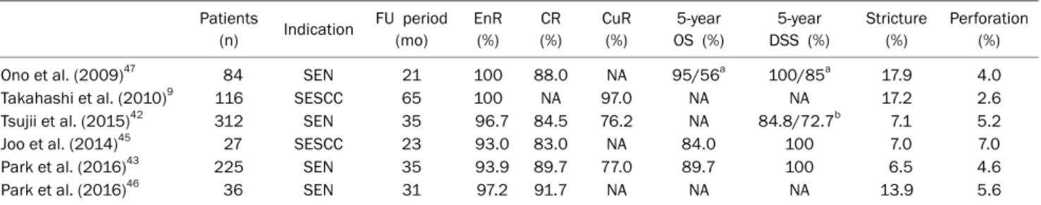
Table 1. Outcomes of Endoscopic Submucosal Dissection for SEN Patients
(n) Indication FU period (mo)
EnR (%)
CR (%)
CuR (%)
5-year OS (%)
5-year DSS (%)
Stricture (%)
Perforation (%)
Ono et al. (2009)47 84 SEN 21 100 88.0 NA 95/56a 100/85a 17.9 4.0
Takahashi et al. (2010)9 116 SESCC 65 100 NA 97.0 NA NA 17.2 2.6
Tsujii et al. (2015)42 312 SEN 35 96.7 84.5 76.2 NA 84.8/72.7b 7.1 5.2
Joo et al. (2014)45 27 SESCC 23 93.0 83.0 NA 84.0 100 7.0 7.0
Park et al. (2016)43 225 SEN 35 93.9 89.7 77.0 89.7 100 6.5 4.6
Park et al. (2016)46 36 SEN 31 97.2 91.7 NA NA NA 13.9 5.6
SEN, superficial esophageal neoplasms; FU, follow-up; EnR, en bloc resection; CR, complete resection; CuR, curative resection; OS, overall survival; DSS, disease specific survival; NA, not available; SESCC, superficial esophageal squamous cell carcinoma.
aM1 and M2 cancer/M3 and SM cancers; bCuR/non-CuR.
이 확인된 경우 5년 누적 원격 전이 발생률이 0.7%로 낮아 내시경적 절제술이 안전하며 효과적일 수 있겠다.41 종합하면 내시경적 절제술로 일괄 완전 절제된 식도암의 병리조직학적 결과 침윤 깊이가 점막근층(M3)을 넘지 않고 미세혈관 혹은 림프관에 암세포 침윤이 없다면 추가 수술 없이 면밀한 경과 관찰을 해볼 수 있다.
4. 내시경 치료 성적
이제까지 식도암 내시경 절제술의 성적은 대부분 일본에서 보고되었으며 대체로 그 결과는 우수하다(Table 1).9,32,41-47 비 교적 짧은 기간이지만 내시경 점막하 박리술 후 632일(중앙 값)을 추적 관찰한 일본의 단일기관 연구는 점막고유층까지만 침범한 식도암의 5년 생존율, 질병특이 5년 생존율을 각각 95%, 100%로 높게 보고하였다.47 반면 점막근판 이상을 침범한 식 도암은 5년 생존율과 질병특이 5년 생존율이 각각 56%, 85%
로 낮았다. 최근에 발표된 일본의 다기관 후향적 보고에서는 비교적 긴 추적 관찰 기간임에도(중앙값 35개월) 214명의 완 치 절제 환자 중 한 명(0.5%)만 식도암으로 사망하였다.42 완 치 절제의 기준이 점막근판으로부터 200 μm 이하의 침윤도 포함하였음을 감안하였을 때 우수한 장기 성적이다.42내시경 절제술을 받은 570명의 식도암 환자를 평균 50개월 추적 관 찰한 연구에서 5년 누적 원격 전이 발생률을 평가하였는데 식도암이 점막고유층에 국한될 때, 점막근판까지 침범할 때, 점막근판으로부터 200 μm 이하의 침윤이 있을 때 그리고 점 막근판으로부터 200 μm 넘는 침윤이 있을 때, 각각 0.4%, 8.7%, 7.7% 그리고 36.2%로 종양의 침범 깊이가 전이 발생의 위험인자임을 확인할 수 있었다. 앞서 언급되었지만 점막고유 층까지만 침범한 식도암의 경우 림프관 침윤이 없을 때는 전 이 위험이 0.7%로 점막고유층에 국한된 식도암의 위험과 비 슷하였다.
국내에서는 2014년 27명 식도암 내시경 점막하 박리술 환 자의 치료 성적이 발표되었다.45 일괄 절제율과 완전 절제율은 각각 93%, 83%였다. 두 명(7%)에서 시술 중 천공이 발생하였
으나 내시경적 봉합과 항생제 치료로 조절되었고, 유의한 출 혈 환자는 없었다. 식도 둘레의 75% 이상이 절제된 7명 환자 중 2명에서 협착이 발생하여 내시경적 풍선 확장술이 필요하 였다. 내시경 점막하 박리술 후 평균 23개월간 추적 관찰하였 고, 5년 생존율, 질병특이 5년 생존율은 각각 84%, 100%로 우수하였다. 하지만 추적 관찰에서 빠져나간 3명 중 SM2 식 도암 환자가 한 명 포함되어 있어서 실제 생존율은 더 낮을 가능성도 있다. 최근에는 국내 단일기관의 식도 내시경 점막 하 박리술 장기 치료 성적도 발표되었다.43 총 225명의 환자, 261개 병소에 대한 자료이며 191개 식도 편평세포암이 포함 되었다. 전체 대상에서 일괄 절제와 완전 절제는 각각 93.9%, 89.7%에서 이루어졌다. 시술 합병증으로 12.6%에서 발생하 였는데, 출혈 1.5%, 천공 4.6% 그리고 협착 6.5%였다. 출혈 환자는 내시경 치료를 받았고, 천공 환자도 내시경 치료를 포 함한 비수술적 치료로 조절이 되었다. 17명의 협착 환자 중 5명은 내시경적 풍선 확장술을 시행 받았다. 병변이 식도 둘 레의 75% 이상을 차지한 경우 45.5%에서 협착이 발생하였 다. 내시경 점막하 박리술 후 35개월(중앙값)간 추적 관찰하였 고, 5년 생존율은 89.7%로 우수하였다. 식도이형성과 M1, M2 식도암은 91.7%, M3 식도암은 80.2% 그리고 SM1 식도 암은 79.1%로 종양의 침범 깊이에 따라 생존율의 차이가 나 는 경향을 확인할 수 있었다(p=0.061). 하지만 저자들은 완치 절제가 된 환자들 중 식도암으로 인한 사망은 없어서 질병특 이 5년 생존율을 100%로 보고하였다.
5. 내시경 치료 합병증
내시경적 절제술 중 급성 출혈이 빈번히 발생하는데, 양이 많지 않아도 쉽게 시야가 나빠져서 시술에 어려움이 발생한 다. 특히 낮은 위치인 7-8시 방향으로는 피가 고이기 때문에 출혈로 시야가 나쁜 상태에서 시술할 때 천공의 위험이 올라 가므로 지혈을 시행한 다음 절개나 박리를 진행해야 한다. 특 히 점막 절개 시에는 점막 밑으로 비쳐 보이는 굵은 혈관을 피하거나 가볍게 지혈을 해주면서 진행하는 것이 이후의 시술
과정을 좀 더 편하게 해줄 수 있다. 점막하 박리 시에는 swift coagulation 모드를 활용하면 작은 혈관에서의 출혈을 대부 분 예방할 수 있으며, 1-2 mm 이상의 큰 혈관이 발견되면 지혈겸자로 잡고 soft coagulation 모드로 예방적 지혈을 해 주면 된다. 박리된 바닥에서 출혈이 있을 때도 겸자를 이용해 서 지혈을 해줘야 하는데, 식도근육층은 위에 비해 매우 얇아 지연 천공의 위험이 있으니 살짝 지혈하는 느낌으로 통전을 해줘야 한다. 식도에서는 지연 출혈이 매우 드물기 때문에 재 출혈을 걱정하기보다는 지연 천공의 위험을 피할 수 있도록 가볍게 지혈을 해야 한다.
앞서 언급된 바와 같이 천공은 식도암 내시경적 절제술의 주 시술 합병증이다.42,43,48 식도암으로 내시경 점막하 박리술 을 시행하였을 때 천공발생률은 0-8%로 보고마다 약간의 차 이가 있다.41,44,45,49
천공을 예방하는 위해서는 점막하 박리 시 근육층이 노출되지 않게 하단부 점막하층의 일부를 남기도록 해야 한다. 그리고 시야가 좋지 않은 7-9시 방향을 박리할 때 조심스럽게 천천히 진행하는 것이 좋으며, 이 부분은 점막 절 개를 특히 충분히 하여 박리 시 점막하층이 충분히 노출될 수 있도록 해야 한다. 또한 전신마취하에 식도 내시경 점막하 박리술을 할 경우 흡인의 위험도 감소하고 급작스러운 움직임 없이 안정적인 상태에서 시술을 진행할 수 있어서 천공의 위 험을 낮출 수 있다.50
식도 내시경 점막하 박리술 후 협착은 병변의 특징 및 예방 법 사용 유무에 따라 발생률이 달라져 약 10% 전후로 발생한 다고 보고되나, 공통적으로 식도 둘레의 75% 이상이 절제된 경우에 주로 발생하는 것으로 되어있다.45,51,52 따라서 이러한 경우 예방적 치료를 시도하는 것이 좋으며, 보통 경구 스테로 이드를 4-8주 복용하거나 내시경적 절제 부위에 스테로이드 를 주입할 수 있다.53-55 절제 범위가 넓은 경우에는 협착 발생 을 완전히 막을 수 없다.56이 경우 풍선 확장술을 시행하거나 일시적으로 식도 스텐트를 삽입해서 치료할 수 있다.57,58하지 만 식도 협착이 반복적으로 발생하여 여러 차례 시술이 필요 한 경우도 많아 내시경적 절제술 전 협착 위험도와 치료 과정 에 대한 충분한 설명이 필요하고, 궁극적으로는 더 효과적인 협착 예방법을 고안해내야 한다.59-61
6. 불완치 절제 후 치료 전략
표재성 식도암에 대해 내시경적 절제술을 시도하였으나 불 완치 절제되었을 때 원칙적으로는 식도 절제술 및 주변 림프 절 곽청술을 시행해야 한다. 특히 점막하 침윤이 있거나 미세 혈관 혹은 림프관에 암세포 침윤이 있을 경우 림프절 전이의 위험이 높기 때문에 수술을 시행하는 것이 권고된다.10 내시경 점막하 박리술 후 추가로 식도 절제술을 시행 받은 표재성 식도암 환자와 바로 수술을 받은 환자의 수술 성적을 비교하
였을 때 차이가 없음을 확인할 수 있었다.62 만약 환자가 수술 을 받기 어렵거나 거부할 경우 철저한 추적 관찰을 해야 하겠 지만 원격 전이가 흔하게 발생하므로 근치적 치료의 기회를 잃을 수 있다.41,42
일본에서 발표된 자료를 보았을 때, 방사선 항암 치료가 표 재성 식도암 치료에 효과적일 수 있음을 보고하였다.63,64 또한 방사선 항암 치료를 내시경적 절제술 후에 시행할 경우 방사 선 치료 단독으로 시행하였을 때보다 국소재발률은 낮추면서 방사선량을 줄여 관련 합병증 위험도 낮출 수 있어서 표재성 식도암에서 수술을 대신할 수 있는 효과적인 치료법이 될 가 능성이 있다.64-66 불완치 절제 후 수술을 거부하여 방사선 항 암 치료를 받은 66명을 51개월(중앙값) 추적 관찰한 결과 2명 (3%)에서 국소재발, 6명(9%)에서 원격 전이가 발생하였고, 17명 (26%)이 사망하였다.67 1년, 3년, 5년 생존율은 각각 98%, 87%
그리고 75%로 양호하게 보고되었다. 36명의 환자는 미세혈 관 혹은 림프관에 암세포 침윤이 있었고, 그중 6명에서 원격 전이가 발생하였으나 침윤이 없었던 나머지 30명 중에서는 원격 전이가 발생하지 않았다. 즉, 미세혈관 혹은 림프관에 암세포 침윤 여부는 방사선 항암 치료 후 원격 전이 발생 여부 를 예측할 수 있는 중요한 예후인자임을 알 수 있다. 하지만 아직 불완치 내시경적 절제술 후 방사선 항암 치료가 수술을 대신할 수 있는지에 대한 충분한 근거가 마련되지 않은 상태 이다. 현재 일본과 한국에서 두 추가 치료법의 성적을 비교하 는 전향적 연구가 진행되고 있어서 그 결과에 따라 향후 방사 선 항암 치료가 새로운 표재성 식도암 치료 전략으로 자리잡 을 수 있을 것으로 기대된다.68
결 론
내시경 검사가 널리 시행되면서 표재성 식도암 발견이 늘 어나고 있다. 내시경 절제술은 식도를 보존할 수 있는 큰 장점 이 있다. 하지만 식도암은 초기에도 전이가 발생할 수 있어 내시경 치료의 대상 선정이 무엇보다도 중요하다. 내시경적 절제술 후 병리조직학적 결과는 향후 재발률을 예측하는 데 가장 정확한 정보를 제공하므로 올바른 해석과 함께 그에 따 른 적절한 치료 방침 결정이 이루어져야 한다. 기술적으로 식 도암 내시경적 절제술은 위에서와 다르며 합병증 발생에서도 차이가 있음을 인지하고 있어야 한다. 절제술 후 협착 발생을 줄일 수 있는 방법을 찾는 것은 향후 내시경적 절제술을 보다 많은 환자에게 적용시킬 수 있게 할 것이다. 내시경적 절제술 후 장기 추적 관찰에 대한 연구도 이루어져야 하겠지만, 불완 치 절제 후 가장 효과적인 치료법이 무엇인가에 대한 연구도 중요한 부분이다.
REFERENCES
1. Lee KS, Oh DK, Han MA, et al. Gastric cancer screening in Korea:
report on the national cancer screening program in 2008.
Cancer Res Treat 2011;43:83-88.
2. Shimizu Y, Takahashi M, Yoshida T, et al. Endoscopic resection (endoscopic mucosal resection/endoscopic submucosal dis- section) for superficial esophageal squamous cell carcinoma:
current status of various techniques. Dig Endosc 2013;25 Suppl 1:13-19.
3. Kumagai Y, Monma K, Kawada K. Magnifying chromoendoscopy of the esophagus: in-vivo pathological diagnosis using an endo- cytoscopy system. Endoscopy 2004;36:590-594.
4. Yoshida T, Inoue H, Usui S, Satodate H, Fukami N, Kudo SE.
Narrow-band imaging system with magnifying endoscopy for super- ficial esophageal lesions. Gastrointest Endosc 2004;59:288-295.
5. Tachibana M, Hirahara N, Kinugasa S, Yoshimura H. Clinicopathologic features of superficial esophageal cancer: results of consecutive 100 patients. Ann Surg Oncol 2008;15:104-116.
6. Ra J, Paulson EC, Kucharczuk J, et al. Postoperative mortality af- ter esophagectomy for cancer: development of a preoperative risk prediction model. Ann Surg Oncol 2008;15:1577-1584.
7. Chang AC, Ji H, Birkmeyer NJ, Orringer MB, Birkmeyer JD.
Outcomes after transhiatal and transthoracic esophagectomy for cancer. Ann Thorac Surg 2008;85:424-429.
8. Connors RC, Reuben BC, Neumayer LA, Bull DA. Comparing out- comes after transthoracic and transhiatal esophagectomy: a 5-year prospective cohort of 17,395 patients. J Am Coll Surg 2007;205:735-740.
9. Takahashi H, Arimura Y, Masao H, et al. Endoscopic submucosal dissection is superior to conventional endoscopic resection as a curative treatment for early squamous cell carcinoma of the esophagus (with video). Gastrointest Endosc 2010;72:255-264, 264.e1-2.
10. Eguchi T, Nakanishi Y, Shimoda T, et al. Histopathological criteria for additional treatment after endoscopic mucosal resection for esophageal cancer: analysis of 464 surgically resected cases.
Mod Pathol 2006;19:475-480.
11. Akutsu Y, Uesato M, Shuto K, et al. The overall prevalence of metastasis in T1 esophageal squamous cell carcinoma: a retro- spective analysis of 295 patients. Ann Surg 2013;257:1032-1038.
12. Choi JY, Park YS, Jung HY, et al. Feasibility of endoscopic resection in superficial esophageal squamous carcinoma. Gastrointest Endosc 2011;73:881-889, 889.e1-e2.
13. Endoscopic Classification Review Group. Update on the paris classification of superficial neoplastic lesions in the digestive tract. Endoscopy 2005;37:570-578.
14. Pyo JH, Byeon SJ, Lee H, et al. Measurement of tumor volume is not superior to diameter for prediction of lymph node metastasis in early gastric cancer with minute submucosal invasion.
Oncotarget 2017;8:113758-113765.
15. Thosani N, Singh H, Kapadia A, et al. Diagnostic accuracy of EUS in differentiating mucosal versus submucosal invasion of super- ficial esophageal cancers: a systematic review and meta-analysis.
Gastrointest Endosc 2012;75:242-253.
16. Lee WC, Lee TH, Jang JY, et al. Staging accuracy of endoscopic
ultrasound performed by nonexpert endosonographers in pa- tients with resectable esophageal squamous cell carcinoma: is it possible? Dis Esophagus 2015;28:574-578.
17. Dhupar R, Rice RD, Correa AM, et al. Endoscopic ultrasound esti- mates for tumor depth at the gastroesophageal junction are in- accurate: implications for the liberal use of endoscopic resection.
Ann Thorac Surg 2015;100:1812-1816.
18. Oyama T, Inoue H, Arima M, et al. Prediction of the invasion depth of superficial squamous cell carcinoma based on microvessel morphology: magnifying endoscopic classification of the Japan Esophageal Society. Esophagus 2017;14:105-112.
19. Ebi M, Shimura T, Yamada T, et al. Multicenter, prospective trial of white-light imaging alone versus white-light imaging followed by magnifying endoscopy with narrow-band imaging for the re- al-time imaging and diagnosis of invasion depth in superficial esophageal squamous cell carcinoma. Gastrointest Endosc 2015;81:1355-1361.e2.
20. Mizumoto T, Hiyama T, Oka S, et al. Diagnosis of superficial esophageal squamous cell carcinoma invasion depth before en- doscopic submucosal dissection. Dis Esophagus 2017 Dec 18.
[Epub ahead of print]
21. Rice TW. Superficial oesophageal carcinoma: is there a need for three-field lymphadenectomy? Lancet 1999;354:792-794.
22. Akutsu Y, Kato K, Igaki H, et al. The prevalence of overall and ini- tial lymph node metastases in clinical T1N0 thoracic esophageal cancer: from the results of JCOG0502, a prospective multicenter study. Ann Surg 2016;264:1009-1015.
23. Li B, Chen H, Xiang J, et al. Prevalence of lymph node metastases in superficial esophageal squamous cell carcinoma. J Thorac Cardiovasc Surg 2013;146:1198-1203.
24. Pech O, May A, Günter E, Gossner L, Ell C. The impact of endo- scopic ultrasound and computed tomography on the TNM stag- ing of early cancer in Barrett's esophagus. Am J Gastroenterol 2006;101:2223-2229.
25. Kato H, Kuwano H, Nakajima M, et al. Comparison between posi- tron emission tomography and computed tomography in the use of the assessment of esophageal carcinoma. Cancer 2002;94:
921-928.
26. Yoon YC, Lee KS, Shim YM, Kim BT, Kim K, Kim TS. Metastasis to regional lymph nodes in patients with esophageal squamous cell carcinoma: CT versus FDG PET for presurgical detection pro- spective study. Radiology 2003;227:764-770.
27. Murata Y, Ohta M, Hayashi K, Ide H, Takasaki K. Preoperative evaluation of lymph node metastasis in esophageal cancer. Ann Thorac Cardiovasc Surg 2003;9:88-92.
28. Kim K, Park SJ, Kim BT, Lee KS, Shim YM. Evaluation of lymph node metastases in squamous cell carcinoma of the esophagus with positron emission tomography. Ann Thorac Surg 2001;71:
290-294.
29. Choi JY, Lee KH, Shim YM, et al. Improved detection of individual nodal involvement in squamous cell carcinoma of the esophagus by FDG PET. J Nucl Med 2000;41:808-815.
30. Ishihara R, Iishi H, Uedo N, et al. Comparison of EMR and endo- scopic submucosal dissection for en bloc resection of early esophageal cancers in Japan. Gastrointest Endosc 2008;68:
1066-1072.
31. Cao Y, Liao C, Tan A, Gao Y, Mo Z, Gao F. Meta-analysis of endo- scopic submucosal dissection versus endoscopic mucosal re- section for tumors of the gastrointestinal tract. Endoscopy 2009;
41:751-757.
32. Kim DH, Jung HY, Gong EJ, et al. Endoscopic and oncologic out- comes of endoscopic resection for superficial esophageal neoplasm. Gut Liver 2015;9:470-477.
33. Pimentel-Nunes P, Dinis-Ribeiro M, Ponchon T, et al. Endoscopic submucosal dissection: European Society of Gastrointestinal Endoscopy (ESGE) guideline. Endoscopy 2015;47:829-854.
34. Japan Esophageal Society. Japanese classification of esoph- ageal cancer, 11th edition: part I. Esophagus 2017;14:1-36.
35. Higuchi K, Tanabe S, Koizumi W, et al. Expansion of the in- dications for endoscopic mucosal resection in patients with su- perficial esophageal carcinoma. Endoscopy 2007;39:36-40.
36. Ajani JA, D'Amico TA, Almhanna K, et al. Esophageal and esoph- agogastric junction cancers, version 1.2015. J Natl Compr Canc Netw 2015;13:194-227.
37. Kim DU, Lee JH, Min BH, et al. Risk factors of lymph node meta- stasis in T1 esophageal squamous cell carcinoma. J Gastroenterol Hepatol 2008;23:619-625.
38. Oyama T, Tomori A, Hotta K, et al. Endoscopic submucosal dis- section of early esophageal cancer. Clin Gastroenterol Hepatol 2005;3(7 Suppl 1):S67-S70.
39. Moriya H, Ohbu M, Kobayashi N, et al. Lymphatic tumor emboli detected by D2-40 immunostaining can more accurately predict lymph-node metastasis. World J Surg 2011;35:2031-2037.
40. Katada C, Muto M, Momma K, et al. Clinical outcome after endo- scopic mucosal resection for esophageal squamous cell carcino- ma invading the muscularis mucosae--a multicenter retro- spective cohort study. Endoscopy 2007;39:779-783.
41. Yamashina T, Ishihara R, Nagai K, et al. Long-term outcome and metastatic risk after endoscopic resection of superficial esoph- ageal squamous cell carcinoma. Am J Gastroenterol 2013;108:
544-551.
42. Tsujii Y, Nishida T, Nishiyama O, et al. Clinical outcomes of endo- scopic submucosal dissection for superficial esophageal neo- plasms: a multicenter retrospective cohort study. Endoscopy 2015;47:775-783.
43. Park HC, Kim DH, Gong EJ, et al. Ten-year experience of esoph- ageal endoscopic submucosal dissection of superficial esoph- ageal neoplasms in a single center. Korean J Intern Med 2016;
31:1064-1072.
44. Probst A, Aust D, Märkl B, Anthuber M, Messmann H. Early esoph- ageal cancer in Europe: endoscopic treatment by endoscopic submucosal dissection. Endoscopy 2015;47:113-121.
45. Joo DC, Kim GH, Park DY, Jhi JH, Song GA. Long-term outcome af- ter endoscopic submucosal dissection in patients with super- ficial esophageal squamous cell carcinoma: a single-center study. Gut Liver 2014;8:612-618.
46. Park JS, Youn YH, Park JJ, Kim JH, Park H. Clinical outcomes of endoscopic submucosal dissection for superficial esophageal squamous neoplasms. Clin Endosc 2016;49:168-175.
47. Ono S, Fujishiro M, Niimi K, et al. Long-term outcomes of endo- scopic submucosal dissection for superficial esophageal squ- amous cell neoplasms. Gastrointest Endosc 2009;70:860-866.
48. Kim GH, Jee SR, Jang JY, et al. Stricture occurring after endo- scopic submucosal dissection for esophageal and gastric tumors. Clin Endosc 2014;47:516-522.
49. Song BG, Min YW, Lee JH, et al. Efficacy and safety of endoscopic submucosal dissection in elderly patients with esophageal squ- amous cell carcinoma. Surg Endosc 2017;31:3905-3911.
50. Yamashita K, Shiwaku H, Ohmiya T, et al. Efficacy and safety of endoscopic submucosal dissection under general anesthesia.
World J Gastrointest Endosc 2016;8:466-471.
51. Jain D, Singhal S. Esophageal stricture prevention after endo- scopic submucosal dissection. Clin Endosc 2016;49:241-256.
52. Shi Q, Ju H, Yao LQ, et al. Risk factors for postoperative stricture after endoscopic submucosal dissection for superficial esoph- ageal carcinoma. Endoscopy 2014;46:640-644.
53. Kataoka M, Anzai S, Shirasaki T, et al. Efficacy of short period, low dose oral prednisolone for the prevention of stricture after cir- cumferential endoscopic submucosal dissection (ESD) for esophageal cancer. Endosc Int Open 2015;3:E113-E117.
54. Wang W, Ma Z. Steroid administration is effective to prevent stric- tures after endoscopic esophageal submucosal dissection: a network meta-analysis. Medicine (Baltimore) 2015;94:e1664.
55. Hashimoto S, Kobayashi M, Takeuchi M, Sato Y, Narisawa R, Aoyagi Y. The efficacy of endoscopic triamcinolone injection for the prevention of esophageal stricture after endoscopic sub- mucosal dissection. Gastrointest Endosc 2011;74:1389-1393.
56. Okamoto K, Matsui S, Watanabe T, et al. Clinical analysis of esophageal stricture in patients treated with intralesional tri- amcinolone injection after endoscopic submucosal dissection for superficial esophageal cancer. Oncology 2017;93 Suppl 1:9-14.
57. Yamasaki T, Tomita T, Takimoto M, et al. Esophageal stricture af- ter endoscopic submucosal dissection treated successfully by temporary stent placement. Clin J Gastroenterol 2016;9:337- 340.
58. Lian JJ, Ma LL, Hu JW, et al. Endoscopic balloon dilatation for be- nign esophageal stricture after endoscopic submucosal dis- section for early esophageal neoplasms. J Dig Dis 2014;15:
224-229.
59. Ohki T, Yamato M, Ota M, et al. Prevention of esophageal stricture after endoscopic submucosal dissection using tissue-en- gineered cell sheets. Gastroenterology 2012;143:582-588.e2.
60. Iizuka T, Kikuchi D, Yamada A, Hoteya S, Kajiyama Y, Kaise M.
Polyglycolic acid sheet application to prevent esophageal stric- ture after endoscopic submucosal dissection for esophageal squamous cell carcinoma. Endoscopy 2015;47:341-344.
61. Yamaguchi N, Isomoto H, Kobayashi S, et al. Oral epithelial cell sheets engraftment for esophageal strictures after endoscopic submucosal dissection of squamous cell carcinoma and air- plane transportation. Sci Rep 2017;7:17460.
62. Kudou M, Shiozaki A, Fujiwara H, et al. Efficacy of additional sur- gical resection after endoscopic submucosal dissection for su- perficial esophageal cancer. Anticancer Res 2017;37:5301-5307.
63. Nemoto K, Yamada S, Nishio M, et al. Results of radiation therapy for superficial esophageal cancer using the standard radio- therapy method recommended by the Japanese Society of Therapeutic Radiology and Oncology (JASTRO) Study Group.
Anticancer Res 2006;26(2B):1507-1512.
64. Motoori M, Yano M, Ishihara R, et al. Comparison between radical esophagectomy and definitive chemoradiotherapy in patients with clinical T1bN0M0 esophageal cancer. Ann Surg Oncol 2012;19:2135-2141.
65. Kawaguchi G, Sasamoto R, Abe E, et al. The effectiveness of endo- scopic submucosal dissection followed by chemoradiotherapy for superficial esophageal cancer. Radiat Oncol 2015;10:31.
66. Ikeda A, Hoshi N, Yoshizaki T, et al. Endoscopic submucosal dis- section (ESD) with additional therapy for superficial esophageal cancer with submucosal invasion. Intern Med 2015;54:2803- 2813.
67. Hamada K, Ishihara R, Yamasaki Y, et al. Efficacy and safety of endoscopic resection followed by chemoradiotherapy for super- ficial esophageal squamous cell carcinoma: a retrospective study. Clin Transl Gastroenterol 2017;8:e110.
68. Kurokawa Y, Muto M, Minashi K, Boku N, Fukuda H; Gastrointes- tinal Oncology Study Group of Japan Clinical Oncology Group (JCOG). A phase II trial of combined treatment of endoscopic mu- cosal resection and chemoradiotherapy for clinical stage I esophageal carcinoma: Japan Clinical Oncology Group Study JCOG0508. Jpn J Clin Oncol 2009;39:686-689.