ISSN 2234-3806 • eISSN 2234-3814
Received: January 28, 2019 Revision received: April 14, 2019 Accepted: June 13, 2019
Corresponding author: Jeong Hwan Shin, M.D.
Department of Laboratory Medicine, Busan Paik Hospital, Inje University College of Medicine, 75 Bokji-ro, Busanjin-gu, Busan 47392, Korea
Tel: +82-51-890-6475 Fax: +82-51-890-8615 E-mail: [email protected] Ann Lab Med 2019;39:537-544
https://doi.org/10.3343/alm.2019.39.6.537
Serotype Distribution and Antimicrobial Resistance of Invasive and Noninvasive Streptococcus pneumoniae Isolates in Korea between 2014 and 2016
Dong-Chul Park , Ph.D.1*, Si Hyun Kim , Ph.D.2*, Dongeun Yong , M.D.3, In Bum Suh , M.D.4, Young Ree Kim , M.D.5, Jongyoun Yi , M.D.6, Wonkeun Song , M.D.7, Sae Am Song , M.D.1, Hee-Won Moon , M.D.8, Hae Kyung Lee , M.D.9, Kyoung Un Park , M.D.10, Sunjoo Kim , M.D.11, Seok Hoon Jeong , M.D.3, Jaehyeon Lee , M.D.12, Joseph Jeong , M.D.13, Yu Kyung Kim , M.D.14, Miae Lee , M.D.15, Jihyun Cho , M.D.16, Jong-Wan Kim , M.D.17, Kyeong Seob Shin , M.D.18, Sang-Hyun Hwang , M.D.19, Jae-Woo Chung , M.D.20, Hye In Woo , M.D.21, Chae Hoon Lee , M.D.22,
Namhee Ryoo , M.D.23, Chulhun L. Chang , M.D.6, Hyun Soo Kim , M.D.7, Jayoung Kim , M.D.24, Jong Hee Shin , M.D.25, Soo Hyun Kim , M.D.25, Mi-Kyung Lee , M.D.26, Seong Gyu Lee , M.D.27, Sook Jin Jang , M.D.28, Kyutaeg Lee , M.D.29, HunSuk Suh , M.D.30, Yong-Hak Sohn , M.D.31, Min-Jung Kwon , M.D.21, Hee Joo Lee , M.D.32, Ki Ho Hong , M.D.33, Kwang-Sook Woo , M.D.34, Chul Min Park , M.D.35, and Jeong Hwan Shin , M.D.1,36
1Department of Laboratory Medicine, Inje University College of Medicine, Busan, Korea; 2Department of Clinical Laboratory Science, Semyung University, Jecheon, Korea; 3Department of Laboratory Medicine and Research Institute of Bacterial Resistance, Yonsei University College of Medicine, Seoul, Korea;
4Department of Laboratory Medicine, Kangwon National University College of Medicine, Chuncheon, Korea; 5Department of Laboratory Medicine, School of Medicine, Jeju National University, Jeju, Korea; 6Department of Laboratory Medicine, Pusan National University School of Medicine, Busan, Korea;
7Department of Laboratory Medicine, Hallym University College of Medicine, Chuncheon, Korea; 8Department of Laboratory Medicine, Konkuk University School of Medicine, Seoul, Korea; 9Department of Laboratory Medicine, Uijeongbu St. Mary’s Hospital, College of Medicine, The Catholic University of Korea, Seoul, Korea; 10Department of Laboratory Medicine, Seoul National University College of Medicine, Seoul, Korea; 11Department of Laboratory Medicine, Gyeongsang National University College of Medicine, Jinju, Korea; 12Department of Laboratory Medicine, Chonbuk National University Medical School and Hospital, Jeonju, Korea; 13Department of Laboratory Medicine, Ulsan University Hospital, University of Ulsan College of Medicine, Ulsan, Korea;
14Department of Laboratory Medicine, School of Medicine, Kyungpook National University, Daegu, Korea; 15Department of Laboratory Medicine, Ewha Womans University College of Medicine, Seoul, Korea; 16Department of Laboratory Medicine, Wonkwang University College of Medicine, Iksan, Korea;
17Department of Laboratory Medicine, Dankook University College of Medicine, Cheonan, Korea; 18Department of Laboratory Medicine, Chungbuk National University College of Medicine, Cheongju, Korea; 19Department of Laboratory Medicine, University of Ulsan College of Medicine and Asan Medical Center, Seoul, Korea; 20Department of Laboratory Medicine, Dongguk University College of Medicine, Ilsan, Korea; 21Department of Laboratory Medicine and Genetics, Samsung Medical Hospital, Sungkyunkwan University School of Medicine, Seoul, Korea; 22Department of Laboratory Medicine, College of Medicine, Yeungnam University, Daegu, Korea; 23Department of Laboratory Medicine, Keimyung University School of Medicine, Daegu, Korea; 24Department of Laboratory Medicine, International St. Mary’s Hospital, College of Medicine, Catholic Kwandong University, Incheon, Korea; 25Department of Laboratory Medicine, Chonnam National University Medical School, Gwangju, Korea; 26Department of Laboratory Medicine, Chung-Ang University College of Medicine, Seoul, Korea; 27Department of Laboratory Medicine, Bundang Jesaeng General Hospital, Seongnam, Korea; 28Department of Laboratory Medicine, College of Medicine, Chosun University, Gwangju, Korea; 29Department of Laboratory Medicine, Cheju Halla General Hospital, Jeju, Korea; 30Department of Laboratory Medicine, Daegu Catholic University School of Medicine, Daegu, Korea; 31Department of Laboratory Medicine, Eulji University School of Medicine, Daejeon, Korea; 32Department of Laboratory Medicine, School of Medicine, Kyung Hee University, Seoul, Korea; 33Department of Laboratory Medicine, Seoul Medical Center, Seoul, Korea; 34Department of Laboratory Medicine, Dong-A University College of Medicine, Busan, Korea; 35Department of Laboratory Medicine, Dongnam Institute of Radiological & Medical Sciences, Busan, Korea; 36Paik Institute for Clinical Research, Inje University College of Medicine, Busan, Korea
2017-03-16 https://crossmark-cdn.crossref.org/widget/v2.0/logos/CROSSMARK_Color_square.svg
Background: Several factors contribute to differences in Streptococcus pneumoniae sero- type distribution. We investigated the serotype distribution and antimicrobial resistance of S. pneumoniae isolated between 2014 and 2016 in Korea.
Methods: We collected a total of 1,855 S. pneumoniae isolates from 44 hospitals between May 2014 and May 2016, and analyzed the serotypes by sequential multiplex PCR. We investigated the distribution of each serotype by patient age, source of the clinical speci- men, and antimicrobial resistance pattern.
Results: The most common serotypes were 11A (10.1%), followed by 19A (8.8%), 3 (8.5%), 34 (8.1%), 23A (7.3%), and 35B (6.2%). The major invasive serotypes were 3 (12.6%), 19A (7.8%), 34 (7.8%), 10A (6.8%), and 11A (6.8%). Serotypes 10A, 15B, 19A, and 12F were more common in patients ≤5 years old, while serotype 3 was more
INTRODUCTION
Streptococcus pneumoniae is an important human pathogen that causes pneumonia, sepsis, and meningitis, especially in children [1-3]. This bacterium has more than 93 serotypes, but only a few cause the majority of pneumonias and invasive pneu- mococcal diseases (IPDs). The serotype distribution differs by patients’ age, geographic region, and time of surveillance; these changes are affected by vaccination trends [4, 5].
Following the introduction of the 7-valent pneumococcal con- jugate vaccine (PCV7, targeting serotypes 4, 6B, 9V, 14, 18C, 19F, and 23F) in children, IPDs caused by PCV7 serotypes de- creased dramatically in many countries [6-10]. However, the use of PCV7 led to an increase in infections with non-vaccine serotypes such as 19A [8, 11-13]. PCV7 has led to extensive changes in serotype distribution in Korea [14, 15]. Since 2010, PCV10 (includes PCV7 plus serotypes 1, 5, and 7F) and PCV13 (includes PCV10 plus serotypes 3, 6A, and 19A) have replaced PCV7 in Korea; national immunization programs (NIPs) have been provided for children since May 2014. Therefore, a survey of serotype distribution is necessary for the design of national strategies following the change in the type of pneumococcal vaccine used.
High rates of drug resistance and the spread of multi-drug re- sistant (MDR) strains of S. pneumoniae constitute serious pub- lic health concerns worldwide [14, 15]. In Korea, high resistance against most antimicrobial agents continues to be observed in pneumococcal diseases [16, 17], although the resistance rate to penicillin has decreased since the change in the CLSI break- points [18]. We aimed to investigate the serotype distribution
and antimicrobial resistance of S. pneumoniae isolated between 2014 and 2016 in Korea
METHODS
Clinical isolatesA total of 1,855 S. pneumoniae isolates were prospectively col- lected from 44 hospitals in Korea between May 2014 and May 2016. All isolates were transported to the Inje University Busan Paik Hospital, Busan, Korea and stored until use at -70°C using 10% skim milk. This study was approved by the Institutional Review Board of Inje University Busan Paik Hospital (No. 14- 0256).
Serotyping by sequential multiplex PCR assay
The serotype of all pneumococcal isolates was determined us- ing sequential multiplex PCR (SM-PCR) according to the rec- ommendations of the U.S. Centers for Disease Control and Pre- vention (CDC) [19]. For DNA extraction, colonies cultured on blood agar plates were mixed with 200 μL of Tris-EDTA buffer solution (Sigma-Aldrich Co., St Louis, MO, USA). This mixture was heated at 100°C for 10 minutes and then promptly placed on a frozen surface (-20°C) for 5 minutes, followed by centrifu- gation at 13,000 rpm. SM-PCR was performed with a PCR pre- mix (AccuPower PCR PreMix, Bioneer Inc., Daejeon, Korea), 1 μL of each primer, 5 μL of DNA template, and distilled water in a final volume of 20 μL. Thermal cycling was conducted in a Veriti96-well thermal cycler (Applied Biosystems, Foster City, CA, USA) under the following conditions: 94°C for 5 minutes;
30 amplification cycles of 94°C for 30 seconds, 54°C for 30 sec- common in patients ≥65 years old compared with the other age groups. The coverage
rates of pneumococcal conjugate vaccine (PCV)7, PCV10, PCV13, and pneumococcal polysaccharide vaccine 23 were 11.8%, 12.12%, 33.3%, and 53.6%, respectively. Of the 1,855 isolates, 857 (46.2%) were multi-drug resistant (MDR), with serotypes 11A and 19A predominant among the MDR strains. The resistance rates against penicillin, cefotax- ime, and levofloxacin were 22.8%, 12.5%, and 9.4%, respectively.
Conclusions: There were significant changes in the major S. pneumoniae serotypes in the community. Non-PCV13 serotypes increased in patients ≤5 years old following the intro- duction of national immunization programs with the 10- and 13-polyvalent vaccines.
Key Words: Streptococcus pneumoniae, Serotype, Antimicrobial resistance, Pneumococ- cal vaccine
* These authors equally contributed to this study.
© Korean Society for Laboratory Medicine This is an Open Access article distributed under the terms of the Creative Commons Attribution Non-Commercial License (http://creativecom- mons.org/licenses/by-nc/4.0) which permits unrestricted non-commercial use, distribution, and reproduction in any medium, provided the original work is properly cited.
onds, and 72°C for 30 seconds; and one cycle of 72°C for 7 minutes. The size of the amplification products was confirmed by electrophoresis on a 2% agarose gel. The Quellung reaction was additionally performed to differentiate serotype 6A from other serotype 6 subtypes using factor antisera (Statens Serum Institute, Copenhagen, Denmark).
Collection of antimicrobial resistance data
The drug resistant results of the pneumococcal isolates were collected from the participating hospitals; the assays were per- formed mainly by Microscan (Siemens Healthcare Diagnostics, Sacramento, CA, USA), the VITEK2 system (bioMérieux, Marcy- l’Étoile, France), and E-test (bioMérieux). The results were inter- preted according to the CLSI guidelines [20]. Separate interpre- tive breakpoints were used to define the resistance of meningeal isolates to penicillin, cefotaxime, and ceftriaxone. An isolate re- sistant to three or more classes of antimicrobial agents was con- sidered MDR. We analyzed serotype prevalence by age group, clinical source, and antimicrobial resistance.
RESULTS
Characteristics of S. pneumoniae isolates
Of the 1,855 isolates, 1,286 (69.3%) were from male patients and 438 (23.6%) were from patients with invasive disease. The most common source of invasive isolates was blood (N =372;
84.9%), followed by cerebrospinal fluid (N=21; 4.8%), pleural fluid (N=13; 3.0%), abscess (N=13; 3.0%), tissue (N=8, 1.8%), and others (N=11; 2.5%). Non-invasive isolates were recovered from respiratory specimens (N=1,253; 88.4%), wounds (N=127;
9.0%), catheter tips (N =16; 1.1%), urine (N =8; 0.6%), and other sites (N=13; 0.9%).
Distribution of pneumococcal serotypes
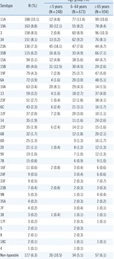
The most common serotype was 11A (10.1%), followed by 19A (8.8%), 3 (8.5%), 34 (8.1%), 23A (7.3%), 35B (6.2%), and 15A (5.1%); these serotypes accounted for 54.2% of the iso- lates (Table 1). Serotypes 23A, 15B, 19A, and 10A were more common in patients ≤5 years old (18.1%, 12.5%, 12.1%, and 8.1%, respectively). In contrast, serotypes 11A, 3, and 34 were much less common in patients ≤5 years old. The frequency of the major serotypes was very similar in patients ≥65 and 6–64 years old. The number of serotypes recovered from ≥65 years and 6–64 years age groups was 35 and 32, respectively, whereas only 20 serotypes were recovered from patients ≤5 years old.
Non-typeable (NT) isolates that were not detected by SM-PCR
Table 1. Distribution of pneumococcal serotypes by patient age (N=1,855)
Serotype N (%)
Age group (%)
≤5 years
(N=248) 6–64 years
(N=673) ≥65 years (N=934)
11A 188 (10.1) 12 (4.8) 77 (11.4) 99 (10.6)
19A 163 (8.8) 30 (12.1) 55 (8.2) 78 (8.4)
3 158 (8.5) 2 (0.8) 60 (8.9) 96 (10.3)
34 151 (8.1) 13 (5.2) 62 (9.2) 76 (8.1)
23A 136 (7.3) 45 (18.1) 47 (7.0) 44 (4.7)
35B 115 (6.2) 16 (6.5) 33 (4.9) 66 (7.1)
15A 94 (5.1) 12 (4.8) 38 (5.6) 44 (4.7)
15B 85 (4.6) 31 (12.5) 30 (4.5) 24 (2.6)
19F 79 (4.3) 7 (2.8) 25 (3.7) 47 (5.0)
6A 72 (3.9) 4 (1.6) 20 (3.0) 48 (5.1)
10A 63 (3.4) 20 (8.1) 29 (4.3) 14 (1.5)
13 59 (3.2) 4 (1.6) 18 (2.7) 37 (4.0)
23F 51 (2.7) 1 (0.4) 12 (1.8) 38 (4.1)
6C 43 (2.3) 6 (2.4) 21 (3.1) 16 (1.7)
12F 37 (2.0) 7 (2.8) 20 (3.0) 10 (1.1)
14 35 (1.9) 11 (1.6) 24 (2.6)
22F 35 (1.9) 6 (2.4) 14 (2.1) 15 (1.6)
6B 32 (1.7) 12 (1.8) 20 (2.1)
6D 25 (1.3) 9 (1.3) 16 (1.7)
20 21 (1.1) 1 (0.4) 8 (1.2) 12 (1.3)
9V 19 (1.0) 7 (1.0) 12 (1.3)
7B 15 (0.8) 6 (0.9) 9 (1.0)
16F 11 (0.6) 2 (0.8) 3 (0.4) 6 (0.6)
24F 9 (0.5) 3 (0.4) 6 (0.6)
33F 9 (0.5) 2 (0.3) 7 (0.7)
23B 7 (0.4) 2 (0.8) 2 (0.3) 3 (0.3)
9N 5 (0.3) 1 (0.1) 4 (0.4)
35A 4 (0.2) 2 (0.3) 2 (0.2)
7F 4 (0.2) 3 (0.4) 1 (0.1)
38 3 (0.2) 1 (0.4) 1 (0.1) 1 (0.1)
17F 3 (0.2) 2 (0.3) 1 (0.1)
5 2 (0.1) 2 (0.3)
8 2 (0.1) 2 (0.3)
18C 2 (0.1) 1 (0.1) 1 (0.1)
4 1 (0.1) 1 (0.1)
Non-typeable 117 (6.3) 26 (10.5) 34 (5.1) 57 (6.1) accounted for 6.3% (N=117) of all isolates. These organisms were more common in children ≤5 years old (10.5%).
The most common serotype among the invasive isolates was 3 (12.6%), followed by 19A (7.8%), 34 (7.8%), 11A (6.8%), 10A (6.8%), and 12F (6.6%) (Table 2). However, serotypes 3,
10A, and 12F were more prevalent among invasive than nonin- vasive isolates (7.3%, 2.3%, and 0.6%, respectively). Serotypes 11A, 23A, and 35B were more common among noninvasive Table 2. Comparison of invasive and noninvasive serotypes by patient age
Serotype Total N (%)
Invasive Noninvasive
Total N (%) ≤5 years 6–64 years ≥65 years Total N (%) ≤5 years 6–64 years ≥65 years
11A 188 (10.1) 30 (6.8) 15 15 158 (11.2) 12 62 84
19A 163 (8.8) 34 (7.8) 6 11 17 129 (9.1) 24 44 61
3 158 (8.5) 55 (12.6) 1 18 36 103 (7.3) 1 42 60
34 151 (8.1) 34 (7.8) 3 13 18 117 (8.3) 10 49 58
23A 136 (7.3) 20 (4.6) 3 10 7 116 (8.2) 42 37 37
35B 115 (6.2) 16 (3.7) 2 4 10 99 (7.0) 14 29 56
15A 94 (5.1) 23 (5.3) 2 10 11 71 (5.0) 10 28 33
15B 85 (4.6) 20 (4.6) 7 8 5 65 (4.6) 24 22 19
19F 79 (4.3) 9 (2.1) 4 5 70 (4.9) 7 21 42
6A 72 (3.9) 13 (3.0) 1 7 5 59 (4.2) 3 13 43
10A 63 (3.4) 30 (6.8) 10 16 4 33 (2.3) 10 13 10
13 59 (3.2) 7 (1.6) 1 3 3 52 (3.7) 3 15 34
23F 51 (2.7) 8 (1.8) 2 6 43 (3.0) 1 10 32
6C 43 (2.3) 8 (1.8) 3 5 35 (2.5) 6 18 11
12F 37 (2.0) 29 (6.6) 5 18 6 8 (0.6) 2 2 4
14 35 (1.9) 16 (3.7) 4 12 19 (1.3) 7 12
22F 35 (1.9) 17 (3.9) 3 7 7 18 (1.3) 3 7 8
6B 32 (1.7) 7 (1.6) 2 5 25 (1.8) 10 15
6D 25 (1.3) 6 (1.4) 3 3 19 (1.3) 6 13
20 21 (1.1) 9 (2.1) 5 4 12 (0.8) 1 3 8
9V 19 (1.0) 6 (1.4) 4 2 13 (0.9) 3 10
7B 15 (0.8) 4 (0.9) 1 3 11 (0.8) 5 6
16F 11 (0.6) 2 (0.5) 1 1 9 (0.6) 2 2 5
24F 9 (0.5) 5 (1.1) 2 3 4 (0.3) 1 3
33F 9 (0.5) 3 (0.7) 1 2 6 (0.4) 1 5
23B 7 (0.4) 2 (0.5) 1 1 5 (0.4) 1 2 2
9N 5 (0.3) 2 (0.5) 2 3 (0.2) 1 2
35A 4 (0.2) 1 (0.2) 1 3 (0.2) 1 2
7F 4 (0.2) 1 (0.2) 1 3 (0.2) 2 1
38 3 (0.2) 3 (0.7) 1 1 1
17F 3 (0.2) 1 (0.2) 1 2 (0.1) 1 1
5 2 (0.1) 1 (0.2) 1 1 (0.1) 1
8 2 (0.1) 2 (0.5) 2
18C 2 (0.1) 2 (0.1) 1 1
4 1 (0.1) 1 (0.2) 1
Non-typeable 117 13 (3.0) 2 5 6 104 (7.3) 24 29 51
Total 1,855 438 48 185 205 1,417 200 488 729
isolates (11.2%, 8.2%, and 7.0%, respectively) than invasive isolates (6.8%, 4.6%, and 3.7%).
Serotypes 10A (20.8%), 15B (14.6%), 19A (12.5%), and 12F (10.4%) were common in patients ≤5 years old, whereas 11A and 3 were rarely observed (0% and 2.1%, respectively).
Among the invasive isolates, serotypes 11A and 3 were com- mon in patients ≥65 years old (7.3% and 17.6%, respectively) and 6–64 years old (8.1% and 9.7%, respectively), while sero- types 10A and 12F were less frequent in patients ≥65 years old (2.0% and 2.9%, respectively) than in those ≤5 years (20.8%
and 10.4%, respectively) and 6–64 years old (8.6% and 9.7%, respectively).
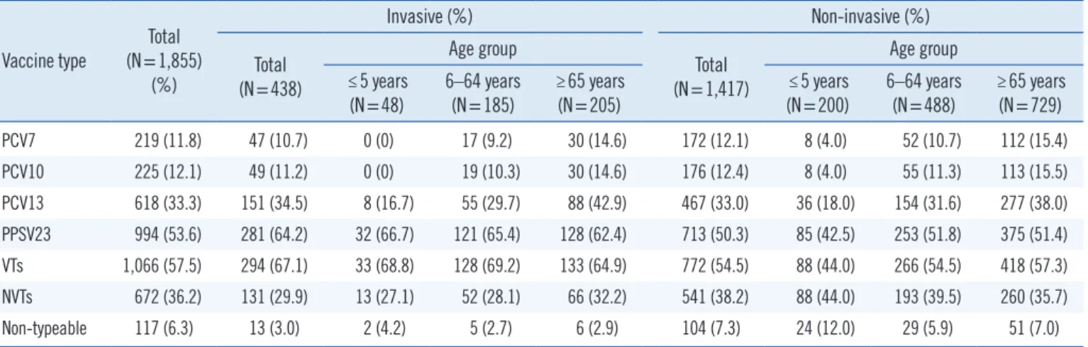
The coverage rates for PCV7, PCV10, PCV13, pneumococcal polysaccharide vaccine 23 (PPSV23), and vaccine serotype (VT) were 11.8%, 12.1%, 33.3%, 53.6%, and 57.5%, respectively
(Table 3). For invasive isolates, the coverage rates of PCV7, PCV10, PCV13, PPSV23, and VTs were 10.7%, 11.2%, 34.5%, 64.2%, and 67.1%, respectively. By age, the coverage rates of PCV7, PCV10, and PCV13 among the invasive isolates were 0%, 0%, and 16.7% in children ≤5 years old and 14.6%, 14.6%, and 42.9% in patients ≥65 years old.
Antimicrobial resistance
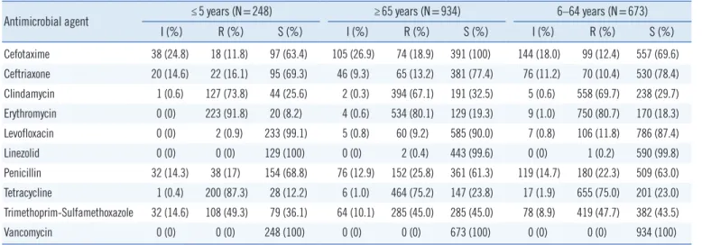
The antimicrobial resistance of the S. pneumoniae isolates is shown in Tables 4 and 5. The resistance rates against penicillin, cefotaxime and levofloxacin were 22.8%, 12.5% and 9.4%, re- spectively. Among the invasive isolates, the resistance rates against cefotaxime, ceftriaxone, and levofloxacin were higher in patients
≥65 years old (7.5%, 5.2%, and 4.9%, respectively) than in patients ≤5 years old (2.6%, 0%, and 0%, respectively).
Table 3. Prevalence of vaccine serotypes by patient age, specimen type, and period
Vaccine type Total (N=1,855)
(%)
Invasive (%) Non-invasive (%)
Total (N=438)
Age group
Total (N=1,417)
Age group
≤5 years
(N=48) 6–64 years
(N=185) ≥65 years
(N=205) ≤5 years
(N=200) 6–64 years
(N=488) ≥65 years (N=729)
PCV7 219 (11.8) 47 (10.7) 0 (0) 17 (9.2) 30 (14.6) 172 (12.1) 8 (4.0) 52 (10.7) 112 (15.4)
PCV10 225 (12.1) 49 (11.2) 0 (0) 19 (10.3) 30 (14.6) 176 (12.4) 8 (4.0) 55 (11.3) 113 (15.5)
PCV13 618 (33.3) 151 (34.5) 8 (16.7) 55 (29.7) 88 (42.9) 467 (33.0) 36 (18.0) 154 (31.6) 277 (38.0)
PPSV23 994 (53.6) 281 (64.2) 32 (66.7) 121 (65.4) 128 (62.4) 713 (50.3) 85 (42.5) 253 (51.8) 375 (51.4) VTs 1,066 (57.5) 294 (67.1) 33 (68.8) 128 (69.2) 133 (64.9) 772 (54.5) 88 (44.0) 266 (54.5) 418 (57.3)
NVTs 672 (36.2) 131 (29.9) 13 (27.1) 52 (28.1) 66 (32.2) 541 (38.2) 88 (44.0) 193 (39.5) 260 (35.7)
Non-typeable 117 (6.3) 13 (3.0) 2 (4.2) 5 (2.7) 6 (2.9) 104 (7.3) 24 (12.0) 29 (5.9) 51 (7.0)
Abbreviations: PCV, pneumococcal conjugate vaccine; PPSV23, pneumococcal polysaccharide vaccine 23; VTs, vaccine serotypes; NVTs, non-vaccine sero- types.
Table 4. Resistance to antimicrobial agents by specimen type
Antimicrobial agent Total (N=1,855) Invasive (N=438) Non-invasive (N=1,417)
I, N (%) R, N (%) S, N (%) I, N (%) R, N (%) S, N (%) I, N (%) R, N (%) S, N (%) Cefotaxime 287 (18.8) 191 (12.5) 1,045 (68.6) 56 (14.7) 31 (8.1) 295 (77.2) 231 (20.2) 160 (14.0) 750 (65.7) Ceftriaxone 142 (10.9) 157 (12.0) 1,006 (77.1) 36 (11.3) 16 (5.0) 268 (83.3) 106 (10.8) 141 (14.3) 738 (74.9) Clindamycin 8 (0.5) 1,079 (69.2) 473 (30.3) 1 (0.3) 260 (66.0) 133 (33.8) 7 (0.6) 819 (70.2) 340 (29.2) Erythromycin 13 (0.7) 1,507 (81.9) 319 (17.3) 2 (0.5) 336 (77.8) 94 (21.8) 11 (0.8) 1,171 (83.2) 225 (16.0) Levofloxacin 12 (0.7) 168 (9.4) 1,604 (89.9) 3 (0.7) 18 (4.4) 390 (94.9) 9 (0.7) 150 (10.9) 1,214 (88.4)
Linezolid 0 (0) 3 (0.3) 1,162 (99.7) 0 (0) 3 (0.9) 325 (99.1) 0 (0) 0 (0) 837 (100)
Penicillin 227 (14.0) 370 (22.8) 1,024 (63.2) 31 (9.0) 73 (21.1) 242 (69.9) 196 (15.4) 297 (23.3) 782 (61.3) Tetracycline 24 (1.4) 1,319 (76.7) 376 (21.9) 5 (1.3) 299 (75.9) 90 (22.8) 19 (1.4) 1,020 (77.0) 286 (21.6) Trimethoprim-Sulfamethoxazole 174 (10.0) 812 (46.9) 746 (43.1) 36 (8.7) 158 (38.3) 219 (53.0) 138 (10.5) 654 (49.6) 527 (40.0)
Vancomycin 0 (0) 0 (0) 1,855 (100) 0 (0) 0 (0) 438 (100) 0 (0) 0 (0) 1,417 (100)
Abbreviations: I, intermediate resistance; R, resistant; S, susceptible.
Fig. 1. Serotype distribution of MDR S. pneumoniae isolates.
Abbreviation: MDR, multi-drug resistant.
25
20
15
10
5
0
11A 19A 15A 19F 23A 15B 13 6A 23F 3 6B 6C 34 35B 9V 10A 14 7B 6D 16F 22F 20 24F 35A 5 12F 4 8 38 17F 18C 23B 33F 7F 9N Serotype
Proportion (%)
Non-MDR MDR Table 5. Resistance to antimicrobial agents by patient age
Antimicrobial agent ≤5 years (N=248) ≥65 years (N=934) 6–64 years (N=673)
I (%) R (%) S (%) I (%) R (%) S (%) I (%) R (%) S (%)
Cefotaxime 38 (24.8) 18 (11.8) 97 (63.4) 105 (26.9) 74 (18.9) 391 (100) 144 (18.0) 99 (12.4) 557 (69.6) Ceftriaxone 20 (14.6) 22 (16.1) 95 (69.3) 46 (9.3) 65 (13.2) 381 (77.4) 76 (11.2) 70 (10.4) 530 (78.4)
Clindamycin 1 (0.6) 127 (73.8) 44 (25.6) 2 (0.3) 394 (67.1) 191 (32.5) 5 (0.6) 558 (69.7) 238 (29.7)
Erythromycin 0 (0) 223 (91.8) 20 (8.2) 4 (0.6) 534 (80.1) 129 (19.3) 9 (1.0) 750 (80.7) 170 (18.3)
Levofloxacin 0 (0) 2 (0.9) 233 (99.1) 5 (0.8) 60 (9.2) 585 (90.0) 7 (0.8) 106 (11.8) 786 (87.4)
Linezolid 0 (0) 0 (0) 129 (100) 0 (0) 2 (0.4) 443 (99.6) 0 (0) 1 (0.2) 590 (99.8)
Penicillin 32 (14.3) 38 (17) 154 (68.8) 76 (12.9) 152 (25.8) 361 (61.3) 119 (14.7) 180 (22.3) 509 (63.0) Tetracycline 1 (0.4) 200 (87.3) 28 (12.2) 6 (1.0) 464 (75.2) 147 (23.8) 17 (1.9) 655 (75.0) 201 (23.0) Trimethoprim-Sulfamethoxazole 32 (14.6) 108 (49.3) 79 (36.1) 64 (10.1) 285 (45.0) 285 (45.0) 78 (8.9) 419 (47.7) 382 (43.5)
Vancomycin 0 (0) 0 (0) 248 (100) 0 (0) 0 (0) 673 (100) 0 (0) 0 (0) 934 (100)
Abbreviations: I, intermediate resistance; R, resistant; S, susceptible.
Of the 1,855 isolates, 857 (46.2%) were MDR, including 11A (17.7%), 19A (15.8%), 19F (7.6%), and 15A (7.6%) (Fig. 1).
The proportion of MDR was extremely high in serotypes 11A (80.9%), 19A (82.8%), 19F (82.3%), 13 (78.0%), 6B (78.1%),
9V (84.2%), and 7B (80.0%). Serotypes 3, 34, and 6A expressed low-level resistance.
DISCUSSION
The prevalence of the common serotypes differed from that in our previous report [16]. Compared with the results from 2011 to 2014, the proportion of non-PCV13 serotypes, such as 11A, 23A, and 15A, remarkably increased. In addition, we confirmed that serotypes 3 and 6A are now less common, whereas there was no change in the prevalence rate of serotype 19A.
The coverage of PCV13 had decreased, whereas the cover- age of PPSV23 had not changed since our previous results from 2011 to 2014 [16]. Surprisingly, the coverage rate of the PCV13 serotype among the invasive isolates was much lower in patients
≤5 years old (16.7%) than in the other age groups (6–64 years old [29.7%] and ≥65 years old [42.9%]). We hypothesize that this change resulted from PCV13 use in children ≤5 years old as the NIPs with PCV13 were provided only for children. In ad- dition, this is associated with the high prevalence of serotype 3 in patients ≥65 years old. The 2014 Korean guidelines recom- mend the administration of PPSV23 or PCV13 to individuals
≥65 years old [21].
Richter, et al. [22] reported a decrease in the prevalence of the PCV13 serotypes in all isolates in the United States from 43.4% (2008–2009) to 27.1% (2012–2013) after the introduc- tion of the PCV13 vaccine. In addition, the prevalence of non- PCV serotypes, such as 11A and 35B, increased among all iso- lates, while that of serotype 3 slightly increased. Interestingly, they observed a decrease in the prevalence of serotype 19A from 22% to 10% of all isolates, which differs from our results.
However, Richter, et al. [23] reported that serotype 19A had not changed between 2010 and 2011. Therefore, we hypothesize that serotype 19A will shortly decrease in Korea. Galanis, et al.
[24] and van der Linden, et al. [25] reported an increase in non- PCV13 serotypes in IPD. We confirmed the increase in non- PCV13 serotypes such as 11A, 23A, and 15A; however, there was no observed increase in serotype 23B. Thus, there is a need for a new pneumococcal vaccine, including non-PCV13 serotypes, to prevent IPDs in children.
Previously, we reported the resistance rate against penicillin as 9.0% from 2008 to 2014 [16] and 10.8% from four univer- sity hospitals in Busan and Gyeongnam in 2015 [17]. In this study, the resistance rate against penicillin among the isolates from 44 hospitals was 22.8%; thus, there was a striking ten- dency towards an increase in penicillin resistance. The resis-
tance rates against cefotaxime, ceftriaxone, and levofloxacin were 12.5%, 12.0%, and 9.4%, respectively, which again are higher than those in a previous report [16]. Our findings suggest that resistance rates are increasing in Korea and elsewhere, high- lighting the need to monitor antimicrobial resistance continually.
There was a strong association between serotype and antimi- crobial resistance. The proportion of MDR S. pneumoniae was extremely high among serotypes 11A, 19A, 19F, 13, 6B, 9V, and 7B. Interestingly, the resistance rate against levofloxacin was quite low in serotypes 19A and 23A. Thus, serotypes show- ing high resistance should be controlled to diminish the risk of severe, even fatal, diseases caused by this organism.
Authors’ Disclosures of Potential Conflicts of Interest
No potential conflicts of interest relevant to this article were re- ported.
Acknowledgements
This research was supported by a grant of the Korea Health Technology R&D Project through the Korea Health Industry De- velopment Institute (KHIDI), funded by the Ministry of Health &
Welfare, Republic of Korea (grant number HI14C1005).
ORCID
Dong-Chul Park https://orcid.org/0000-0001-9392-420X Si Hyun Kim https://orcid.org/0000-0003-0713-7985 Dongeun Yong https://orcid.org/0000-0002-1225-8477 In Bum Suh https://orcid.org/0000-0001-7012-0305 Young Ree Kim https://orcid.org/0000-0003-2454-8815 Jongyoun Yi https://orcid.org/0000-0001-9098-3765 Wonkeun Song https://orcid.org/0000-0001-5056-9033 Sae Am Song https://orcid.org/0000-0002-3574-1621 Hee-Won Moon https://orcid.org/0000-0001-9509-6073 Hae Kyung Lee https://orcid.org/0000-0001-8545-9272 Kyoung Un Park https://orcid.org/0000-0002-2402-7633 Sunjoo Kim https://orcid.org/0000-0001-8099-8891 Seok Hoon Jeong https://orcid.org/0000-0001-9290-897X Jaehyeon Lee https://orcid.org/0000-0003-3211-8903 Joseph Jeong https://orcid.org/0000-0002-1407-9376 Yu Kyung Kim https://orcid.org/0000-0002-4699-8502 Miae Lee https://orcid.org/0000-0001-9140-3814 Jihyun Cho https://orcid.org/0000-0002-6504-7892
Jong-Wan Kim https://orcid.org/0000-0003-1995-4253 Kyeong Seob Shin https://orcid.org/0000-0002-1680-1510 Sang-Hyun Hwang https://orcid.org/0000-0003-3201-5728 Jae-Woo Chung https://orcid.org/0000-0002-4867-9708 Hye In Woo https://orcid.org/0000-0001-8851-7028 Chae Hoon Lee https://orcid.org/0000-0001-7722-9004 Namhee Ryoo https://orcid.org/0000-0001-8383-709X Chulhun L. Chang https://orcid.org/0000-0001-9117-4919 Hyun Soo Kim https://orcid.org/0000-0002-7026-6715 Jayoung Kim https://orcid.org/0000-0003-2977-1813 Jong Hee Shin https://orcid.org/0000-0001-9593-476X Soo Hyun Kim https://orcid.org/0000-0001-9739-711X Mi-Kyung Lee https://orcid.org/0000-0003-1824-476X Seong Gyu Lee https://orcid.org/0000-0002-7838-5065 Sook Jin Jang https://orcid.org/0000-0003-0286-2483 Kyutaeg Lee https://orcid.org/0000-0003-0729-688X HunSuk Suh https://orcid.org/0000-0001-6364-5525 Yong-Hak Sohn https://orcid.org/0000-0002-1539-9304 Min-Jung Kwon https://orcid.org/0000-0002-2372-0700 Hee Joo Lee https://orcid.org/0000-0002-5527-7125 Ki Ho Hong https://orcid.org/0000-0002-5700-9036 Kwang-Sook Woo https://orcid.org/0000-0002-3674-8534 Chul Min Park https://orcid.org/0000-0002-9707-2216 Jeong Hwan Shin https://orcid.org/0000-0003-3960-6969
REFERENCES
1. Kalin M, Ortqvist A, Almela M, Aufwerber E, Dwyer R, Henriques B, et al. Prospective study of prognostic factors in community-acquired bac- teremic pneumococcal disease in 5 countries. J Infect Dis 2000;182:
840-7.
2. O’Brien KL, Wolfson LJ, Watt JP, Henkle E, Deloria-Knoll M, McCall N, et al. Burden of disease caused by Streptococcus pneumoniae in chil- dren younger than 5 years: global estimates. Lancet 2009;374:893-902.
3. Walker CLF, Rudan I, Liu L, Nair H, Theodoratou E, Bhutta ZA, et al.
Global burden of childhood pneumonia and diarrhoea. Lancet 2013;
381:1405-16.
4. Kim SH, Song JH, Chung DR, Thamlikitkul V, Yang Y, Wang H, et al.
Changing trends in antimicrobial resistance and serotypes of Strepto- coccus pneumoniae isolates in Asian countries: an Asian Network for Surveillance of Resistant Pathogens (ANSORP) study. Antimicrob Agents Chemother 2012;56:1418-26.
5. Konradsen HB and Kaltoft MS. Invasive pneumococcal infections in Denmark from 1995 to 1999: epidemiology, serotypes, and resistance.
Clin Diagn Lab Immunol 2002;9:358-65.
6. Whitney CG, Farley MM, Hadler J, Harrison LH, Bennett NM, Lynfield R, et al. Decline in invasive pneumococcal disease after the introduction of protein-polysaccharide conjugate vaccine. N Engl J Med 2003;348:1737- 46.
7. Kaplan SL, Barson WJ, Lin PL, Romero JR, Bradley JS, Tan TQ, et al.
Early trends for invasive pneumococcal infections in children after the introduction of the 13-valent pneumococcal conjugate vaccine. Pediatr
Infect Dis J 2013;32:203-7.
8. Moore MR, Gertz RE Jr, Woodbury RL, Barkocy-Gallagher GA, Schaff- ner W, Lexau C, et al. Population snapshot of emergent Streptococcus pneumoniae serotype 19A in the United States, 2005. J Infect Dis 2008;
197:1016-27.
9. Centers for Disease Control and Prevention (CDC). Direct and indirect effects of routine vaccination of children with 7-valent pneumococcal conjugate vaccine on incidence of invasive pneumococcal disease-Unit- ed States, 1998-2003. MMWR Morb Mortal Wkly Rep 2005;54:893-7.
10. Hsu HE, Shutt KA, Moore MR, Beall BW, Bennett NM, Craig AS, et al.
Effect of pneumococcal conjugate vaccine on pneumococcal meningi- tis. N Engl J Med 2009;360:244-56.
11. Choi EH, Kim SH, Eun BW, Kim SJ, Kim NH, Lee J, et al. Streptococcus pneumoniae serotype 19A in children, South Korea. Emerg Infect Dis 2008;14:275-81.
12. Miller E, Andrews NJ, Waight PA, Slack MP, George RC. Herd immunity and serotype replacement 4 years after seven-valent pneumococcal conjugate vaccination in England and Wales: an observational cohort study. Lancet Infect Dis 2011;11:760-8.
13. Lepoutre A, Varon E, Georges S, Dorléans F, Janoir C, Gutmann L, et al.
Impact of the pneumococcal conjugate vaccines on invasive pneumo- coccal disease in France, 2001-2012. Vaccine 2015;33:359-66.
14. Klugman KP. Pneumococcal resistance to antibiotics. Clin Microbiol Rev 1990;3:171-96.
15. Jacobs MR, Koornhof HJ, Robins-Browne RM, Stevenson CM, Vermaak ZA, Freiman I, et al. Emergence of multiply resistant pneumococci. N Engl J Med 1978;299:735-40.
16. Kim SH, Bae IK, Park D, Lee K, Kim NY, Song SA, et al. Serotype distri- bution and antimicrobial resistance of Streptococcus pneumoniae iso- lates causing invasive and noninvasive pneumococcal diseases in Ko- rea from 2008 to 2014. Biomed Res Int 2016;2016:6950482.
17. Kim SH, Song SA, Yi J, Song D, Chang CL, Park DC, et al. Distribution and antimicrobial resistance of Streptococcus pneumoniae at four uni- versity hospitals in Busan and Gyeongnam. Ann Clin Microbiol 2016;
19:48-53.
18. CLSI. Performance standards for antimicrobial susceptibility testing.
18th ed. Informational supplement M100-S18. Wayne, PA: Clinical and Laboratory Standards Institute. 2008.
19. Brito DA, Ramirez M, de Lencastre H. Serotyping Streptococcus pneu- moniae by multiplex PCR. J Clin Microbiol 2003;41:2378-84.
20. CLSI. Performance standards for antimicrobial susceptibility testing M100, 27th ed. Wayne, PA: Clinical and Laboratory Standards Institute.
2017.
21. Choi WS, Choi JH, Kwon KT, Seo K, Kim MA, Lee SO, et al. Revised adult immunization guideline recommended by the Korean society of infectious diseases, 2014. Infect Chemother 2015;47:68-79.
22. Richter SS, Diekema DJ, Heilmann KP, Dohrn CL, Riahi F, Doern GV.
Changes in pneumococcal serotypes and antimicrobial resistance after introduction of the 13-valent conjugate vaccine in the United States.
Antimicrob Agents Chemother 2014;58:6484-9.
23. Richter SS, Heilmann KP, Dohrn CL, Riahi F, Diekema DJ, Doern GV.
Evaluation of pneumococcal serotyping by multiplex PCR and quellung reactions. J Clin Microbiol 2013;51:4193-5.
24. Galanis I, Lindstrand A, Darenberg J, Browall S, Nannapaneni P, Sjö- ström K, et al. Effects of PCV7 and PCV13 on invasive pneumococcal disease and carriage in Stockholm, Sweden. Eur Respir J 2016;47:1208- 18.
25. van der Linden M, Perniciaro S, Imöhl P. Increase of serotypes 15A and 23B in IPD in Germany in the PCV13 vaccination era. BMC Infect Dis 2015;15:207.