ISSN 2234-3806 • eISSN 2234-3814
https://doi.org/10.3343/alm.2018.38.1.46
Evaluation of Allplex Respiratory Panel 1/2/3 Multiplex Real-Time PCR Assays for the Detection of Respiratory Viruses with Influenza A Virus subtyping
Jaehyeon Lee, M.D.1,3, Hye Soo Lee, M.D.2,3, Yong Gon Cho, M.D.2,3, Sam Im Choi, M.D.2,3, and Dal Sik Kim, M.D.2,3
Department of Laboratory Medicine1, Chonbuk National University Hospital, Jeonju; Department of Laboratory Medicine2, Chonbuk National University Medical School, Jeonju; Research Institute of Clinical Medicine of Chonbuk National University-Biomedical Research Institute of Chonbuk National University Hospital3, Jeonju, Korea
The Allplex Respiratory Panel 1/2/3 (All16) is a multiplex PCR assay for detecting 16 re- spiratory viruses with influenza A virus (FluA) subtyping, and the first clinical assay based on multiple detection temperatures. We compared the results between All16 and Anyplex II RV16 (Any16) in 426 clinical samples. Samples showing discrepancies between the two tests were further tested using monoplex PCR. FluA subtyping based on the hemaggluti- nin type results of All16, which yielded H1, H3, and non-H1/H3, was compared with the results of the BioFire FilmArray respiratory panel. The positive and negative percent agree- ments and kappa value for each virus between All16 and Any16 ranged from 54.5-100.0%, 84.7-100.0%, and 0.57-1.00, respectively. FluA subtype results from All16 for 26 sam- ples were consistent with those from FilmArray. Good agreement was observed between the two methods, except when analyzing human enterovirus (kappa value 0.70), and the All16 showed reliable FluA subtyping results. For parainfluenza virus 3, the All16 was more sensitive than Any16. When testing 28 samples simultaneously, the mean test time and hands-on time were 4.3 and 0.5 hours, respectively in All16. In conclusion, All16 showed reliable performance, but further studies are needed regarding human enterovirus analysis.
Key Words: Respiratory Virus, Multiplex real-time PCR, Reverse transcriptase-PCR, Influ- enza A subtyping, Evaluation
Received: March 20, 2017 Revision received: April 27, 2017 Accepted: September 16, 2017 Corresponding author: Dal Sik Kim Department of Laboratory Medicine, Chonbuk National University Medical School, 20 Geonji-ro, Dukjin-gu, Jeonju 54907, Korea
Tel: +82-63-250-1793 Fax: +82-63-250-1200 E-mail: [email protected]
© Korean Society for Laboratory Medicine This is an Open Access article distributed under the terms of the Creative Commons Attribution Non-Commercial License (http://creativecom- mons.org/licenses/by-nc/4.0) which permits unrestricted non-commercial use, distribution, and reproduction in any medium, provided the original work is properly cited.
Detecting pathogens in respiratory infections is crucial for diag- nosis, patient management, and to avoid improper antibiotic treat- ment and unnecessary laboratory testing. However, the gold- standard methods for detecting viral infections, non-molecular methods, have critical limitations such as the requirement of la- bor-intensive and complex procedures and time-consuming steps [1, 2]. Therefore, in many instances, cell culture is no lon- ger considered the gold-standard method, and molecular assays have become the standard of care for diagnosis [1, 3]. Multiplex real-time PCR is useful for diagnosing respiratory viral infections because the process time is faster than that of viral culture, it detects multiple pathogenic viruses simultaneously, and it is reli-
able [3].
The Allplex Respiratory Panel 1/2/3 (All16, Seegene, Seoul, Republic of Korea), which detects 16 respiratory viruses simul- taneously with influenza A virus (FluA) subtyping, represents the first clinical assay based on multiple detection temperature (MuDT), which enables the detection of multiple targets in sin- gle-channel multiplexing without melting curve analysis via real- time PCR [4]. It covers adenovirus (AdV); coronavirus 229E (229E), coronavirus NL63 (NL63), and coronavirus OC43 (OC43); FluA and influenza B virus (FluB); human bocavirus 1/2/3/4 (HBoV);
human enterovirus (HEV); human metapneumovirus (MPV);
human rhinovirus A/B/C (HRV); parainfluenza virus 1 (PIV1),
2017-03-16 https://crossmark-cdn.crossref.org/widget/v2.0/logos/CROSSMARK_Color_square.svg
parainfluenza virus 2 (PIV2), parainfluenza virus 3 (PIV3), and parainfluenza virus 4 (PIV4); and respiratory syncytial virus A (RSVA) and respiratory syncytial virus B (RSVB). Additionally, it is a one-step PCR assay and is therefore much simpler than other commercial multiplex PCR assays for respiratory viruses.
Thus, we evaluated the performance of All16 compared with Anyplex II RV16 (Any16, Seegene), which also detects 16 vi- ruses without FluA subtyping. Additionally, we analyzed the test time and hands-on time from the start of nucleic acid extraction until result acquisition.
We examined 426 nasopharyngeal swab samples submitted for respiratory multiplex PCR in Chonbuk National University Hospital, Korea. For comparison with Any16, 250 samples show- ing negative results and 150 samples showing positive results in Any16, totaling 400 consecutive clinical samples from January to June 2016, were tested except samples with insufficient vol- ume. For further evaluation of FluA subtyping, 26 more samples with FluA positivity by Any16 from January to February 2017 were tested. All samples (including 226 pediatric samples and
174 adult samples, median patient age: 7 years; range: 0.1-94 years), were nasopharyngeal swabs (eNAT, Copan, Brescia, It- aly). Nucleic acids were extracted and prepared for PCR using the STARMag 48×8 Virus Cartridge Kit (Seegene) and MICRO- LAB Nimbus IVD (Hamilton, Reno, NV, USA). All samples were first tested with Any16 followed by All16, according to the man- ufacturer’s instructions. Samples showing discrepancies between the two tests were further analyzed using monoplex real-time re- verse transcription-PCR (RT-PCR), with the same primer pairs as for All16 and Any16, and the FluA genotyping results of All16 were compared with hemagglutinin gene sequencing results.
FluA subtyping results of the 26 samples with FluA positivity in All16 were compared with the results of the BioFire FilmArray respiratory panel (FilmArray, BioFire Diagnostics, Salt Lake City, UT, USA).
We also evaluated cross-reactivity with 24 common respira- tory pathogens received from the Chonbuk National University Hospital Branch of the National Culture Collection for Pathogens (See Supplemental Table S1). The mean test time and hands-
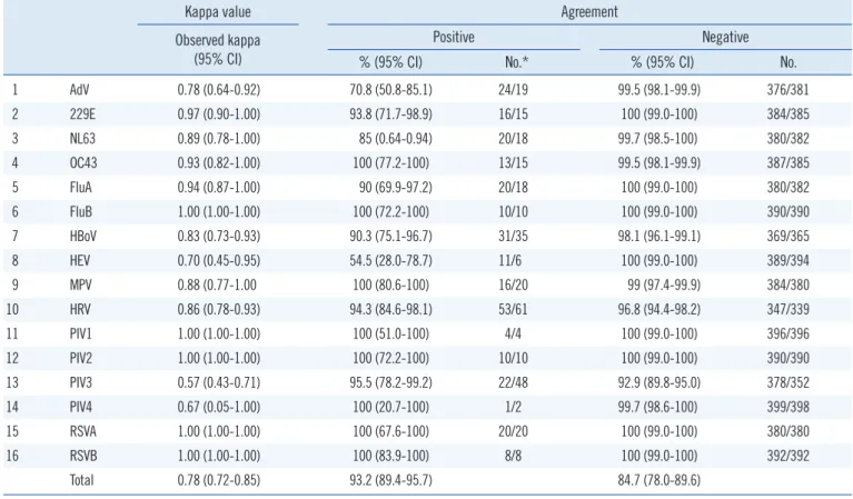
Table 1. Comparison of Allplex Respiratory Panel 1/2/3 and Anyplex II RV16 in the detection of a single respiratory virus
Kappa value Agreement
Observed kappa (95% CI)
Positive Negative
% (95% CI) No.* % (95% CI) No.
1 AdV 0.78 (0.64-0.92) 70.8 (50.8-85.1) 24/19 99.5 (98.1-99.9) 376/381
2 229E 0.97 (0.90-1.00) 93.8 (71.7-98.9) 16/15 100 (99.0-100) 384/385
3 NL63 0.89 (0.78-1.00) 85 (0.64-0.94) 20/18 99.7 (98.5-100) 380/382
4 OC43 0.93 (0.82-1.00) 100 (77.2-100) 13/15 99.5 (98.1-99.9) 387/385
5 FluA 0.94 (0.87-1.00) 90 (69.9-97.2) 20/18 100 (99.0-100) 380/382
6 FluB 1.00 (1.00-1.00) 100 (72.2-100) 10/10 100 (99.0-100) 390/390
7 HBoV 0.83 (0.73-0.93) 90.3 (75.1-96.7) 31/35 98.1 (96.1-99.1) 369/365
8 HEV 0.70 (0.45-0.95) 54.5 (28.0-78.7) 11/6 100 (99.0-100) 389/394
9 MPV 0.88 (0.77-1.00 100 (80.6-100) 16/20 99 (97.4-99.9) 384/380
10 HRV 0.86 (0.78-0.93) 94.3 (84.6-98.1) 53/61 96.8 (94.4-98.2) 347/339
11 PIV1 1.00 (1.00-1.00) 100 (51.0-100) 4/4 100 (99.0-100) 396/396
12 PIV2 1.00 (1.00-1.00) 100 (72.2-100) 10/10 100 (99.0-100) 390/390
13 PIV3 0.57 (0.43-0.71) 95.5 (78.2-99.2) 22/48 92.9 (89.8-95.0) 378/352
14 PIV4 0.67 (0.05-1.00) 100 (20.7-100) 1/2 99.7 (98.6-100) 399/398
15 RSVA 1.00 (1.00-1.00) 100 (67.6-100) 20/20 100 (99.0-100) 380/380
16 RSVB 1.00 (1.00-1.00) 100 (83.9-100) 8/8 100 (99.0-100) 392/392
Total 0.78 (0.72-0.85) 93.2 (89.4-95.7) 84.7 (78.0-89.6)
*No.: Number of results for each virus in AnyplexII RV 16/Allplex Respiratory Panel 1/2/3.
Abbreviations: AdV, adenovirus; 229E, coronavirus 229E; NL63, coronavirus NL63; OC43, coronavirus OC43; FluA, influenza A virus; FluB, influenza B vi- rus; HBoV, human bocavirus 1/2/3/4; HEV, human enterovirus; MPV, human metapneumovirus; HRV, human rhinovirus A/B/C; PIV1, parainfluenza virus 1;
PIV2, parainfluenza virus 2; PIV3, parainfluenza virus 3; PIV4, parainfluenza virus 4; RSVA, respiratory syncytial virus A; RSVB, respiratory syncytial virus B.
on time from the start of nucleic acid extraction until result ac- quisition were compared.
Statistical analyses were conducted using Microsoft Excel (2010) with Analyse-it (Ver. 4.65, Analyse-it Software, Ltd, Leeds, UK).
Inter-rater agreement statistics (kappa values) were obtained to compare the detection of respiratory viruses between All16 and Any16, and we analyzed the sensitivity and specificity based on the monoplex PCR results. This study was exempted by the In- stitutional Review Board of Chonbuk National University Hospi- tal (IRB No. CUH 2015-11-029) with low ethical load, not speci- fying the request of informed consent to the patient because all the studies were done after data was anonymized.
A total of 256 and 250 samples were positive in All16 and Any16, respectively. When a sample was positive for more than one virus, regardless of the virus type or discrepancy, the posi- tive percent agreement between All16 and Any16 was 93.2%
(95% confidence interval [CI], 89.4–95.7), and the negative percent agreement was 84.7% (95% CI, 78.0–89.6). The kappa value for the two methods was 0.78 (95% CI, 0.72–0.85). Re- sults for each virus are summarized in Table 1. Eighteen sam- ples in All16 were found to be H1 or H3 (14 and 4 samples, re-
spectively) by FluA subtyping, and these results were consistent with the sequencing results targeting the hemagglutinin gene of FluA. Further comparison of FluA subtyping with the FilmArray findings showed consistent results for another 26 samples with H3 subtyping.
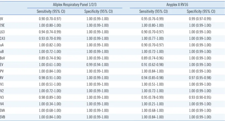
Seventy-four samples showed discrepant results between the two assays, and 68 discrepant samples showed positive results with only one method. Two samples showed discrepancies be- tween the two methods, and the results for four samples were completely different between the assays. For further analysis, we performed monoplex real-time RT-PCR for samples with dis- crepant results, specifically All16-positive and Any16-negative samples; this analysis yielded 83.6% (46/55) consistency with the All16 results. For samples with discrepancies involving All16 negativity and Any16 positivity, rate of consistency with the Any16 results was 34.6% (9/26). The discrepancies for each virus are described in Table 2. Additionally, we analyzed the sensitivity and sensitivity of each assay after resolving discrepant results (Table 3); the analytical specificity test of All16 for 24 bacteria showed all negative results.
When 28 samples were analyzed simultaneously, the test time Table 2. Confirmations of discordant results between Allplex Respiratory Panel 1/2/3 (All16) and Anyplex II RV16
Virus All16(P)*/Any16(P)† No.
All16(P)/Any16(N)‡ All16(N)§/Any16(P)
All16(N)/
Any16(N) No. No.
No. of positive samples in
monoplex PCR No.
No. of positive samples in monoplex PCR
1 AdV 17 2 1 7 2 374
2 229E 15 0 0 1 0 383
3 NL63 17 1 0 3 1 379
4 OC43 13 2 1 0 0 385
5 FluA 18 0 0 2 0 380
6 FluB 10 0 0 0 0 390
7 HBoV 28 7 3 3 1 362
8 HEV 6 0 0 5 4 389
9 MPV 16 4 4 0 0 380
10 HRV 50 11 10 3 0 336
11 PIV1 4 0 0 0 0 396
12 PIV2 10 0 0 0 0 390
13 PIV3 21 27 26 1 0 351
14 PIV4 1 1 1 0 0 398
15 RSVA 8 0 0 0 0 392
16 RSVB 20 0 0 0 0 380
*Positive results in Allplex Respiratory Panel 1/2/3; †Positive results in Anyplex II RV16; ‡Negative results in Allplex Respiratory Panel 1/2/3; §Negative results in Anyplex II RV16
Abbreviations: see Table 1.
Table 3. Sensitivity and specificity of Allplex Respiratory Panel 1/2/3 (All16) and Anyplex II RV16
Allplex Respiratory Panel 1/2/3 Anyplex II RV16
Sensitivity (95% CI) Specificity (95% CI) Sensitivity (95% CI) Specificity (95% CI)
AdV 0.90 (0.70-0.97) 1.00 (0.99-1.00) 0.95 (0.76-0.99) 0.99 (0.97-0.99)
229E 1.00 (0.80-1.00) 1.00 (0.99-1.00) 1.00 (0.80-1.00) 1.00 (0.99-1.00)
NL63 0.94 (0.74-0.99) 1.00 (0.99-1.00) 0.90 (0.70-0.97) 1.00 (0.99-1.00)
OC43 0.93 (0.70-0.99) 1.00 (0.99-1.00) 1.00 (0.77-1.00) 1.00 (0.99-1.00)
FluA 1.00 (0.82-1.00) 1.00 (0.99-1.00) 0.90 (0.70-0.97) 1.00 (0.99-1.00)
FluB 1.00 (0.72-1.00) 1.00 (0.99-1.00) 1.00 (0.72-1.00) 1.00 (0.99-1.00)
HBoV 0.89 (0.74-0.96) 1.00 (0.99-1.00) 0.89 (0.74-0.96) 1.00 (0.99-1.00)
HEV 1.00 (0.61-1.00) 0.99 (0.94-1.00) 0.91 (0.62-0.98) 1.00 (0.99-1.00)
MPV 1.00 (0.84-1.00) 1.00 (0.99-1.00) 1.00 (0.84-1.00) 1.00 (0.99-1.00)
HRV 0.98 (0.91-1.00) 1.00 (0.99-1.00) 0.94 (0.85-0.98) 0.97 (0.95-0.98)
PIV1 1.00 (0.51-1.00) 1.00 (0.99-1.00) 1.00 (0.51-1.00) 1.00 (0.99-1.00)
PIV2 1.00 (0.72-1.00) 1.00 (0.99-1.00) 1.00 (0.72-1.00) 1.00 (0.99-1.00)
PIV3 0.98 (0.89-1.00) 1.00 (0.99-1.00) 0.95 (0.78-0.99) 0.93 (0.90-0.95)
PIV4 1.00 (0.34-1.00) 1.00 (0.99-1.00) 1.00 (0.21-1.00) 1.00 (0.99-1.00)
RSVA 1.00 (0.68-1.00) 1.00 (0.99-1.00) 1.00 (0.68-1.00) 1.00 (0.99-1.00)
RSVB 1.00 (0.84-1.00) 1.00 (0.99-1.00) 1.00 (0.84-1.00) 1.00 (0.99-1.00)
Abbreviations: see Table 1; CI, confidence interval.
and hands-on time showed mean times of 4.3 and 0.5 hour, re- spectively. The mean times required for DNA extraction and PCR preparation using MICROLAB Nimbus IVD and PCR itself were 2.4 and 1.9 hours, respectively, and both analyses required less time than Any16.
Both multiplex RT-PCR assays, All16 and Any16, are based on tagging oligonucleotide cleavage and extension technology, but All16 utilizes MuDT technology. Multiplex real-time PCR typi- cally requires either multiple fluorescence channels or a melting curve analysis step after amplification [5, 6]. The MuDT tech- nology saves time because the melting curve analysis step is unnecessary. A 2.2-hour reduction in the test time for All16 is achieved, regardless of the sample number, compared with Any- 16; this finding reflects the application of MuDT and a one-step process, including PCR and cDNA synthesis, in All16 compared with the two-step analysis, including PCR amplification after cDNA synthesis, used in Any16. Reduction of the hands-on time, work- load, and test time because of the simple one-step analysis with MuDT used in All16 is helpful in clinical laboratories.
Results obtained using the newly developed All16 test showed good agreement with Any16 results. For each virus, the positive and negative percent agreement showed good results. However, slight discrepancies were observed for HEV and PIV3; discrep- ancies in HEV results might reflect low viral loads or primer com-
petition. Any16 detected more positive results for HEV than that by All16. The analytical sensitivity of All16 is reported as 50 cop- ies/μL, except for when PIV1 is analyzed, yielding a sensitivity of 10 copies/reaction, and when PIV4 and MPV are analyzed, with a sensitivity of 103 copies/reaction [7]. In contrast, the sensitivity of Any16 under the same conditions is approximately 6 copies/
μL [8]. Based on the manufacturer’s package inserts, the ana- lytical sensitivities of Any16 and All16 are 50 copies/reaction and 100 copies/reaction, respectively. Therefore, HEV results were consistent with the package inserts. Additionally, Any16 involves a two-step analysis with PCR following cDNA synthesis.
Separation of the cDNA synthesis step from PCR might have in- fluenced HEV RNA purity or extraction, but other RNA virus re- sults were inconsistent with this. Therefore, we speculate that the results were likely due to primer competition.
HRV results from All16 were more sensitive and specific than results from Any16 in this study. Discrepancies in results were likely due to broader primer coverage for various HRV genotypes according to the primer information provided by the manufac- turer (data not shown).
Regarding PIV3, All16 showed more positive cases than Any16, and most discrepant results were confirmed as positive (96%, 26/27) via monoplex PCR. In this respect, Any16 might miss many cases of PIV3, which infects the distal airways and causes
pneumonia and bronchiolitis [9, 10]. Further studies on the de- tection and clinical investigations of HEV and PIV3 infections should be conducted.
Recently, FluA subtyping has become more important because of the emergence of avian and swine FluA [11]; however, there are few relevant clinically available assays. We observed good agreement on FluA subtyping results between All16 and FilmArray.
Since rapid reporting of viral diseases is critical, rapid PCR as- says have been introduced, but these assays have limited through- put. However, according to our analysis, All16 can be performed twice per day without requiring more workers. Therefore, if many samples need to be run, All16 could be advantageous, especially compared with Any16.
The present study has certain limitations. Some samples con- tained viruses at low titers, hindering statistical analyses for cer- tain viruses, such as PIV1 and PIV4, and could not reflect the entire range of seasonal variations in each viral infection.
In conclusion, All16 showed good agreement with Any16. Its FluA subtyping and reduced test time and workload are useful in clinical laboratories. However, further investigations, such as clinical studies of analyses of HEV and PIV3, are needed.
REFERENCES
1. Ginocchio CC. Detection of respiratory viruses using non-molecular based methods. J Clin Virol 2007;40 Suppl 1:S11-4.
2. Mahony JB, Blackhouse G, Babwah J, Smieja M, Buracond S, Chong S, et al. Cost analysis of multiplex PCR testing for diagnosing respiratory virus infections. J Clin Microbiol 2009;47:2812-7.
3. Jorgensen JH, Pfaller MA, et al. eds., American Society for Microbiolo- gy. Manual of clinical microbiology. 11th ed. Washington, DC: ASM Press, 2015:1432-5.
4. Lee YJ, Kim D, Lee K, Chun JY. Single-channel multiplexing without melt- ing curve analysis in real-time PCR. Sci Rep 2014;4:7439.
5. Fu G, Miles A, Alphey L. Multiplex detection and SNP genotyping in a single fluorescence channel. PLoS One 2012;7:e30340.
6. Huang Q, Liu Z, Liao Y, Chen X, Zhang Y, Li Q. Multiplex fluorescence melting curve analysis for mutation detection with dual-labeled, self-quen- ched probes. PLoS One 2011;6:e19206.
7. Huh HJ, Kim JY, Kwon HJ, Yun SA, Lee MK, Lee NY, et al. Performance evaluation of Allplex respiratory panels 1, 2, and 3 for detection of respi- ratory viruses and influenza A virus subtypes. J Clin Microbiol 2017;55:
479-84.
8. Huh HJ, Park KS, Kim JY, Kwon HJ, Kim JW, Ki CS, et al. Comparison of the Anyplex II RV16 and Seeplex RV12 ACE assays for the detection of respiratory viruses. Diagn Microbiol Infect Dis 2014;79:419-21.
9. Fry AM, Curns AT, Harbour K, Hutwagner L, Holman RC, Anderson LJ.
Seasonal trends of human parainfluenza viral infections: United States, 1990-2004. Clin Infect Dis 2006;43:1016-22.
10. Porter DD, Prince GA, Hemming VG, Porter HG. Pathogenesis of hu- man parainfluenza virus 3 infection in two species of cotton rats: Sig- modon hispidus develops bronchiolitis, while Sigmodon fulviventer de- velops interstitial pneumonia. J Virol 1991;65:103-11.
11. Morens DM, Folkers GK, Fauci AS. The challenge of emerging and re- emerging infectious diseases. Nature 2004;430:242-9.
Supplemental Data Table S1. Analytical specificity* of Bacteria for the Allplex Respiratory Panel 1/2/3
Pathogen name Strain
Acinetobacter baumannii KBN06P05033
Actinomyces odontolyticus KBN06P04619
Arcanobacterium haemolyticum KBN06P04505
Burkholderia cepacia KBN06P04105
Corynebacterium amycolatum KBN06P04532
Corynebacterium striatum KBN06P04558
Cryptococcus neoformans KBN06P04554
Enterobacter aerogenes KBN06P04590
Enterobacter asburiae KBN06P04918
Enterobacter cloacae KBN06P04972
Enterococcus faecalis KBN06P04871
Enterococcus faecium KBN06P04561
Escherichia coli KBN06P04939
Klebsiella pneumoniae KBN06P05065
Proteus vulgaris KBN06P05036
Pseudomonas aeruginosa KBN06P05035
Ralstonia mannitolilytica KBN06P04541
Staphylococcus aureus KBN06P04893
Staphylococcus cohnii KBN06P04930
Staphylococcus hominis KBN06P04937
Staphylococcus lugdunensis KBN06P04858
Staphylococcus sciuri KBN06P04931
Staphylococcus simulans KBN06P04874
Streptococcus pneumoniae KBN06P04517
*Samples were incubated on blood agar plates for 24 hours at 35°C under 5% CO2 conditions. We prepared a 0.5-McFarland standard solution from the colonies, and all tests were performed with 100-fold dilutions following the same procedures as used for sample processing.