INTRODUCTION
A fibrous pseudotumor is a benign fibroproliferative tumor arising from the paratesticular tissues. The majority of the re- ported cases show an involvement of the tunica vaginalis and the others are associated with the epididymis, spermatic cord and other surrounding connective tissues (1-3). To the best of our knowledge, there are only rarely reports on the experiences in diagnosing with both ultrasonography (US) and magnetic resonance imaging (MRI), even more rarely are reports on diag- nosing with dynamic contrast-enhanced MRI. In this article, we present a case of fibrous pseudotumor of the tunica vaginalis presenting as painless palpable masses on the right scrotum in a
33-year-old man, with US and MRI findings including the dy- namic study.
CASE REPORT
A 33-year-old man presented to our institution with painless palpable masses on the right scrotum for 2 years. The patient did not have any history of trauma or infection. Laboratory studies were unremarkable. On physical examination, the right scrotum was swollen and a small non-tender hard mass was palpated.
An US with 5- to 12-MHz linear array transducer (iU22, Phil- ips Healthcare, Eindhoven, The Netherlands) revealed multiple well-demarcated, round to oval, iso- to slightly hypoechoic nod-
J Korean Soc Radiol 2014;71(2):75-79 http://dx.doi.org/10.3348/jksr.2014.71.2.75
Received January 28, 2014; Accepted June 18, 2014 Corresponding author: Jae Ho Cho, MD
Department of Radiology, College of Medicine, Yeungnam University, 170 Hyeonchung-ro, Nam-gu, Daegu 705-717, Korea.
Tel. 82-53-620-3043 Fax. 82-53-653-5484 E-mail: [email protected]
This is an Open Access article distributed under the terms of the Creative Commons Attribution Non-Commercial License (http://creativecommons.org/licenses/by-nc/3.0) which permits unrestricted non-commercial use, distri- bution, and reproduction in any medium, provided the original work is properly cited.
A fibrous pseudotumor is a rare benign fibroproliferative disease of the paratesticu- lar tissues, usually originating from the tunica vaginalis. A 33-year-old man with a painless palpable mass on the right scrotum visited our institution. On ultrasonog- raphy (US), multiple well-defined isoechoic nodular lesions with a small amount of vascularity were seen along the tunica vaginalis. Some of the nodules showed pos- terior shadowing. On magnetic resonance imaging (MRI), the lesions showed dark signal intensity on the T2-weighted image and slightly low signal intensity on the T1-weighted image. The dynamic contrast-enhanced MRI showed a mild and pro- gressive enhancement which was persistent on the delayed phase. After surgical excision, the mass was pathologically confirmed as fibrous pseudotumor. Herein we report a case of fibrous pseudotumor of the tunica vaginalis, which was diag- nosed with both US and MRI findings.
Index terms Fibrous Pseudotumor Paratesticular Tunica Vaginalis Ultrasound
Magnetic Resonance
Ultrasonographic and Magnetic Resonance Imaging Findings of Fibrous Pseudotumor of the Tunica Vaginalis: A Case Report
1 고환초막에 발생한 섬유성 가성종양의 초음파 및 자기공명영상 소견:증례 보고1
Jae Beom Hong, MD
1, Jae Ho Cho, MD
1, Mi-Jin Kim, MD
2Departments of 1Radiology, 2Pathology, College of Medicine, Yeungnam University, Daegu, Korea
cord (2-4). First reported by Balloch in 1904, a fibrous pseudo- tumor is a benign reactive process of fibrous proliferation in the paratesticular tissue (1-4). It has been known by several syn- onyms including inflammatory pseudotumor, pseudofibroma- tous periorchitis, chronic proliferative periorchitis, fibrous me- sothelioma, and reactive periorchitis (1, 4). It is most likely that the etiology of this entity is closely related to an inflammatory process such as epididymitis, infected hydrocele, previous sur- gery or trauma (1). Although a fibrous pseudotumor is rare and comprises about 6% of paratesticular masses, it is the second most common mass involving paratesticular tissues, second only to the adenomatoid tumor of the epididymis (1, 2). It has a peak incidence in the third decade of life, but can occur at any age (4, 5). Generally, the size of the tumor ranges from 0.5 to 8 cm in maximal diameter, with the largest one reported to be 25 cm (4). While intratesticular tumors are more likely to arise from the right side, paratesticular tumors have no reported predilec- tion of laterality (1, 2).
The patients usually present with a palpated single or multi- ple, painless, firm scrotal nodules. Less commonly, the lesion may also appear as a diffuse thickening of the testicular capsule (3, 5). Approximately 30% are related to a history of prior trau- ma or infection, supporting the reactive pathogenesis of the le- sion (2, 3, 5). Nearly 50% of the cases represent a hydrocele or hematocele as the most frequently associated findings (2-5).
On microscopic examination, the fibrous pseudotumor shows proliferative fibroblasts and other inflammatory cells intermixed with collagen bundles in the dense hyalinized fibrous tissue (1, 2, 5). Small capillary vessels or calcifications may also be present within the lesion. Immunohistochemical staining is usually pos- itive for smooth muscle-specific actin, vimentin, common mus- cle actin and negative for keratin, desmin and S-100 protein (4).
An US examination with a high-frequency linear probe is the first modality of choice for the detection and evaluation of the scrotal lesion. On gray-scale US, fibrous pseudotumors appear as single or multiple, hypoechoic or hyperechoic nodules, de- pending on the amount of collagen, fibroblasts or calcifications (2, 3, 5, 6). The lesion may show posterior shadowing without calcification due to the dense fibrous and collagenous content.
On color Doppler US, a small to moderate amount of vasculari- ty can be seen within the lesion (5). Our case appeared as multi- ple isoechoic nodular lesions in the right scrotum with minimal ular lesions along the tunica vaginalis in the right scrotal sac
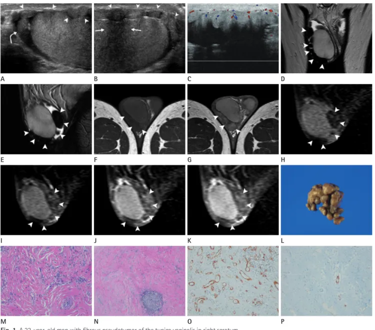
(Fig. 1A, B). The largest one measured about 1.5 cm in size.
Some nodules showed posterior shadowing without any calcifi- cation. On Doppler study, multifocal small areas of color flow signal were noted (Fig. 1C). A small amount of fluid collection was also seen in the right tunical sac. Both testes and epididymi- des were normally well seen.
On MRI (Intera, 1.5-T, Philips Medical Systems, Best, The Netherlands), multiple sharply demarcated, round to oval nod- ules were seen in the right scrotal sac. The lesions showed a mark- edly low signal intensity (SI) on T2-weighted image (T2WI), a slightly low SI on T1-weighted image (T1WI) and a mild en- hancement on the contrast-enhanced T1WI (Fig. 1D-G). A dy- namic contrast-enhanced study with sagittal fat suppression T1WI was performed. Four post-contrast images were obtained at 30, 90, 180, and 300 seconds after the administration of the contrast agent. The lesions showed a mild and progressive en- hancement persisting through the delayed phase (Fig. 1H-K). The provisional diagnosis was a fibrous pseudotumor of the testicular tunic and the differential diagnosis included a mesothelioma.
The patient underwent a surgical excision and the intraopera- tive findings revealed up to 1 cm sized multiple irregular shaped hard nodules attached to the testis and epididymis without direct invasion. A total excision of the mass was performed with some part of the tunica vaginalis preserving testis and epididymis.
Pathologically, the gross specimen showed the lesion in the right scrotal tunica vaginalis measuring 5.7 × 2.7 × 2.9 cm in size.
Multiple firm consistent nodules were attached to the surface of the tunica vaginalis (Fig. 1L). Microscopically, the lesion showed abundant sclerotic and fibrous tissue surrounding vascular struc- tures and largely hypocellular tissue with clusters of inflammato- ry cells (Fig. 1M, N). An immunohistochemical study was per- formed and the tumor cells were unusually not reactive for smooth muscle actin, desmin, CD34, and S-100 protein (Fig. 1O, P). An additional stain for immunoglobulin G4 (IgG4), done to rule out an IgG-related disease was also negative. Finally, the le- sion was pathologically confirmed as fibrous pseudotumor.
DISCUSSION
Paratesticular tumors originate in the intrascrotal surround- ing structures such as testicular tunic, epididymis or spermatic
and thus a better depiction of the fibrous nature of the mass (7).
It also provides additional information of adjacent tissues with a wider field of view. The mass usually shows homogeneous low vascularity and some nodules showed posterior shadowing
without any calcification.
A MRI may help to provide a better tissue characterization
Fig. 1. A 33-year-old man with fibrous pseudotumor of the tunica vaginalis in right scrotum.
A, B. Longitudinal scan of gray-scale ultrasonography (US) at the level of upper (A) and lower poles (B) of the testis shows multiple iso- to slightly hypoechoic nodular lesions (arrowheads) attached at the tunica vaginalis on the anterior aspect of the testis and minimal amount of flu- id collection in the tunical sac. One of the nodules shows posterior shadowing without calcification (arrows). Epididymal head is normally well seen (curved arrow).
C. On color Doppler US, small amount of vascularity is noted in the lesion.
D-G. Multiple nodular lesions (arrowheads) at the tunica vaginalis show homogeneous dark signal intensity (SI) on coronal and sagittal T2- weighted image (D, E), slightly low SI on axial T1-weighted image (T1WI) (F), and mild enhancement on contrast-enhanced axial T1WI (G).
H-K. On dynamic contrast-enhanced study with sagittal fat suppressed T1WI, the lesion shows mild and progressive enhancement which per- sists through the delayed phase (arrowheads). Pre-contrast (H), arterial (after 30 seconds, I), venous (after 90 seconds, J), and delayed phase (af- ter 300 seconds, K) images are presented.
L. Pathologically, the gross specimen of the lesion consists of right scrotal tunica vaginalis with multiple dark brownish firm nodules on the surface.
M. Microscopically, the lesion shows abundant sclerotic and fibrous tissue surrounding vascular structures (hematoxylin and eosin stain, × 100).
N. The lesion also shows largely hypocellular tissue with a cluster of inflammatory cells (hematoxylin and eosin stain, × 40).
O, P. On immunohistochemical stain for actin (O) and desmin (P), the tumor cells reveal negative reaction (immunohistochemistry, × 100).
M I E A
N J F B
O K G C
P L H D
the rarity of this tumor. US and MR imaging facilitate an early recognition of the intactness of testis and the establishment of an appropriate treatment plan (2, 7). An orchiectomy is recom- mended in some rare cases of diffuse band-like fibrous tissue surrounding the entire testis (4).
In summary, we report a case of fibrous pseudotumor arising from the tunica vaginalis of the right scrotum. Characteristic imaging findings on US and MRI are helpful in the detection and diagnosis of the fibrous pseudotumor, thus avoiding an un- necessary treatment such as a radical orchiectomy.
REFERENCES
1. Bruijnes E, Laddé BE, Dabhoiwala NF, Stukart RA. Fibrous pseudotumor of the tunica vaginalis testis. Urol Int 1984;
39:314-317
2. Grebenc ML, Gorman JD, Sumida FK. Fibrous pseudotumor of the tunica vaginalis testis: imaging appearance. Abdom Imaging 1995;20:379-380
3. Tobias-machado M, Corrêa Lopes Neto A, Heloisa Simardi L, Borrelli M, Wroclawski ER. Fibrous pseudotumor of tuni- ca vaginalis and epididymis. Urology 2000;56:670-672 4. Parker PM, Pugliese JM, Allen RC Jr. Benign fibrous pseu-
dotumor of tunica vaginalis testis. Urology 2006;68:427.
e17-427.e19
5. Krainik A, Sarrazin JL, Camparo P, Vincendeau S, Houlgatte A, Cordoliani YS. Fibrous pseudotumor of the epididymis:
imaging and pathologic correlation. Eur Radiol 2000;10:
1636-1638
6. al-Otaibi L, Whitman GJ, Chew FS. Fibrous pseudotumor of the epididymis. AJR Am J Roentgenol 1997;168:1586 7. Cassidy FH, Ishioka KM, McMahon CJ, Chu P, Sakamoto K,
Lee KS, et al. MR imaging of scrotal tumors and pseudotu- mors. Radiographics 2010;30:665-683
8. Patel MD, Silva AC. MRI of an adenomatoid tumor of the tunica albuginea. AJR Am J Roentgenol 2004;182:415-417 9. Hertzberg BS, Kliewer MA, Hertzberg MA, Distell BM. Epi- didymal leiomyoma: sonographic features. J Ultrasound Med 1996;15:797-799
10. Fields JM, Russell SA, Andrew SM. Case report: ultrasound appearances of a malignant mesothelioma of the tunica vaginalis testis. Clin Radiol 1992;46:128-130
SI relative to the testis on both T1- and T2WIs due to dense fi- brotic tissue (2, 3, 6, 7). Typically, the mass shows a minimal en- hancement on the contrast-enhanced image, but the enhance- ment can be variable depending on the capillary network in the lesion (5, 7). In our case, the mass showed dark SI on T2WI, slightly low SI on T1WI and a mild enhancement on the con- trast-enhanced T1WI. Although reported cases including dy- namic study are limited, the lesion is generally thought to show slow but persistent enhancement due to its fibrous nature as presented in our case (7).
The radiologic differential diagnosis includes adenomatoid tumor, leiomyoma, lipoma, and mesothelioma. An adenoma- toid tumor is a benign tumor of mesothelial origin and the most common tumor of the epididymis, accounting for 30% of all pa- ratesticular tumors. The US shows a well-defined iso- or hy- poechoic ovoid lesion with or without a cystic portion (8). The MRI shows an intermediate SI on T1WI, a slightly low SI on T2WI and a variable enhancement (7). The leiomyoma is the second most common neoplasm of the epididymis. On US, multiple recurrent narrow areas of shadow suggesting transition zones and whorl-shaped echo pattern are characteristic findings (9). The lipoma tends to be a well-defined homogeneous hyper- echoic mass without internal vascularity on US, but may be rare- ly hypoechoic or heterogeneous in echotexture. A characteristic of fat tissue is a homogeneous high SI without enhancement on both T1 and T2WIs on MRI. The fat suppression technique or the in- and out-of-phase imaging may also help defining the fat component within the tumor (7). As the tunica vaginalis is a layer of reflected peritoneum, a malignant mesothelioma can also occur, especially in individuals with a history of asbestos exposure. Suggestive US findings are multiple heterogeneous or hyperechoic nodules within the enlarging hydrocele space and a hyperemia of the involved tunica vaginalis (10). On MRI, the le- sion is iso- to hyperintense on T1WI, hyperintense on T2WI and it is well enhanced. Although fibrous pseudotumor can oc- cur as both single and multiple lesions, the multiplicity of the le- sion helps to narrow the differential diagnosis into fibrous pseu- dotumor and malignant mesothelioma (7-10).
The treatment of the fibrous pseudotumor includes the exci- sion of the mass and involved testicular tunic, sparing the testis (1-3, 6). However, a radical orchiectomy has been frequently performed in many cases despite the benign nature because of
고환초막에 발생한 섬유성 가성종양의 초음파 및 자기공명영상 소견: 증례 보고1
홍재범
1· 조재호
1· 김미진
2섬유성 가성종양은 고환주위 조직에서 발생하는 드문 양성 섬유증식성 질환으로, 주로 고환초막에서 발생한다. 33세 남 자가 우측 고환에 무통성 종물을 주소로 본원에 내원하였다. 초음파 검사에서, 혈류신호를 보이는 고환과 비슷한 에코의, 경계가 좋은 구형 또는 난형의 다발성 결절들이 고환초막에 관찰되었고, 일부 결절은 후방그림자를 나타내었다. 자기공명 영상에서, T2-강조영상에서 저신호강도, T1-강조영상에서 약간 저신호강도로 관찰되었고, 역동적 조영증강 T1-강조영 상에서 약하고 점진적으로 조영증강되어 지연기까지 지속되었다. 수술 후 종괴는 섬유성 가성종양으로 확진되었다. 저자 들은 초음파 및 자기공명영상 검사를 통해 진단된 고환초막에서 발생한 섬유성 가성종양의 1예를 보고하고자 한다.
영남대학교 의과대학 1영상의학과학교실, 2병리과학교실