서론
시신경척수염(neuromyelitis optica; NMO)은 시신경과 척수 를 침범하는 드문 염증성 탈수초성 질환이다. 1870년에 Albutt가 시력저하와 척수 병변이 서로 연관성이 있음을 인지한 이후 1880년에 Erb가 처음으로 NMO란 용어를 사용하였으며 Devic 과 그의 제자 Gault가 시신경과 급성척수염이 동반된 16명의 환자를 보고한 이후 보고자의 이름을 따서 Devic’s disease라고 알려져 왔다(1). NMO는 처음에 단발성 질환으로 알려졌으나 이후 여러 보고들에서 재발이 가능한 질환으로 보고되었고(2), 특히 NMO가 재발성인 경우 다발성 경화증(multiple sclerosis;
MS)의 아형 중의 하나인지 아시아에서 흔한 재발성의 optic- spinal MS (OSMS)와 유사한 질환인지 논란이 계속되어 왔다 (3-5).
2004년 Lennon 등(6, 7)에 의해 혈청 자가항체(NMO-
IgG)가 NMO의 진단에 있어서 특징적 표지자로 제시되면서 classic MS와 달리 심한 척수손상을 보이는 NMO가 하나의 독 특한 질환이라는 주장이 대두되었고, 최근에는 임상소견 및 MRI, cerebrospinal fluid (CSF) 소견을 보조적으로 이용하는 Wingerchuk 등(8)의 기준이 NMO를 진단하는 데 널리 이용되 고 있다.
저자들은 Wingerchuk 등(8)의 진단기준에 따라 NMO로 진단 된 11명의 환자를 대상으로 임상소견, 뇌척수액 검사소견 및 진 단에 도움이 될 수 있는 뇌와 척수 영상소견을 보고하고자 한다.
대상과 방법
2001년 1월 1일부터 2010년 3월 31일까지 본원 및 타기관에 서 NMO로 진단받은 환자들의 의무기록과 추적관찰 기간 동 안 촬영한 brain, orbital, spine MRI 소견을 후향적으로 분석하
Received May 10, 2012; Accepted February 22, 2013 Corresponding author: Chang June Song, MD Department of Radiology, Chungnam National University College of Medicine, Chungnam National University Hospital, 282 Munhwa-ro, Jung-gu, Daejeon 301-721, Korea.
Tel. 82-42-280-7834 Fax. 82-42-253-0061 E-mail: [email protected]
This is an Open Access article distributed under the terms of the Creative Commons Attribution Non-Commercial License (http://creativecommons.org/licenses/by-nc/3.0) which permits unrestricted non-commercial use, distri- bution, and reproduction in any medium, provided the original work is properly cited.
Purpose: To report the magnetic resonance (MR) imaging features of the spinal
cord and brain in patients of neuromyelitis optica (NMO).Materials and Methods: Between January 2001 and March 2010, the MR images
(spinal cord, brain, and orbit) and the clinical and serologic findings of 11 NMO pa- tients were retrospectively reviewed. The contrast-enhancement of the spinal cord was performed (20/23). The presence and pattern of the contrast-enhancement in the spinal cord were classified into 5 types.Results: Acute myelitis was monophasic in 8 patients (8/11, 72.7%); and optic neu-
ritis preceded acute myelitis in most patients. Longitudinally extensive cord lesion (average, 7.3 vertebral segments) was involved. The most common type was the dif- fuse and subtle enhancement of the spinal cord with a multifocal nodular, linear or segmental intense enhancement (45%). Most of the brain lesions (5/11, 10 lesions) were located in the brain stem, thalamus and callososeptal interphase. Anti-Ro au- toantibody was positive in 2 patients, and they showed a high relapse rate of acute myelitis. Anti-NMO IgG was positive in 4 patients (4/7, 66.7%).Conclusion: The imaging findings of acute myelitis in NMO may helpful in making
an early diagnosis of NMO which can result in a severe damage to the spinal cord, and to make a differential diagnosis of multiple sclerosis and inflammatory diseases of the spinal cord such as toxocariasis.Index terms
Neuromyelitis Optica Magnetic Resonance Imaging SpineBrain
Sjögren Syndrome
Magnetic Resonance Imaging Features of Neuromyelitis Optica
1시신경척수염의 자기공명영상 소견
1Sun Kyoung You, MD
1, Chang June Song, MD
1, Woon Ju Park, MD
1, In Ho Lee, MD
1, Eun-Hee Son, MD
2Departments of 1Radiology, 2Neurology, Chungnam National University College of Medicine, Chungnam National University Hospital, Daejeon, Korea J Korean Soc Radiol 2013;68(5):375-383
http://dx.doi.org/10.3348/jksr.2013.68.5.375
(fluorescent antinuclear antibody)에서 specked pattern을, 그 리고 안구 증상과 구강 증상을 보였던 환자가 1명 있었다.
영상의학적 소견
Orbital, spine, and brain MRI에 대한 영상분석은 뇌신경 영 상전문의 1명과 영상의학과 전공의 1명이 분석하여 척수 병변 의 조영증강 pattern과 뇌 병변의 유무 및 침범부위에 대해 토 의하여 결론을 얻었다. 여러 병원에서 NMO로 진단받은 환자들 의 영상을 모았기 때문에 영상들은 다양한 기종으로 촬영되었 으며, 영상지표도 병원마다 차이가 있었으나, 7명(63%)의 환자 가 포함되어 있는 기관의 지표를 명시하였다.
Spinal cord image는 T1-, T2-weighted image 각각 axial, sagittal image와 galdolinium-DTPA를 이용한 조영증강 image 를 얻었다(Signa® EXCITETM 1.5, GE Medical Systems, Mil- waukee, WI, USA or Signa® HDxTM, GE Medical Systems, Mil- waukee, WI, USA). 척수 병변의 종축방향의 범위와 조영증강 양상을 분석하기 위해 sagittal image를 5가지의 type으로 분 류하여 평가하였다(Fig. 1).
Brain MRI는 5 mm thickness로 T1, T2 axial, coronal, sa- gittal image를 얻었으며(Signa® EXCITETM 1.5, GE Medical Systems, Milwaukee, WI, USA or Signa® HDxTM, GE Medical Systems, Milwaukee, WI, USA), 그외 추가적으로 fluid atten- uated inversion recovery image와 galdolinium-BOPTA를 이 였다. 발병 당시의 나이, 성별, CSF study, anti-NMO IgG 검사
결과, 그리고 다른 자가면역항체와 Sjogren’s syndrome 관련 증 상 및 혈액학적 검사결과 등이 포함되었다. 분석의 대상이 되었 던 11명의 환자는 9명이 여자, 2명이 남자로 여자(81.8%)가 많 았다. 연령분포는 20~61세로 평균 39.5세였다.
진단기준
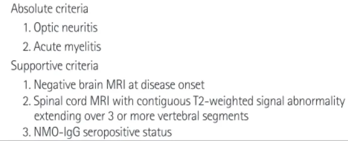
NMO는 2006년 Wingerchuk 등(8)이 제시한 진단 기준(Table 1)에 부합되는 환자들을 포함하였다.
Sjogren’s syndrome의 진단은 American-European Con- sensus Group의 classification criteria를 따랐다(9). 11명 중 1 명이 Primary Sjogren’s syndrome으로 진단받았다. 정확히 Sjogren’s syndrome으로 진단받지는 못하였으나 anti-Ro (SSA) autoantibody positive 소견을 보이고, 형광성항핵항체검사 Table 1. Proposed Diagnostic Criteria for Neuromyelitis Optica
Absolute criteria 1. Optic neuritis 2. Acute myelitis Supportive criteria
1. Negative brain MRI at disease onset
2. Spinal cord MRI with contiguous T2-weighted signal abnormality extending over 3 or more vertebral segments
3. NMO-IgG seropositive status
Note.-Diagnosis requires absolute criteria plus at least 2 of the 3 support- ive criteria. Data from Wingerchuk et al. (8).
Fig. 1. Type of enhancement pattern of spinal cord on sagittal image.
Type A. No enhancement.
Type B. Subtle diffuse heterogenous contrast-enhancement without solitary or multiple strong contrast-enhancing foci.
Type C. Subtle diffuse enhancement with strong solitary nodular- or tumefactive peripheral rim-enhancing foci.
Type Da. Subtle diffuse enhancement with multiple posterior nodular enhancement foci.
Type Db. Subtle diffuse enhancement with multiple posterior continuous longitudinal enhancement foci.
Type Dc. Subtle diffuse enhancement with multiple central nodular enhancement foci.
Type E. Diffuse strong contrast enhancement without disruption.
46.3세(32~61세)였고, 여자가 3명, 남자가 1명이었다. Anti- NMO IgG 항체 음성 환자들의 평균나이는 42.3세(31~59세) 였으며, 3명 모두 여자였다.
Anti-Ro (SSA) 자가면역 항체는 11명 중 2명에서 양성결과 를 보였다. 모두 여성이었으며, 각각 37세, 50세였다.
결과
11명의 임상정보와 anti-NMO IgG 항체유무, 급성척수염과 용한 image를 얻었다. Brain lesion은 brain stem, thalamus, cor-
pus callosum, 3rd ventricle 주위, lateral ventricle 주위, aque- ductal area, 그리고 white matter로 나누어 침범 유무 및 조영 증강 여부를 확인하였다.
혈청검사
Anti-NMO IgG 항체 검사는 간접 면역형광검사법을 통해 이루어졌으며, 11명 중에서 7명이 검사를 시행하였다. Anti- NMO IgG 항체 양성 환자는 4명(57.1%)이었으며 평균나이는 Table 2. Summary of Clinical and Imaging Findings of 11 Patients with NMO
Case Age Sex Anti-NMO IgG
Acute Myelitis Optic Neuritis
Location Enhancement
Type Number of
Attacks Location Number of Attacks
1 59 F (-) C6 to T8 E 1 L 2
L
2 32 M (+) C6 to T9 E 1 Both 2
R
3 50 F (+) C4 to T6 Dc 5 L 1
T1 to T10 Db
C1 to C2 Db
MO to C4 Db
T4 to T6 Db
4 61 F (+) C2 to C6 B 1 R 6
L R R R R
5 18 F × T2 to T4 Dc 18 R 3
Whole spinal cord Da R
T1 to T9 E L
C6 to T11 Dc
T2 to T12 Da
C5 to C6 Da
C1 to C6 B
C2 to C5 C
C1 C
6 31 F (-) MO to T1 B 1 R 2
R
7 37 F (-) T5 to T9 B 5 L 9
Both R
8 45 F × Whole spinal cord × 1 L 3
Both
9 22 F × Lower MO to C6 × 1 L 1
10 42 F (+) C4 to T6 E 1 L 2
R
11 35 M (-) C3 to T1 × 1 L 2
Both Note.-L = left, NMO = neuromyelitis optica, MO = medulla oblongata, R = right, × = do not exam
여 광범위한 영역의 침범을 보이고 있었다. 발생 부위는 경추 척 수부터 척수원추까지 척수 전장를 침범한 2예를 포함하여 경 추 척수와 흉추 척수를 동시에 침범한 경우가 9예(39.1%), 경 추만 침범한 경우가 7예(31.8%), 흉추만 침범한 경우가 7예 (31.8%)였다.
척수 병변의 조영증강은 spine MRI를 시행한 23예 중 20예 에서 시행하였다. 모든 예에서 척수병변에 diffuse heteroge- nous contrast-enhancement를 보여 급성척수염에 합당한 소견 이었다. 조영증강 양상은 B형이 4예, C형이 2예, D형이 10예, E형이 4예로 대부분 D형(50%)과 B형(20%) 또는 E형 (20%)의 조영증강 양상을 보였다. 가장 흔한 조영증강 양상은 D형이었으며 Da가 3예, Db 4예, Dc가 3예였다(Figs. 2-5).
뇌 병변은 11명 중 5명에서 관찰되었다. 시신경염 혹은 급성 척수염과 뇌 병변 간의 시간 간격은 4예에서 각각 17개월, 19 개월, 38개월, 75개월로 평균 37.3개월(0~75개월)이었으며 1 예에서는 척수염과 뇌 병변이 동시에 발생하였다. 뇌 병변이 시 신경염이나 급성척수염을 선행한 경우는 없었다. 병변들은 조영 증강 되지 않았으며, 관련하여 특이적 증상을 보이는 경우도 없 었다. 뇌 병변이 있었던 5명의 환자에서의 Anti-NMO IgG, Anti-Ro (SSA) 항체 유무 및 구체적 뇌 병변의 위치는 Table 3 와 같다. Anti-NMO IgG 항체 유무에 따라 뇌 병변 분포에 뚜 렷한 차이는 없었다. 다만 anti-NMO IgG 항체 결과 여부와 무 관하게 anti-Ro (SSA) 항체 양성 환자 두 명(case number 3 &
7) 모두에서 뇌량과 뇌백질에 병변을 보였다(Fig. 6). 그리고 2 명의 환자 모두에서 각각 5회의 급성척수염 재발이 있었다.
시신경염은 11명의 환자에서 총 32회 발생하여 재발률은 평 시신경염의 유무 및 침범부위와 재발횟수 등은 Table 2에 표기
하였다. 급성척수염은 11명 중 8명(72.7%)에서 재발 없이 단발 성이었다. 척수염이 재발되었던 3명 중 2명에서 5회, 1명에서 18회의 재발을 보였다. 척수염은 평균 7.3 척추 분절을 침범하
Fig. 2. Type B. 1st attack of acute myelitis in patient 7. Contrasted en- hanced T1-weighted sagittal image shows subtle diffuse heteroge- neous contrast enhancement. There are no solitary or multiple nodular enhancement.
Fig. 3. Type C. 10th attack of acute myelitis in patient 5.
A. T2-weighted image shows high signal intensity in the cervical cord at the level of C1-2 (arrow).
B, C. Contrasted enhanced T1-weighted sagittal (arrow) and axial image (arrow) shows tumefactive peripheral rim enhancing nodular lesion at C1-2.
A B C
균 2.9회였다(Fig. 7). 9명의 환자에서 시신경염은 재발성이었 으나 2명의 환자에서는 재발이 없었다. 좌측과 우측을 동시에 침범한 경우가 4명에서 있었으며, 재발을 포함하여 좌측과 우 측 모두 침범했던 경우는 7명에서 볼 수 있었다. 좌측과 우측이 각각 10회, 12회로 좌우 발병의 차이는 없었다. 시신경염은 11 명 중 9명에서 급성척수염에 선행하였으며, 1명의 환자는 시신 경염이 급성척수염과 동시에 발생했고, 나머지 한 명은 급성척 수염이 시신경염에 선행하여 발생하였다. 시신경염과 급성척수 염 발병 시간 간격은 평균 40.9개월(0~108개월)이었다.
고찰
1970년대까지만 하더라도 NMO는 단발성이 특징적 소견으 로 알려졌으며, 이는 재발하는 특징을 가지는 동양의 OSMS와 의 감별점이었다. 하지만 1990년대 NMO의 진단에 있어 NMO 의 재발성이 고려되기 시작하였으며, 2006년 새로운 진단기준
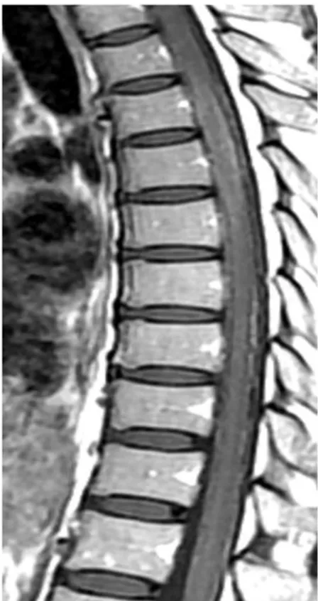
Fig. 5. Type E. Acute myelitis in patient 2, without recurrence of acute myelitis. Contrast-enhanced T1-weighted sagittal image shows subtle diffuse contrast-enhancement in whole spinal cord (arrows) with dif- fuse strong contrast-enhancement.
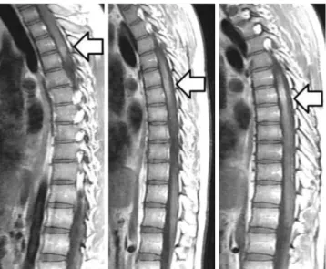
Fig. 4. Type D acute myelitis in patient 3 and 5.
A, B. Type Da. At 5th attack of aucte myelitis in patient 5, contrast-enhanced T1-weighted sagittal image shows multifocal nodular (arrows) and continuous longitudinal enhancement (empty arrows in B) mainly along the posterior aspect of cervical and upper thoracic spinal cord.
C, D. Type Db. At 5th attack of acute myelitis in patient 3, contrast-enhanced T1-weighted sagittal image shows multiple continuous longitudinal enhancement (arrows) along the spinal cord at C1 to C2 (C). Simultaneously the similar lesion were along the spinal cord at T4-T6 (arrows) (D).
E, F. Type Dc. At 1st attack of acute myelitis in patient 3, contrast-enhanced T1-weighted sagittal and axial image shows nodular enhancement (arrow) in central portion of thoracic spinal cord at the level of T4.
A B C D E F
Table 3. Location of Brain Lesions; 5 of 11 Patients Patient
Number Anti-NMO
Antibody Anti-Ro (SSA)
Autoantibody Enhancement Brain Lesion
BS Tha CC 3rd 4th Aque Lat WM
2 + - - o o
3 + + - o o
5 × - - o o
6 - - - o
7 - + - o o o
Note.-Aque = periaqueductal, BS = brain stem, CC = corpus callosum, Lat = lateral ventricle, T = thalamus, WM = white matter, 3rd = 3rd ventricle, 4th = 4th ventricle, × = do not exam
ter에서 비특이적 병변들이 보인다고 보고된 바 있다(18). 최근 NMO와 MS의 척수 병변에 대해 MRI 축상영상을 비교하여 발 표한 연구에 따르면 NMO는 연속적이며 중심부를 침범하는 양 상을, MS는 주변부를 부분적으로 침범한다고 하였다(19). 본 연구에서는 11명의 환자에서 척수 병변 침범이 평균 7.3 분절 침범을 보여 NMO의 척수 병변의 특징으로 제시되었던 3개 이 상의 척수 분절 침범, 즉 long extensive 척수 병변은 본 연구에 서도 크게 다르지 않은 결과를 보였다. 척수의 조영증강 pat- tern은 D type이 10회(50%)로 빈도수로만 본다면, 가장 흔한 type이었다.
Spinal visceral larva migrans of Toxocara canis가 횡단성 척 수염을 mimicking 할 수 있지만, 상대적으로 짧은 분절을 침범 하고, 이동성을 보이는 특징이 있어(20) NMO와의 감별이 어렵 지 않을 것으로 생각된다.
1999년 Wingerchuk 등이 제시한 진단기준을 따르면 시신경 과 척수염 이외의 증상을 보이는 경우 NMO 진단을 배제할 수 있었다. 하지만 무증상 뇌 병변이 NMO 환자에서 흔하게 보고 되었고(21), 증상이 있는 뇌 병변도 진단에서 배제하지 않는 진 단기준이 제시되었으며(8), 최근에는 NMO에 특이적인 뇌 병 변이 있다는 주장과 함께 그 병변이 뇌에서의 aquaporin 4 분포 와 관련이 있으며, periaqueductal, hypothalamic and periven- tricular lesion들이 특징적 위치라고 제시되고 있다(21, 22). 본 연구에서도 뇌 병변을 보이는 경우에 있어 periaqueductal area, thalamus, corpus callosum, internal capsule, midbrain의 cere- bral pedencle에 분포하고 있어, 그간 보고되었던 NMO에서의 특징적 뇌 병변의 분포(21, 22)에 합당하다고 볼 수 있었다.
Anti-NMO IgG 양성 결과와 급성횡단성 척수염의 높은 재 발률이 관계가 있다는 보고들이 있지만(13, 14), 본 연구에서는 anti-NMO IgG 항체 양성 환자 4명 중 1명에서 척수염이 5회 이 제시되면서 NMO와 OSMS 간의 구분이 모호해졌으며, 일부
저자는 NMO와 OSMS를 같은 질환으로 보아야 한다고 주장했 다(3). 하지만 아직도 NMO와 OSMS와의 구분은 명확하지 않 으며(10-14) 저자는 그간 OSMS로 발표되었던 보고들에 MS 환자와 NMO 환자가 모두 포함되어 있을 가능성이 높아, OSMS 환자와 NMO 환자의 소견을 비교할 때 공통점과 그렇 지 않은 점들이 혼재하는 것으로 생각된다.
한국에서는 MS 유병률에 대한 대단위 연구가 보고된 적은 없으나, 동양에서 MS가 낮은 유병률을 보이는 것과 더불어 한 국에서도 그 발생빈도가 낮을 것으로 예상된다(10, 15). 한국인 에서 보이는 MS의 임상 소견과 영상 소견에 대해 분석한 2006 년 보고를 보면(16) 한국의 MS는 서양보다는 동양의 환자들에 서 보이는 MS와 더 일치되는 특징을 보인다고 하였다(16, 17).
따라서 한국에서도 OSMS의 빈도가 높을 것으로 예상해 볼 수 있다.
NMO 환자의 급성기 동안 시행한 spine MRI에서 cord swell- ing과 multiple level involvement, diffuse enhancement를 보이 며, brain MRI에서는 supratentorial, frontoparietal white mat-
B
B
C D
A
A
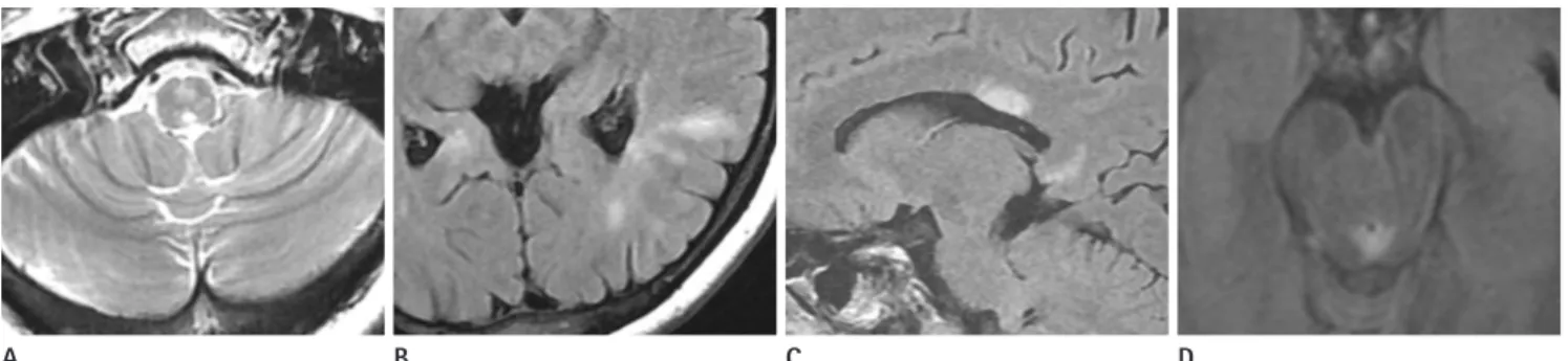
Fig. 6. Variable located high signal intensity lesions of the brain on fluid attenuated inversion recovery (FLAIR) or T2-weighted image in patient 7 (A-C) and patient 6 (D).
A. T2-weighted axial image shows focal high signal intensity (SI) in medulla oblongata.
B. FLAIR axial image shows high SI in left periventricular white matter.
C. FLAIR sagittal image shows edematous white matter lesions involving the corpus callosum, particularly the splenium.
D. FLAIR axial image shows high signal intensity lesion in periaqueductal area of midbrain.
Fig. 7. In patient 2, optic neuritis at first attack. T2-weighted axial im- age (A) shows high-signal intensity foci in the minimally expanded left optic nerve. These lesion enhances following intravenous contrast administration on fat saturated T1-weighted image (B).
syndrome). Neurology 1999;53:1107-1114
3. Lucchinetti CF, Mandler RN, McGavern D, Bruck W, Gleich G, Ransohoff RM, et al. A role for humoral mechanisms in the pathogenesis of Devic’s neuromyelitis optica. Brain 2002;125(Pt 7):1450-1461
4. Galetta SL, Bennett J. Neuromyelitis optica is a variant of multiple sclerosis. Arch Neurol 2007;64:901-903
5. Weinshenker BG. Neuromyelitis optica is distinct from multiple sclerosis. Arch Neurol 2007;64:899-901
6. Lennon VA, Wingerchuk DM, Kryzer TJ, Pittock SJ, Lucchi- netti CF, Fujihara K, et al. A serum autoantibody marker of neuromyelitis optica: distinction from multiple sclerosis.
Lancet 2004;364:2106-2112
7. Lennon VA, Kryzer TJ, Pittock SJ, Verkman AS, Hinson SR.
IgG marker of optic-spinal multiple sclerosis binds to the aquaporin-4 water channel. J Exp Med 2005;202:473-477 8. Wingerchuk DM, Lennon VA, Pittock SJ, Lucchinetti CF,
Weinshenker BG. Revised diagnostic criteria for neuromy- elitis optica. Neurology 2006;66:1485-1489
9. Vitali C, Bombardieri S, Jonsson R, Moutsopoulos HM, Al- exander EL, Carsons SE, et al. Classification criteria for Sjögren’s syndrome: a revised version of the European cri- teria proposed by the American-European Consensus Group. Ann Rheum Dis 2002;61:554-558
10. Kira J. Multiple sclerosis in the Japanese population. Lan- cet Neurol 2003;2:117-127
11. Nakashima I, Fujihara K, Miyazawa I, Misu T, Narikawa K, Nakamura M, et al. Clinical and MRI features of Japanese patients with multiple sclerosis positive for NMO-IgG. J Neurol Neurosurg Psychiatry 2006;77:1073-1075
12. Tanaka K, Tani T, Tanaka M, Saida T, Idezuka J, Yamazaki M, et al. Anti-aquaporin 4 antibody in selected Japanese mul- tiple sclerosis patients with long spinal cord lesions. Mult Scler 2007;13:850-855
13. Chong H, Kermode A, Tan C. The role of anti-aquaporin-4 antibody in asian patients with multiple sclerosis: confu- sions and controversies. Neurology Asia 2007;12:135-139 14. Matsuoka T, Matsushita T, Kawano Y, Osoegawa M, Ochi H,
Ishizu T, et al. Heterogeneity of aquaporin-4 autoimmuni- ty and spinal cord lesions in multiple sclerosis in Japanese.
Brain 2007;130(Pt 5):1206-1223
15. Park KH, Kim SW, Kim SK. Multiple sclerosis in Busan Ko- 재발하였으며, 나머지 3명에서는 재발 없이 1회의 척수염을 보
이고 있어, 재발률과의 관계는 뚜렷하지 않았다.
5회의 척수염 재발을 보였던 두 명의 환자에서 anti-Ro (SSA) 항체 양성이었고, 11명의 환자 중 8예(72.2%)에서 척수염의 재발이 없었던 것과 비교하여 anti-Ro (SSA) 항체 양성 환자에 서 급성척수염 재발률이 높음을 알 수 있었다. 이는 재발성 횡단 척수염 환자에서 anti-Ro (SSA) 항체 비율이 높았다는 결과를 보였던(23) 연구와 비슷한 결과를 보인 것으로 볼 수 있었다.
NMO는 systemic lupus erythematosus 또는 Sjogren’s syn- drome과 같은 자가면역질환이 동반될 수 있는 것으로 알려져 있으나, 그 관계는 명확히 정립된 바가 없다(24, 25). 최근 한 국에서 Sjogren’s syndrome 환자에서 뇌 또는 척수 병변을 보 일때 NMO 환자에서 보이는 것과 비슷한 양상을 보이고, anti- NMO IgG 항체 양성을 보이기도 한다는 보고가 있다(26-28).
이러한 경우 뇌나 척수 병변이 Sjogren’s syndrome보다 NMO 에 의해 발생한 것으로 의심해 보아야 하는데, steroid 치료를 하는 Sjogren’s syndrome과 달리 NMO라고 생각되는 경우에 는 조기에 공격적 면역치료가 필요하기 때문이다(28).
본 연구가 가지는 제한점으로는, 첫째, 임상기록에 대한 검토 가 후향적으로 이루어졌다는 점과 두 번째로는 연구대상에 대 한 대조군이 없다는 점이다. 세 번째로 모든 환자에서 anti- NMO IgG 항체와 anti-Ro (SSA) 항체 검사가 시행되지 않았 다는 점이고, 네 번째로 증상이 발생하였을 당시 모든 예에서 영상의학적 검사를 시행하지는 않았다는 점이다.
결론적으로 NMO 환자에서 급성척수염은 평균 7개 이상의 척추 분절을 침범하고 있었으며, 조영증강 양상은 미만성의 미 세한 조영증강과 함께 여러 부위에서 결절정, 선상, 분절성의 강한 조영증강을 보이는 경우가 대부분이었다. 시신경염은 대 부분의 환자에서 척수염에 선행하여 발생하였다. Anti-NMO IgG 항체는 보고된 것과 유사한 양성률을 보였으며 anti-Ro (SSA) 항체는 양성률이 높지 않았으나 재발성 환자에서 양성 인 경우가 많았다. 이상의 급성척수염의 영상 소견은 심한 척수 손상을 초래할 수 있는 NMO의 조기진단과 다발성경화증, toxocariasis 등 염증성 질환과의 감별진단에 도움을 줄 수 있을 것으로 생각된다.
참고문헌
1. Devic E. Myélite subaiguë compliquée de névrite optique.
Bull Med (Paris) 1894;8:1033-1034
2. Wingerchuk DM, Hogancamp WF, O’Brien PC, Weinshenker BG. The clinical course of neuromyelitis optica (Devic’s
sions localized at sites of high aquaporin 4 expression.
Arch Neurol 2006;63:964-968
23. Hummers LK, Krishnan C, Casciola-Rosen L, Rosen A, Mor- ris S, Mahoney JA, et al. Recurrent transverse myelitis as- sociates with anti-Ro (SSA) autoantibodies. Neurology 2004;62:147-149
24. Pittock SJ, Lennon VA, de Seze J, Vermersch P, Homburger HA, Wingerchuk DM, et al. Neuromyelitis optica and non organ-specific autoimmunity. Arch Neurol 2008;65:78-83 25. Koo YS, Yoo JK, Kwon DY, Park MH, Koh SB, Kim BJ, et al.
Neuromyelitis optica with positive anti-Ro and anti-La antibodies. J Korean Neurol Assoc 2009;27:446-448 26. Cho J, Kim S, Kim J, Chu C, Lee M, Shin H, et al. Two cases
of primary sjogren’s syndrome presenting as relapsing-re- mitting multiple sclerosis. J Korean Neurol Assoc 2004;22:
410-413
27. Min JH, Kim HJ, Kim BJ, Lee KW, Sunwoo IN, Kim SM, et al. Brain abnormalities in Sjogren syndrome with recurrent CNS manifestations: association with neuromyelitis optica.
Mult Scler 2009;15:1069-1076
28. Kim SM, Waters P, Vincent A, Kim SY, Kim HJ, Hong YH, et al. Sjogren’s syndrome myelopathy: spinal cord involve- ment in Sjogren’s syndrome might be a manifestation of neuromyelitis optica. Mult Scler 2009;15:1062-1068 rea clinical features and prevalence. J Korean Neurol Assoc
1983;1:29-36
16. Lee SS, Sohn EH, Nam SW. Preliminary studies on the clin- ical features of multiple sclerosis in Korea. J Clin Neurol 2006;2:231-237
17. Cho YJ, Jeon BS, Kim YH, Chang KH. Clinical features and outcomes from diagnostic work-up in definite multiple sclerosis. J Korean Neurol Assoc 1999;17:823-828
18. O’Riordan JI, Gallagher HL, Thompson AJ, Howard RS, Kingsley DP, Thompson EJ, et al. Clinical, CSF, and MRI findings in Devic’s neuromyelitis optica. J Neurol Neuro- surg Psychiatry 1996;60:382-387
19. Lalan S, Khan M, Schlakman B, Penman A, Gatlin J, Hern- don R. Differentiation of neuromyelitis optica from multi- ple sclerosis on spinal magnetic resonance imaging. Int J MS Care 2012;14:209-214
20. Lee IH, Kim ST, Oh DK, Kim HJ, Kim KH, Jeon P, et al. MRI findings of spinal visceral larva migrans of Toxocara canis.
Eur J Radiol 2010;75:236-240
21. Pittock SJ, Lennon VA, Krecke K, Wingerchuk DM, Lucchi- netti CF, Weinshenker BG. Brain abnormalities in neuro- myelitis optica. Arch Neurol 2006;63:390-396
22. Pittock SJ, Weinshenker BG, Lucchinetti CF, Wingerchuk DM, Corboy JR, Lennon VA. Neuromyelitis optica brain le-
시신경척수염의 자기공명영상 소견
1유선경
1· 송창준
1· 박운주
1· 이인호
1· 손은희
2목적: 시신경척수염 환자들의 척수와 뇌에서 보이는 자기공명영상 소견을 보고하고자 한다.
대상과 방법: 2001년 1월부터 2010년 3월까지, 시신경척수염으로 진단받은 11명의 의무기록과 brain, orbital, spine MRI 를 후향적으로 분석하였다. 23예 중 20예에서 척수 병변에 대해 조영증강 영상을 얻었으며, 조영증강 유무 및 그 양상을 5가지 type으로 분류하였다.
결과: 급성척수염은 11명 중 8명(72.7%)에서 단발성이었고 대부분에서 시신경염이 척수염에 선행하였다. 척수 병변은 평균 적으로 7.3개의 vertebral segment를 침범하는 광범위한 분포를 보였다. 미미한 조영증강을 보이는 척수 내에 여러 개의 결 절형, 선형, 또는 분절형의 강한 조영증강을 보이는 것이 가장 흔하였다(45%). 뇌 병변(5/11, 10개)의 대부분은 brainstem, thalamus, callososeptal interphase에 위치하였다. Anti-Ro autoantibody는 2명의 환자에서 양성이었고, 공통적으로 높은 급성척수염의 재발률을 보였다. Anti-NMO IgG는 7명 중 4명에서 양성(66.7%)이었다.
결론: 시신경척수염 환자들의 척수 자기공명영상 소견은 심한 척수손상을 초래할 수 있는 시신경척수염의 조기진단과 다 발성경화증, toxocariasis 등의 염증성 질환과의 감별진단에 도움을 줄 수 있을 것으로 생각된다.
충남대학교 의과대학 충남대학교병원 1영상의학과, 2신경과