149
Copyrights © 2013 The Korean Society of Radiology
INTRODUCTION
Dysplasia epiphysealis hemimelica (DEH) or Trevor’s disease is a rare non-hereditary developmental bone dysplasia charac- terized by an osteocartilaginous tumor arising from an epiphysis (1). The general prevalence has been reported be 1 in 1000000 and the etiology is unknown. Most patients are first seen be- tween the ages of 2 and 14 years and three times more common in boys (2). DEH predominantly occurs in the lower limb with the talus, distal tibia, distal fibula, distal femur, proximal tibia and tarsal bones in the order of the frequency (3).
CASE REPORT
Case 1
A 4-year-old boy presented to the orthopedics clinic, with in- cidentally found mass on the right knee with no symptoms.
There was no history of trauma or previous joint swelling. The patient had normal gait and normal range of motion of the knee. Radiographs of the right knee showed multiple ossifica-
tions in the postero-medial aspect of the distal femoral epiphy- sis. The metaphyses of the femur and tibia were normal (Fig.
1A). Ultrasound and magnetic resonance imaging (MRI) re- vealed the presence of asymmetric epiphyesal cartilaginous overgrowth, which contained multiple ossifications (Fig. 1B-E).
It was decided to observe the patient with no surgical interven- tion since no pain and normal range of movement of the knee.
During the past eight years, serial follow-up radiographs showed that the epiphysis of the distal femur had been gradually normalized (Fig. 2) and the last follow-up MRI showed normal bone contour and signal intensity in the distal femur with disap- pearance of the DEH findings (Fig. 3).
Case 2
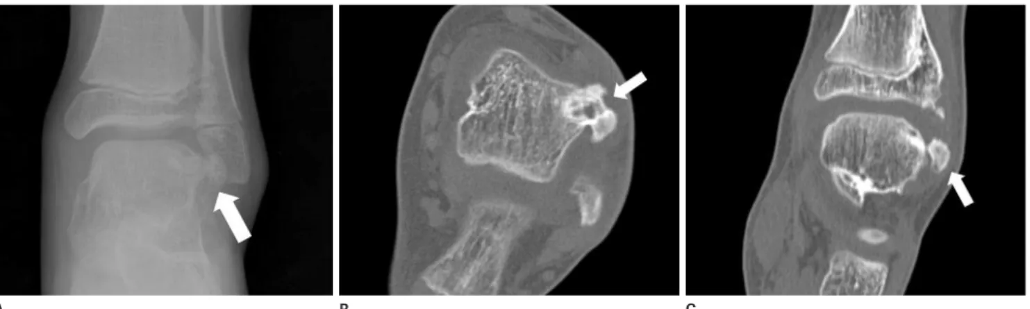
A 10-year-old boy presented to the orthopedic clinic, with an- kle pain and mass on the anterolateral aspect of the left ankle, which was palpable for a month. There was a history of left an- kle sprain about 8 months ago. Physical examination revealed a palpable hard mass (1 × 1 cm in size) of the anterior lateral as- pect of the talus. Pain was induced on full dorsiflexion. Radio-
Case Report
pISSN 1738-2637 / eISSN 2288-2928 J Korean Soc Radiol 2013;69(2):149-152 http://dx.doi.org/10.3348/jksr.2013.69.2.149
Received March 27, 2013; Accepted April 14, 2013 Corresponding author: Kil Ho Cho, MD Department of Diagnostic Radiology, College of Medicine, Yeungnam University,
170 Hyeonchung-ro, Nam-gu, Daegu 705-717, Korea.
Tel. 82-53-620-3045 Fax. 82-53-653-5484 E-mail: [email protected]
This is an Open Access article distributed under the terms of the Creative Commons Attribution Non-Commercial License (http://creativecommons.org/licenses/by-nc/3.0) which permits unrestricted non-commercial use, distri- bution, and reproduction in any medium, provided the original work is properly cited.
Trevor’s disease, also known as dysplasia epiphysealis hemimelica, is a rare develop- mental disorder presented with epiphyseal overgrowth involving one or multiple epiphyses. Here we report the radiologic findings of two cases of dysplasia epiphy- sealis hemimelica in a 4-year-old boy in the knee without symptom and a 10-year- old boy in the ankle with pain. The former was observed for eight years and the lat- ter was treated with surgical resection.
Index terms Trevor’s Disease
Dysplasia Epiphysealis Hemimelica Osteocartilage Overgrowth
Case Report of Imaging Analyses of the Dysplasia Epiphysealis Hemimelica (Trevor’s Disease)
편측성 골단 이형성증(트레버 질환)의 영상소견: 2예 보고
Jang Ho Suh, MD, Kil Ho Cho, MD
Department of Diagnostic Radiology, College of Medicine, Yeungnam University, Daegu, Korea
Case Report of Imaging Analyses of the Dysplasia Epiphysealis Hemimelica (Trevor’s Disease)
150
J Korean Soc Radiol 2013;69(2):149-152 jksronline.orgmelica (Fig. 5). Surgical resection was repeated for the recurred mass with similar pathological results as before.
DISCUSSION
The clinical manifestations of Trevor’s disease can be varied.
The most common presenting complaints are from a painless deformity around a joint to a painful joint with mechanical symptoms (1). The typical radiographic finding is asymmetric epiphyseal cartilaginous overgrowth, containing multiple ossifi- cation centers. The patterns of the epiphyseal chondral calcifica- tion are variable namely stippled, irregular or dense. The epiph- yseal calcified spots of the lesion are often multi-centric, which are gradually enlarged with mineralization and become conflu- ent with the main epiphysis. CT is useful for the detection of small foci of early calcification or ossification within the carti- laginous mass and can identify cortical and medullary continu- ity between the DEH lesion and the adjacent bone. MRI is the technique of choice to identify the dimensions of the unossified cartilage mass, the extent of epiphyseal involvement and the sta- graph and computed tomography (CT) of the ankle showed ex-
cessive bony overgrowth from the anterolateral aspect of the talus with soft tissue swelling (Fig. 4). The abnormal over-growth portion of the talus was excised and for pathological examination which indicated dysplasia epiphysealis hemimelica. Two years later, the patient revisited with ankle pain. Radiograph and CT of the ankle revealed recurrence of the dysplasia epiphysealis hemi- Fig. 1. 4-year-old man with Trevor’s disease in the right distal femur.
A. Initial oblique (A) radiograph shows multiple calcific foci in the postero-medial aspect of the epiphysis of the right distal femur (arrow).
B. Sagittal sonography (B) shows epiphyseal cartilaginous overgrowth, containing multiple echogenic foci (arrow).
C-E. Axial T2-weight image (C), axial T1-weight image (D) and axial enhanced T1-weight image (E) show the presence of asymmetric epiphyseal cartilaginous overgrowth (small arrows), which contain multiple ossifications, in the postero-medial aspect of the distal femur (arrows).
Fig. 2. 4-year-old man with Trevor’s disease in the right distal femur. Follow-up oblique radiographs after 12 months (A), 24 months (B), 54 months (C) and 8 years (D) show that calcific foci are maturated and becomes confluent with the epiphysis of the distal femur (arrows).
Fig. 3. 4-year-old man with Trevor’s disease in the right distal femur.
Follow-up after 8 years, axial T2 weight image (A) and coronal T1 weight image (B) show normal bone contour and signal intensity in the distal femur with disappearance of the dysplasia epiphysealis hemimelica findings.
A
A
A
B C
B
B
D
C
E
D
Case Report of Imaging Analyses of the Dysplasia Epiphysealis Hemimelica (Trevor’s Disease) Jang Ho Suh, et al
151
jksronline.org J Korean Soc Radiol 2013;69(2):149-152
eight years and the other was surgically managed. In particular, there is significant that first case was confirmed by radiologic image that DEH findings gradually became confluent with the main epiphysis during observation for eight years and finally made normal bone contour and density without deformity.
REFERENCES
1. Kuo RS, Bellemore MC, Monsell FP, Frawley K, Kozlowski K.
Dysplasia epiphysealis hemimelica: clinical features and management. J Pediatr Orthop 1998;18:543-548
2. Shinozaki T, Ohfuchi T, Watanabe H, Aoki J, Fukuda T, Tak- agishi K. Dysplasia epiphysealis hemimelica of the proxi- mal tibia showing epiphyseal osteochondroma in an adult.
Clin Imaging 1999;23:168-171
3. Rosero VM, Kiss S, Terebessy T, Köllö K, Szöke G. Dysplasia epiphysealis hemimelica (Trevor’s disease): 7 of our own cases and a review of the literature. Acta Orthop 2007;78:
tus of the epiphysis (4).
Differential diagnoses include myositis ossificans, infection, tumoral calcinosis, synovial chondromatosis, loose bodies, vas- cular or parasitic calcification on radiography. Biopsy is not nec- essary; however, if imaging results are not conclusive, biopsy should be performed to exclude chondrosarcoma and osteosar- coma (5).
Management options for the treatment of Trever’s disease in- clude simple observation or surgical excision. By considering pain or deformity, appropriate treatment should be performed.
As in our first case, asymptomatic DEH can be observed be- cause there is no known risk of malignant transformation. Prog- nosis and symptoms depend on the site and size of the lesion and the degree of incongruity of the involved site. The recur- rence rate of the deformity is reported to be high (6).
The two cases presented here were precisely diagnosed by CT or MRI prior to the management. Since the different symptoms and deformities of the each patient, the first was observed for
Fig. 4. 10-year-old man with Trevor’s disease in the left talus. Initial AP radiograph (A), unenhanced axial (B) and coronal (C) CT show irregular ossified mass, arising talus, in the antero-lateral aspect of the talus (arrows).
Fig. 5. 10-year-old man with Trevor’s disease in the left talus. Follow-up study after 2 years due to recurred ankle pain, oblique radiograph (A), unenhanced axial (B) and coronal (C) CT show recurred mass in the antero-lateral aspect of the talus (arrows).
A
A
B
B
C
C
Case Report of Imaging Analyses of the Dysplasia Epiphysealis Hemimelica (Trevor’s Disease)
152
J Korean Soc Radiol 2013;69(2):149-152 jksronline.orgsealis hemimelica of the knee. Skeletal Radiol 1997;26:
226-229
6. Keret D, Spatz DK, Caro PA, Mason DE. Dysplasia epiphyse- alis hemimelica: diagnosis and treatment. J Pediatr Orthop 1992;12:365-372
856-861
4. Tyler PA, Rajeswaran G, Saifuddin A. Imaging of dysplasia epiphysealis hemimelica (Trevor’s disease). Clin Radiol 2013;68:415-421
5. Lang IM, Azouz EM. MRI appearances of dysplasia epiphy-
편측성 골단 이형성증(트레버 질환)의 영상소견: 2예 보고
서장호 · 조길호
트레버 질환 혹은 편측성 골단 이형성증은 성장기 뼈 골단 골연골의 과성장이 하나 혹은 여러 골단에 발생하는 드문 질환 이다. 저자는 증상없이 내원한 4세 남자 환아의 오른쪽 무릎의 증례와 좌측 발목통증이 있던 10세 남자 환아 등 2예를 영 상소견과 함께 보고한다. 전자는 보존적 치료를, 후자는 수술적 치료를 시행하였다.
영남대학교 의과대학 영상의학과