Percutaneous Retrieval of Embolized Amplatzer Septal Occluder after Treatment of Double Atrial Septal Defect: A Case Report
Embolization of the occlusion device after percutaneous closure of atrial septal defect (ASD) is a potential disastrous complication. The usual site of embolization is the right side of the heart including pulmonary artery, but the device embolization to the extracardiac aorta is extremely rare. Here, we report a successful percutaneous retrieval case of the embolized Amplatzer Septal Occluder (ASO) to the descending thoracic aorta after the successful deployment of two ASO devices in a patient with double ASD. Competition between the two devices to obtain a stable position may be an explanation for the migration of ASO.
Keywords: Heart Septal Defects, Atrial; Septal Occluder Device; Device Removal Jae Yeong Cho,1,3 Kye Hun Kim,1,3*
Hyun Ju Yoon,1,3 Hyun Ju Seon,2,3 Youngkeun Ahn,1 Myung Ho Jeong,1 Jeong Gwan Cho,1 and Jong Chun Park1,3
1Department of Cardiovascular Medicine, 2Radiology,
3Translational Research Center on Aging, Chonnam National University Hospital, Gwangju, Korea Received: 15 September 2014
Accepted: 16 January 2015 Address for Correspondence:
Kye Hun Kim, MD
Department of Cardiovascular Medicine, Chonnam National University Hospital, 42 Jebong-ro, Dong-gu, Gwangju 501-757, Korea
Tel: +82.62-220-6977, Fax: +82.62-223-3105 E-mail: [email protected]
http://dx.doi.org/10.3346/jkms.2015.30.9.1361 • J Korean Med Sci 2015; 30: 1361-1366
INTRODUCTION
Surgical closure has been the gold standard for the treatment of adult patients with atrial septal defect (ASD) (1). Since the first attempt of transcatheter device closure of ASD in 1970s, how- ever, it has become an effective alternative therapy to surgical repair in patients with single ASD of secundum type (2, 3). As the improvement of devices, percutaneous closure of ASD with multiple defects has also been attempted and closed success- fully (4, 5).
Although percutaneous device closure of ASD is known to be safe, several potential complications may develop (6-8). Embo- lization of the occlusion device after percutaneous closure of ASD is one of the most disastrous complication even though it is rare. The usual site of embolization is the right side of the heart including pulmonary artery, but the device embolization to the extracardiac aorta is an extremely rare complication (8). Embo- lization of Amplatzer Septal Occluder (ASO) to the ascending aorta, aortic arch, or abdominal aorta in a patient with single ASD has been described so far (8).
Here, we report a successful percutaneous retrieval case of the embolized ASO to the descending thoracic aorta after the successful deployment of two ASO devices in a patient with dou- ble ASD with review of the literature.
CASE DESCRIPTION
A 27-yr-old male presented with mild dyspnea. Physical exami- nations were non-specific except for the fixed splitting of sec- ond heart sound on auscultation. Electrocardiography showed normal sinus rhythm with right atrial enlargement and right bundle branch block. Transthoracic (TTE) and transesopha- geal echocardiography (TEE) revealed dilated right ventricle and atrium (Fig. 1A), abnormal left to right shunt flow through maximally 10.1 mm (superior rim) and 14.3 mm (inferior rim)- sized two ASDs of secundum type (Fig. 1B), and the calculated ratio of pulmonary (Qp) to systemic blood flow (Qs) was 1.8.
Around superior defect showed the 9.3 mm sized-aortic rim, and another defect was seen inferiorly with 8.2 mm-sized inter- vening septum between them. Postero-inferior rim was mea- sured as 13.6 mm, and no defect was shown posterior to the su- perior one with 29 mm-sized floppy posterior rim. Under the TEE and fluoroscopic guidance, percutaneous device closure of double ASD was performed using ASO. Balloon-sizing diam- eter of each ASD was measured as 13.5 mm and 15 mm using stop-flow technique, and thereby 14 mm- and 16 mm-sized ASOs were placed in each defect in order and deployed one by one subsequently and successfully (Fig. 1C). Final TEE revealed good apposition of two ASO devices with minimal residual shunt flow (Fig. 1D).
CASE REPORT
Cardiovascular Disorders
The patient experienced sudden and transient chest discom- fort on the next day. On the third day, pre-discharge follow up TTE was showed no visualization of one of the deployed ASO devices, which was located in postero-inferior rim (Fig. 1D), with de novo left to right shunt (Fig. 1E), and the other ASO showed stable position. The embolized ASO was identified in the descen- ding thoracic aorta on careful TTE examination and chest CT angiography (Fig. 2). Because TTE showed decreased amounts
of shunt through the remained ASD with decreased size of the right ventricle, percutaneous retrieval of the embolized ASO was planned without performing ASD closure. After 10-french catheter was introduced through the femoral artery, the embo- lized ASO was successfully retrieved by snaring the screw on the right atrial disc of ASO (Fig. 3). The patient have not had any symptoms and events for 1 yr of clinical follow up.
A B
C D
E F
Fig. 1. Transesophageal echocardiography revealed about 10.1 mm (antero-superior rim, long arrow) and 14.3 mm (postero-inferior rim, short arrow)-sized double atrial septal defects (ASD) (A) with shunt flow from left atrium to right atrium (B). After successful closure of ASD, two Amplatzer Septal Occluders (ASOs) were identified in superior (long arrow) and inferior rim (short arrow) of the interatrial septum (C) without residual defects (D). At third day, one of the two ASOs was not seen in the inferior defect (short arrow) due to device embolization and the other device in superior defect showed stable position (E). De novo left-to-right shunt developed between both atria (F).
Cho JY, et al. • Embolization of Amplatzer Device in Double Atrial Septal Defect
DISCUSSION
As the percutaneous transcatheter device closure has been ac- cepted and widely used for the treatment of ASD, several device related safety issues or unexpected complications such as atrial perforation, thrombus formation, hemopericardium associated with erosion of aorta, device embolization have also been de- scribed (6-8). Embolization of the occlusion device after percu- taneous closure of ASD is one of the most disastrous complica- tion even though it is rare (8). The incidence of device emboli- zation after device closure of ASD is varied from 0.01% to 0.55%
(8, 9). In a recent study of 284 cases of ASD device closure, de- vice embolization was reported up to 1.4% (10). In the present case, ASO was embolized to the descending thoracic aorta. Ac- cording to the previous study, the usual site of embolization is the right side of the heart including pulmonary artery, but the device embolization to the extracardiac aorta, as shown in the
present case, is very rare (9).
Device embolization after percutaneous ASD closure usually developed during the procedure or immediate peri-procedural period (8-10), but late embolization has been described also (11, 12). Considering clinical symptoms of the patient, device embolization might be developed at the second hospital day and identified at the third hospital day after successful ASD clo- sure in the present case.
Device embolization was treated by percutaneous retrieval by using gooseneck snare in the present case. In the early peri- od of transcatheter occlusion of ASD, device embolization is usually treated by surgery (10-12), but more recently it is usually treated by percutaneously by using snares or biopsy forceps (9, 13-15). Therefore, it would be a reasonable therapeutic strategy to try percutaneous retrieval of the embolized device initially before performing surgical removal.
The proposed mechanism or predisposing conditions of de- Fig. 2. Transthoracic echocardiography revealed the embolized Amplatzed Septal Occluder (ASO) in the descending aorta on parasternal long (A) and modified short axis view (B). Chest computed tomographic angiography revealed one deployed ASO in interatrial septum and the other ASO in the descending thoracic aorta on axial (C) and sagital view (D). Arrow indicates embolized ASO and arrow head indicates deployed ASO in interatrial septum.
A B
C D
A
C
B
D Fig. 3. (A) Aortogram revealed the embolized Amplatzed Septal Occluder (ASO) in the descending thoracic aorta (arrow head) and another ASO in the interatrial septum (arrow).
After successful snaring of the screw on right atrial disc of ASO (B), ASO was successfully retrieved into the 10-french catheter (C, D).
vice embolization are as follows; undersized device, inadequate or floppy rim, and operator-related technical issues such as poor experience or device malposition or excessive tension (8, 16). However, these predisposing risk factors for device emboli- zation were not identified, and thus other risk factors might be involved in the present case (Fig. 4). The authors suggested that competitive movement between the two devices to obtain their stable positions, especially in the overlapped site of the devices, would be a possible explanation of the device embolization in
the present case.
Percutaneous treatment strategy of multiple ASDs has been constantly evolving. Mehta et al. (17) studied in 28 patients with multiple ASD and found that the ability of the Helex Septal Oc- cluder (HSO) devices to overlap or sandwich each other may make it an ideal choice particularly when multiple devices need to be implanted. Song et al. (18) reported that 2 occluders are necessary for the distance of two ASDs more than 7 mm, but a single occluder is sufficient for those 7 mm and less. Since the
Cho JY, et al. • Embolization of Amplatzer Device in Double Atrial Septal Defect
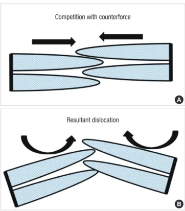
Fig. 4. A proposed mechanism of the embolization of Amplatzer Septal Occluder (ASO).
(A) Two ASO devices compete with each other along cardiac movement. (B) Counter- force between two devices results in dislocation and embolization of less stable ASO.
Competition with counterforce
Resultant dislocation
A
B
intervening septum between two defects was measured as 8.2 mm, it would be appropriate to choose two-device closure in the present case. However, interventional cardiologist who un- dergoes device closure for ASD with multiple defects should keep in mind and closely monitor the possibility of late device embolization even though initial transcatheter closures of ASDs are successful. To the best of our knowledge, the present case was the first report on the late embolization of one Amplatzer device after the successful deployment of 2 ASO devices for dou- ble ASDs. Considering the present case, multiple ASDs or im- plantation of multiple devices should be added as one of the important risk factors for device embolization.
DISCLOSURE
The authors have no conflicts of interest to disclose.
AUTHOR CONTRIBUTION
Conception and coordination of the study: Cho JY, Kim KH. De- sign of ethical issues: Cho JY, Yoon HJ, Park JC. Acquisition of data: Cho JY, Kim KH, Yoon HJ, Seon HJ. Data review: Cho JY, Youngkeun Ahn, Jeong MH, Cho JG. Manuscript preparation:
Cho JY, Kim KH. Manuscript approval: all authors.
ORCID
Jae Yeong Cho http://orcid.org/0000-0002-9393-2821 Kye Hun Kim http://orcid.org/0000-0002-6885-1501 Hyun Ju Yoon http://orcid.org/0000-0003-1285-3660 Hyun Ju Seon http://orcid.org/0000-0003-1146-8875 Youngkeun Ahn http://orcid.org/0000-0003-2022-9366 Myung Ho Jeong http://orcid.org/0000-0003-4173-1494 Jeong Gwan Cho http://orcid.org/0000-0001-7855-4490 Jong Chun Park http://orcid.org/0000-0002-1637-7991 REFERENCES
1. Hopkins RA, Bert AA, Buchholz B, Guarino K, Meyers M. Surgical patch closure of atrial septal defects. Ann Thorac Surg 2004; 77: 2144-9; author reply 9-50.
2. King TD, Thompson SL, Steiner C, Mills NL. Secundum atrial septal de- fect. Nonoperative closure during cardiac catheterization. JAMA 1976;
235: 2506-9.
3. Du ZD, Hijazi ZM, Kleinman CS, Silverman NH, Larntz K, Amplatzer Investigators. Comparison between transcatheter and surgical closure of secundum atrial septal defect in children and adults: results of a multi- center nonrandomized trial. J Am Coll Cardiol 2002; 39: 1836-44.
4. Pedra CA, Fontes-Pedra SR, Esteves CA, Assef J, Fontes VF, Hijazi ZM.
Multiple atrial septal defects and patent ductus arteriosus: successful out- come using two Amplatzer septal occluders and Gianturco coils. Cathet Cardiovasc Diagn 1998; 45: 257-9.
5. Cao Q, Radtke W, Berger F, Zhu W, Hijazi ZM. Transcatheter closure of multiple atrial septal defects. Initial results and value of two- and three- dimensional transoesophageal echocardiography. Eur Heart J 2000; 21:
941-7.
6. Berger F, Vogel M, Alexi-Meskishvili V, Lange PE. Comparison of results and complications of surgical and Amplatzer device closure of atrial sep- tal defects. J Thorac Cardiovasc Surg 1999; 118: 674-8; discussion 8-80.
7. Krumsdorf U, Ostermayer S, Billinger K, Trepels T, Zadan E, Horvath K, Sievert H. Incidence and clinical course of thrombus formation on atrial septal defect and patient foramen ovale closure devices in 1,000 consecu- tive patients. J Am Coll Cardiol 2004; 43: 302-9.
8. Moore J, Hegde S, El-Said H, Beekman R 3rd, Benson L, Bergersen L, Holzer R, Jenkins K, Ringel R, Rome J, et al. Transcatheter device closure of atrial septal defects: a safety review. JACC Cardiovasc Interv 2013; 6:
433-42.
9. Levi DS, Moore JW. Embolization and retrieval of the Amplatzer septal occluder. Catheter Cardiovasc Interv 2004; 61: 543-7.
10. Amanullah MM, Siddiqui MT, Khan MZ, Atiq MA. Surgical rescue of embolized amplatzer devices. J Card Surg 2011; 26: 254-8.
11. Son JW, Park JS. Subacute, silent embolization of amplatzer atrial septal defect closure device to the pulmonary artery. J Cardiovasc Ultrasound 2012; 20: 201-4.
12. Mashman WE, King SB, Jacobs WC, Ballard WL. Two cases of late em- bolization of Amplatzer septal occluder devices to the pulmonary artery following closure of secundum atrial septal defects. Catheter Cardiovasc Interv 2005; 65: 588-92.
13. Chan KT, Cheng BC. Retrieval of an embolized amplatzer septal occlud- er. Catheter Cardiovasc Interv 2010; 75: 465-8.
14. Pala S, Açar G, Tigen K, Kirma C. Percutaneous retrieval of an interatrial septal occluder device embolized into the aortic arch. Turk Kardiyol Dern Ars 2010; 38: 502-4.
15. Guimaraes M, Denton CE, Uflacker R, Schonholz C, Selby B Jr, Hannegan C. Percutaneous retrieval of an Amplatzer septal occluder device that had migrated to the aortic arch. Cardiovasc Intervent Radiol 2012; 35: 430-3.
16. Misra M, Sadiq A, Namboodiri N, Karunakaran J. The ‘aortic rim’ recount:
embolization of interatrial septal occluder into the main pulmonary ar- tery bifurcation after atrial septal defect closure. Interact Cardiovasc Tho- rac Surg 2007; 6: 384-6.
17. Mehta S, Hill JA, Qureshi AM, Latson LA, Prieto LR. Helex device closure of multiple atrial septal defects. Catheter Cardiovasc Interv 2014; 84: 204- 10.
18. Song ZY, He GX, Shu MQ, Hu HY, Tong SF, Ran BL, Liu JP, Li YH, Jing T.
Clinical efficiency and safety analysis of transcatheter closure of multiple atrial septal defects in adults. Clin Cardiol 2009; 32: 130-4.