This is an Open Access article distributed under the terms of the Creative Commons Attribution Non-Commercial License (http://creativecommons.org/licenses/by-nc/4.0/) which permits unrestricted non-commercial use, distribution, and reproduction in any medium, provided the original work is properly cited.
Copyright © 2019. Anatomy & Cell Biology
Introduction
The mandibular division of the trigeminal nerve (cranial nerve V) is a mixed nerve containing somatic afferent and special visceral efferent fibers that run downward through the foramen ovale. The lingual nerve (LN) is one of the terminal branches of the mandibular division and supplies afferent fibers to the mucosa of the floor of the mouth, lingual gin- giva of the mandible, and mucosa of the anterior two-thirds of the tongue [1, 2]. In the pterygomandibular space, the LN receives the chorda tympani branch of the facial nerve (CN VII) posteriorly, runs along the LN, and innervates the sense of taste from the anterior two-thirds of the tongue. The LN has communicating branches (anterior, middle, and posterior parts of the tongue) with the hypoglossal nerve [3]. The LN
passes through and under the attachment on the mandible of the superior pharyngeal constrictor and pterygomandibular raphe and travels close to the lingual plate and lingual crest near the lower third molar [4]. There are studies describing the relationship of the LN and the mandible in formalin fixed cadavers [5-9]. Reliability of the measurements in the location of the LN in the fixed cadavers is doubtful as the position of the LN could change before and after tissue fixation. Interest- ingly, Iwanaga [10] demonstrated that the LN moved based on tongue position in fresh frozen cadavers. LN injury is one of the major complications of wisdom tooth extraction [11].
Therefore, an improved anatomical knowledge of the LN in the lower third molar area is critical to avoiding unnecessary complications for dentists and oral surgeons. Therefore, this study aims to observe the diameter of the LN at the level of the third lower molar using fresh frozen cadavers.
Materials and Methods
Twenty sides (right, 10; left, 10) from 10 fresh frozen adult cadaveric Caucasian heads were used for the present study.
The specimens were derived from three males and seven fe-
Original Article
https://doi.org/10.5115/acb.2019.52.2.140 pISSN 2093-3665 eISSN 2093-3673
Corresponding author:
Joe Iwanaga
Seattle Science Foundation, 550 17th Ave, James Tower, Suite 600, Seattle, WA 98122, USA
Tel: +1-2067326500, Fax: +1-2067326599, E-mail: joei@seattlesciencefoundation.org
An anatomical study of the lingual nerve in the lower third molar area
Shogo Kikuta
1,2, Joe Iwanaga
1,2,3, Jingo Kusukawa
2, R. Shane Tubbs
1,41Seattle Science Foundation, Seattle, WA, USA, 2Dental and Oral Medical Center, Kurume University School of Medicine, Kurume, 3Division of Gross and Clinical Anatomy, Department of Anatomy, Kurume University School of Medicine, Kurume, Japan, 4Department of Anatomical Sciences, St. George’s University, St. George’s, Grenada, West Indies
Abstract: The lingual nerve (LN) is a branch of the mandibular division of the trigeminal nerve, and its injury is one of the major complications during oral surgery. This study aims to investigate the anatomy of the LN in the lower third molar area.
Twenty sides from ten fresh-frozen adult cadaveric Caucasian heads were examined to measure the diameter of the LN. The mean diameter of the LN was 2.20±0.37 mm (range, 1.61–2.95 mm). There were no statistically significant differences in the measurements between sexes, sides, or tooth status (dentulous or edentulous). Understanding the anatomical features of the LN is essential for performing any surgical procedure in the oral region.
Key words: Lingual nerve, Mandibular nerve, Anatomy, Fresh cadaver, Wisdom tooth Received November 27, 2018; Revised December 29, 2018; Accepted January 2, 2019
Anatomical study of the lingual nerve
https://doi.org/10.5115/acb.2019.52.2.140
Anat Cell Biol 2019;52:140-142
141
www.acbjournal.org
males with age at death ranging from 57 to 93 years (average age, 71.5±12.7 years). The number of the dentulous and eden- tulous mandibles were both five. The mucosa of the oral floor at the level of the lower third molar was incised anteroposte- riorly and the loose connective tissue was bluntly dissected to expose the LN. Then, the diameter of the LN at the level of the third lower molar was measured. The relationship between the LN, mucosa, and submandibular gland was noted. All dissec- tion and measurements were performed by one oral surgeon.
A microcaliper (Mitutoyo, Kanagawa, Japan) was used for all measurements. This study was not required to be approved by our ethical committee as this was a cadaveric study. No previ- ous scar was observed in the floor of the oral cavity or tongue.
All quantitative measurements were documented as the mean±standard deviation. To compare data, one-way analysis of variance was used with Scheffé’s post hoc test and Fisher exact test. Statistical significance was set at P<0.05.
Results
On all sides, the main trunk of the LN was identified at the level of the lower third molar just below the mucosa and above the submandibular gland (Fig. 1). The diameter of the LN ranged from 1.61 to 2.95 mm (mean, 2.20±0.37 mm). No statistically significant differences in the measurements were observed between males (n=3) and females (n=7) (P>0.05).
The mean diameter of the LN was 2.18±0.34 mm (range, 1.62–2.60 mm) on right sides and 2.22±0.40 (range, 1.79–2.95 mm) on left sides. There was no statistically significant dif-
ference (P>0.05) between right and left sides. The mean diameters of the LN in the dentulous and edentulous man- dibles were 2.17±0.43 mm (n=10; range, 1.62–2.95 mm) and 2.23±0.30 (n=10; range, 1.89–2.88 mm), and there was no sta- tistically significant difference (P>0.05). There were no major anatomical variations of the LN during dissections.
Discussion
The LN arises from the posterior division of the mandibu- lar nerve under the foramen oval and into the infratemporal fossa [12]. It then travels under the mandibular attachment of the superior pharyngeal constrictor and pterygomandibu- lar raphe and approaches the lower third molar. Kiesselbach and Chamberlain [5], in a study of 34 adult cadaveric heads, described that the LN contacted the lingual plate in 62% and existed at or above the alveolar crest in 17.6% at the region of the third lower molar. Miloro et al. [13], using high-resolution magnetic resonance imaging (HR-MRI), reported that the LN contacted the lingual plate in 25% and existed at or above the alveolar crest in 10% of patients at the region of the third lower molar. Behnia et al. [4], in 430 fresh cadaveric heads, also reported that the LN contacted the lingual plate in 22.3%
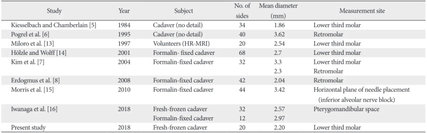
of patients and existed above and below the lingual crest in 14.5% and 85.8% at the region of the third lower molar, re- spectively. There are several pieces of literature that described the diameter of the LN at the third lower molar [5, 7, 13, 14].
Kiesselbach and Chamberlain [5] showed that the diameter of the nerve was 1.86 mm in 34 adult cadaveric heads. Miloro et al. [13] studied using HR-MRI that the diameter was 2.54 mm (range, 1.58–3.13 mm) in 10 volunteers (mean age, 24.7 years;
range, 21–35 mm). Hölzle and Wolff [14] reported that the diameter was 2.7±0.3 mm (range, 1.9–3.6) in 68 specimens (34 formalin-fixed cadaveric heads). Kim et al. [7] also clari- fied that the diameter was 3.3±0.6 mm (range, 2.2–4.4) in 32 specimens (hemi-sectioned formalin-fixed cadaveric heads).
In our study, the diameter of the LN in the fresh frozen cadav- ers was 2.20±0.37 mm (range, 1.61–2.95). However, several authors reported that the mean diameter of the LN at the retromolar area was 3.62±1.0 mm (range, 2.5–4.5) [6], 2.3±0.4 mm (range, 1.5–3.5) [7], and 2.04±0.4 mm (range, 1.42–2.96) [8] in formalin-fixed cadavers. At the horizontal plane of nee- dle placement, the mean diameter of the LN in formalin-fixed cadavers was 3.42±0.38 mm (range, 2.55–4.15) [15]. At the pterygomandibular space, the mean diameter of the LN was 2.57±0.44 mm in fresh frozen cadavers and 2.97±0.48 mm Fig. 1. Lingual nerve in the left lower third molar area (arrow).
Anat Cell Biol 2019;52:140-142 Shogo Kikuta, et al
142
www.acbjournal.org https://doi.org/10.5115/acb.2019.52.2.140
in formalin fixed cadavers [16]. There is a discrepancy in the mean diameter of the LN in the literature (Table 1). However, Hölzle and Wolff [14] concluded that the influence of forma- lin fixation on the shape of the LN was minimal by compar- ing its shape between formalin fixed and fresh cadavers. By contrast, Iwanaga et al. [16] reported that the mean diameter of the LN had a statistically significant difference between fresh-frozen and formalin-fixed cadavers. Despite some dis- crepancy between these studies, there is no doubt that the LN is a risk factor during oral surgery.
In conclusion, this study observed the detailed diameter of the LN using fresh frozen cadavers. A better anatomical un- derstanding of the LN may help minimize LN injury, which is a severe complication of oral procedures.
Acknowledgements
The authors thank those who donated their bodies for ana- tomical education and research.
References
1. Heasman PA, Beynon AD. Quantitative diameter analysis of lin- gual nerve axons in man. J Dent Res 1986;65:1016-9.
2. Zur KB, Mu L, Sanders I. Distribution pattern of the human lin- gual nerve. Clin Anat 2004;17:88-92.
3. Iwanaga J, Watanabe K, Saga T, Tabira Y, Nakamura M, Fisahn C, Tubbs RS, Kusukawa J, Yamaki KI. Communicating branches be- tween lingual and hypoglossal nerve: observation using Sihler's staining technique. Surg Radiol Anat 2017;39:741-5.
4. Behnia H, Kheradvar A, Shahrokhi M. An anatomic study of the lingual nerve in the third molar region. J Oral Maxillofac Surg 2000;58:649-51.
5. Kiesselbach JE, Chamberlain JG. Clinical and anatomic observa-
tions on the relationship of the lingual nerve to the mandibular third molar region. J Oral Maxillofac Surg 1984;42:565-7.
6. Pogrel MA, Renaut A, Schmidt B, Ammar A. The relationship of the lingual nerve to the mandibular third molar region: an ana- tomic study. J Oral Maxillofac Surg 1995;53:1178-81.
7. Kim SY, Hu KS, Chung IH, Lee EW, Kim HJ. Topographic anato- my of the lingual nerve and variations in communication pattern of the mandibular nerve branches. Surg Radiol Anat 2004;26:
128-35.
8. Erdogmus S, Govsa F, Celik S. Anatomic position of the lingual nerve in the mandibular third molar region as potential risk fac- tors for nerve palsy. J Craniofac Surg 2008;19:264-70.
9. Shimoo Y, Yamamoto M, Suzuki M, Yamauchi M, Kaketa A, Kasahara M, Serikawa M, Kitamura K, Matsunaga S, Abe S.
Anatomic and histological study of lingual nerve and its clinical implications. Bull Tokyo Dent Coll 2017;58:95-101.
10. Iwanaga J. The clinical view for dissection of the lingual nerve with application to minimizing iatrogenic injury. Clin Anat 2017;30:467-9.
11. Standring S. Gray’s anatomy: the anatomical basis of clinical practice. 41st ed. London: Elsevier Health Sciences; 2015.
12. Joo W, Funaki T, Yoshioka F, Rhoton AL Jr. Microsurgical anato- my of the infratemporal fossa. Clin Anat 2013;26:455-69.
13. Miloro M, Halkias LE, Slone HW, Chakeres DW. Assessment of the lingual nerve in the third molar region using magnetic reso- nance imaging. J Oral Maxillofac Surg 1997;55:134-7.
14. Hölzle FW, Wolff KD. Anatomic position of the lingual nerve in the mandibular third molar region with special consideration of an atrophied mandibular crest: an anatomical study. Int J Oral Maxillofac Surg 2001;30:333-8.
15. Morris CD, Rasmussen J, Throckmorton GS, Finn R. The ana- tomic basis of lingual nerve trauma associated with inferior alveolar block injections. J Oral Maxillofac Surg 2010;68:2833-6.
16. Iwanaga J, Choi PJ, Vetter M, Patel M, Kikuta S, Oskouian RJ, Tubbs RS. Anatomical study of the lingual nerve and inferior alveolar nerve in the pterygomandibular space: complications of the inferior alveolar nerve block. Cureus 2018;10:e3109.
Table 1. The diameter of the lingual nerve in each measurement site
Study Year Subject No. of
sides
Mean diameter
(mm) Measurement site
Kiesselbach and Chamberlain [5] 1984 Cadaver (no detail) 34 1.86 Lower third molar
Pogrel et al. [6] 1995 Cadaver (no detail) 40 3.62 Retromolar
Miloro et al. [13] 1997 Volunteers (HR-MRI) 20 2.54 Lower third molar
Hölzle and Wolff [14] 2001 Formalin- fixed cadaver 68 2.7 Lower third molar
Kim et al. [7] 2004 Formalin-fixed cadaver 32 3.3 Lower third molar
2.3 Retromolar
Erdogmus et al. [8] 2008 Formalin-fixed cadaver 42 2.04 Retromolar
Morris et al. [15] 2010 Formalin-fixed cadaver 44 3.42 Horizontal plane of needle placement (inferior alveolar nerve block)
Iwanaga et al. [16] 2018 Fresh-frozen cadaver 32 2.57 Pterygomandibular space
Formalin-fixed cadaver 12 2.97
Present study 2018 Fresh-frozen cadaver 20 2.20 Lower third molar
HR-MRI, high-resolution magnetic resonance imaging.