Progression of Compression Type Femoral Neck Stress Fracture
5
0
0
전체 글
(2) 이승림 외. 진행 양상을 보인 압박형 대퇴 경부 피로 골절. 상 우측 대퇴 경부 내하방에 피질골의 연속성이 소실된 소견. 검사상 우측 대퇴 경부의 하방 뿐만 아니라 상방 피질골에. 없이 형성된 내측 가골이 관찰되었다(Fig. 1). 골 스캔 검사상. 연하여 형성된 내측 가골이 추가로 발견되었고(Fig. 9), MRI. 우측 대퇴 경부의 흡수가 증가되어 있었으며(Fig. 2), magnetic. 검사상에서 내하방 피질골 뿐만 아니라 외상방 피질골까지. resonance imaging (MRI) 검사상 내하방 피질골에서 해면골로. 이어진 골절선이 관찰되어(Fig. 10), 불안정 골절로 진행되었. 진행하고 있으나 외상방 피질골까지는 도달하지 않은 골절선. 다고 판단하여 유관나사못을 이용한 수술적 내고정을 시행하. 이 관찰되었다(Fig. 3). 압박형 대퇴 경부 피로 골절로 진단되어. 였다(Fig. 11).. 입원 하에 체중부하 금지 및 침상 안정가료의 보존적 치료를 예정하였다. 동통은 완화되는 소견이었으나, 2주 간격으로. 고. 찰. 시행한 추시 단순 방사선 검사상 대퇴 경부의 내측 가골이 점점 뚜렷해지면서 외상방 피질골을 향해 진행되는 소견이. 피로 골절은 정상 골에 반복되는 비정상적인 응력이 발생하. 관찰되었으며(Fig. 4), 6주 후 시행한 추시 MRI 검사상 내하방 피질골에서의 골절선이 외상방 피질골까지 진행되었다(Fig. 5). 진단 7주 후에 추가적인 골절선의 진행이나 전위를 우려하 여 수술적 치료로서 3개의 유관 나사를 이용한 내고정을 시행 하였다(Fig. 6).. 증례 2 19세 남자로 약 2개월 전 구보 훈련 도중 특별한 외상력 없이 우측 고관절 동통이 발생하였으며 운동시 심해지는 양상 이 지속되어 내원하였다. 단순 방사선 사진상 우측 대퇴 경부 내하방 피질골에 연하여 형성된 내측 가골이 관찰되었으며 (Fig. 7), 골 스캔 검사 역시 양성이었다(Fig. 8). 압박형 대퇴 경부 피로 골절이 의심되어 입원 하에 체중부하 금지 및 침상 안정가료의 보존적 치료를 시행하였다. 3주 후 추시 방사선 Fig. 2. A scintigram of the same patient demonstrate increased activity in the right femoral neck.. Fig. 1. A 20-year-old male with a gradual onset of right groin pain. An anteroposterior radiograph of the right hip shows haziness of internal callus at inferomedial portion of right femoral neck (arrow).. Fig. 3. MRI shows that fracture line involves inferomedial cortex of right femoral neck (arrow).. 제28권 제2호 2010. 145.
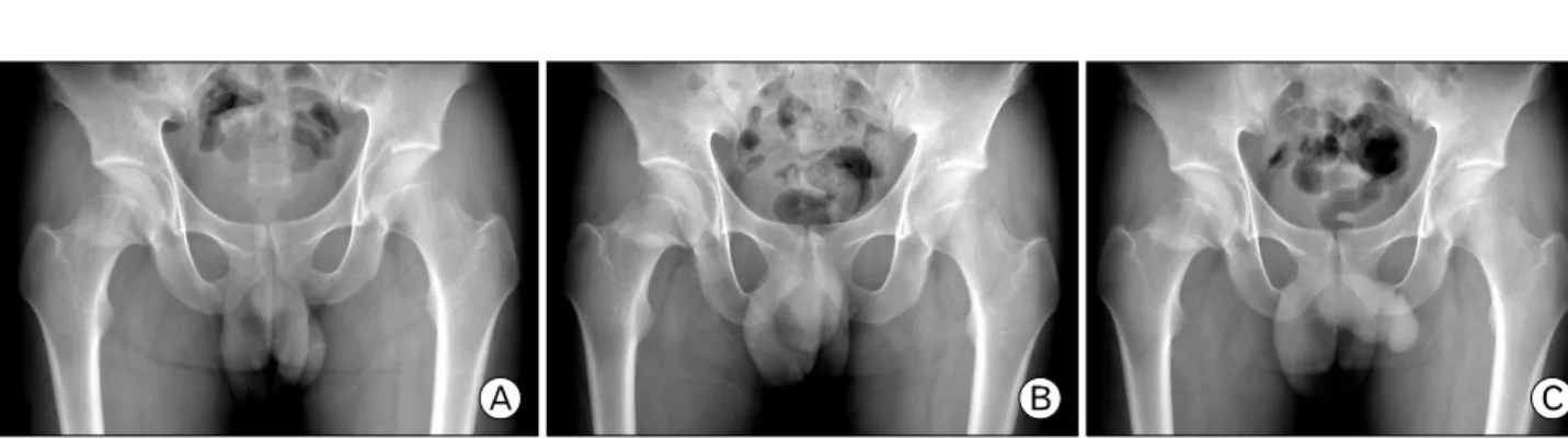
(3) SR Yi, et al. Progression of Compression Type Femoral Neck Stress Fracture. Fig. 4. Serial radiographs reviewed every 2 weeks after initial radiographs show that internal callus of the femoral neck becomes clear and progresses to superolateral cortex. (A) 2weeks after initial radiographs, (B) 4 weeks later, (C) 6 weeks later.. Fig. 5. MRI shows that fracture line of the inferomedial cortex progresses and involves in the superior cortex (arrow).. Fig. 7. Initial anteroposterior radiograph of the right hip shows compression type stress fracture manifested by sclerosis at inferomedial portion of the femoral neck (arrow).. 0.4-11% 정도에서 드물게 발생한다고 보고되고 있다5). 1). Devas 는 대퇴 경부의 피로 골절을 방사선 사진 소견과 임상적 결과를 기준으로 분류하였는데, 신장력의 결과로 대퇴 경부 외상방 표면에 발생하는 신장형과 대퇴 경부 내하방에 가골의 형성이 발견되는 압박형의 두 가지 형태로 나누었다. 이후 Blickenstaff and Morris6)는 세 가지 형태로 분류하였는데 제1형은 골절선의 소견 없이 대퇴경부 내하방에 골막 반응 혹은 가골 형성의 소견이 보이는 경우, 제 2형은 대퇴 경부나 Fig. 6. Seven weeks after initial radiographs, surgical fixation was done using three cannulated screws.. 경판(calcar)을 가로지르는 비전위 골절, 제3형은 전위된 완전 골절의 경우로 분류하였다. Fullerton and Snowdy7)는 신장형, 압박형, 그리고 전위된 골절의 세가지 범주로 분류하였는데,. 여 발생하거나 비정상 골에 반복되는 정상적인 응력이 발생하 3,4). 신장형은 대퇴 경부 외상방에서 나타나 전위의 위험이 증가되. 여 나타날 수 있다 . 운동 선수에서 가장 흔한 피로 골절의. 어 있어 수술적 내고정을 필요로 하며, 압박형은 Blickenstaff. 위치는 경골, 중족골 및 족근골이며 대퇴 경부 피로 골절은. and Morris 의 제1형과 Devas 의 압박형과 일치하고, 전위의. 146. 대한스포츠의학회지. 6). 1).
(4) 이승림 외. 진행 양상을 보인 압박형 대퇴 경부 피로 골절. Fig. 10. MRI shows that fracture line involves both superolateral and inferomedial cortex (arrow).. Fig. 8. A scintigram of the same patient demonstrate increased activity in the right femoral neck.. Fig. 11. Surgical fixation was done using three cannulated screws.. Fig. 9. Three weeks after initial radiographs, there is another fracture line at the superolateral portion of femoral neck (arrow).. 고 하였으며, Blickenstaff and Morris6)는 제1형 골절에 대해 통증이 소실되고 전 고관절 운동이 가능할 때까지 절대 침상안 정가료 후 약 1주간의 간격으로 추시 방사선 촬영을 하면서 악화되지 않는 소견을 확인하고 목발을 이용한 부분 체중. 위험이 적어 보존적 치료가 우선이며, 심지어 전 체중 부하를 1,3). 7). 부하를 허용하였고 통증 없이 보행이 가능한 범위 안에서. 하여도 치유가 가능하다고 하였다 . Fullerton and Snowdy 는. 체중부하를 늘려나가는 방법으로 평균 9.5주의 치료기간이. 4년 간 1,049개의 피로 골절에서 평균 나이 22세의 49명의. 필요하였다고 하였다.. 환자에서 54개의 대퇴 경부의 피로 골절을 대상으로 전향적. 저자들도 압박형 대퇴 경부 피로 골절 환자에서 체중부하. 연구를 수행하였으며, 이 연구에서 골 스캔 검사에서만 확인되. 금지 및 침상 안정 가료로 추적 방사선 상 이상 소견 없이. 는 신장형 대퇴 경부 피로 골절을 통증이 없어질 때까지 체중. 일반적인 경과대로 유합되는 결과를 경험하였으나 위 2예에. 부하를 금지하고 추시 방사선 검사를 시행하면서 비수술적으. 서는 보존적 치료 중에 추시 방사선 사진 및 MRI 상 골절선의. 로 치료하였다. 압박형 대퇴 경부 피로 골절뿐만 아니라 신장. 확대 및 전경부 침범 등의 진행 소견이 관찰되어 내고정술이. 형 피로 골절에서도 보존적 치료 도중에 전위된 사례는 없었다. 필요한 경우로 판단하였다2). 대퇴 경부 피로 골절에서 전위가. 제28권 제2호 2010. 147.
(5) SR Yi, et al. Progression of Compression Type Femoral Neck Stress Fracture. 발생할 시 대퇴 골두 무혈성 괴사, 부정 유합, 불유합 및 골관절 8,9). 염 등의 합병증이. 발생할 수 있고 그 예후가 불량하므로. 압박형 대퇴 경부 피로 골절이라 하더라도 정기적인 추적 방사선 검사를 통한 적극적인 추시 경과 관찰이 필요하며, 골절선의 연장 혹은 확대가 발견되는 경우에는 전위 발생으로 인한 합병증 발생을 막기 위해 조기 수술적 치료를 고려하는 것이 바람직할 것으로 생각된다.. 참 고 문 헌 1. Devas MB. Stress fractures of the femoral neck. J Bone Joint Surg Br 1965;47:728-738. 2. Egol KA, Koval KJ, Kummer F, Frankel VH. Stress fractures of the femoral neck. Clin Orthop Relat Res 1998;348:72-78. 3. Thorne DA, Datz FL. Pelvic stress fracture in female runners. Clin Nucl Med 1986;11:828-829.. 148. 대한스포츠의학회지. 4. Aslam N, Gwilym S, Natarajan R. Femoral neck stress fracture in a sanitary worker. Eur J Emerg Med 2004;11:220222. 5. Lee CH, Huang GS, Chao KH, Jean JL, Wu SS. Surgical treatment of displaced stress fractures of the femoral neck in military recruits: a report of 42 cases. Arch Orthop Trauma Surg 2003;123:527-533. 6. Blickenstaff LD, Morris JM. Fatigue fracture of the femoral neck. J Bone Joint Surg Am 1966;48:1031-1047. 7. Fullerton LR Jr, Snowdy HA. Femoral neck stress fractures. Am J Sports Med 1988;16:365-377. 8. Skinner HB, Cook SD. Fatigue failure stress of the femoral neck. A case report. Am J Sports Med 1982;10:245-247. 9. Johansson C, Ekenman I, Tornkvist H, Eriksson E. Stress fractures of the femoral neck in athletes. The consequence of a delay in diagnosis. Am J Sports Med 1990;18:524-528..
(6)
수치
관련 문서
Bilateral femoral neck fractures without trauma in young male patients is ex- tremely rare and only few cases have been reported in the literature; it is usually associated with
Hip fractures are classified into four major types accord- ing to anatomical location: femoral head fractures, femoral neck fractures, intertrochanteric fractures and subtrochan-
Pathologic Fracture in Radiation-Induced Osteosarcoma Misdiagnosed as Delayed Femoral Neck Fracture.. 전영수 • 한정수* • 곽상준*
(B) T1-weighted and proton MR images show a diffuse area of signal intensity changes (asterisk) on the right femoral head and neck with fracture line (arrowhead).. Slight
(C) A reconstruction nail was inserted for fixation of the femoral neck and shaft fracture; the radiolucent line appeared to decrease in size after insertion of two 6.0-mm
We investigated the relationship between decreased eGFR and outcomes of bipolar hemiarthroplasty for displaced femoral neck fracture in elderly patients and identified
Hip arthroplasty is more commonly performed in elderly patients with displaced femoral neck fractures than internal fixation because of more significant improvements in pain and
However, several more recent cases cannot be explained by this injury mechanism. For example, a femoral neck fracture may occur when the hip joint is abducted, extended, or
