http://e-jbm.org/
197
Copyright © 2017 The Korean Society for Bone and Mineral Research
This is an Open Access article distributed under the terms of the Creative Commons Attribution Non-Commercial Li- cense (http://creativecommons.org/licenses/by-nc/4.0/) which permits unrestricted non-commercial use, distribu- tion, and reproduction in any medium, provided the original work is properly cited.
Atraumatic Bilateral Fracture of the Femoral Neck in Young Male Patient with Suspected Osteomalacia
Byung-Ho Yoon, Min-Soo Kwon
Department of Orthopaedic Surgery, Inje University College of Medicine, Seoul Paik Hospital, Seoul, Korea
In this report, we describe the case of a healthy 37-year-old male patient without a his- tory of disease, who developed atraumatic bilateral fracture of the femoral neck. Radio- logical and blood investigations revealed osteopenia and severe vitamin D deficiency (7.42 ng/mL), respectively, but patient had no apparent risk factors for insufficiency frac- ture. Bilateral osteosynthesis was obtained using cannulated screws and laboratory find- ings improved after vitamin D supplementation. This case highlights the effect of vita- min D deficiency on demineralization in a young male patient.
Key Words: Femur neck, Fractures, Bone, Osteomalacia, Vitamin D deficiency
INTRODUCTION
Vitamin D, a hormone-like nutrient, is a key nutrient to maintain optimal bone health.[1] Vitamin D deficiency is characterized by impaired bone mineralization, which can cause rickets in children, and precipitate and exacerbate osteopenia, osteoporosis, and fractures in adults.[2]
Bilateral femoral neck fractures without trauma in young male patients is ex- tremely rare and only few cases have been reported in the literature; it is usually associated with a specific underlying disease.[3] We managed a young patient with atraumatic bilateral fracture in the femoral neck secondary to vitamin D defi- ciency. This report highlights the effect of 25-hydroxy-vitamin D (25[OH]D) defi- ciency on bone demineralization in young adults. The patient was informed that data concerning the case would be submitted for publication, and he provided consent.
CASE
A 37-year-old, 183-cm-tall, 69 kg man visited our hospital’s emergency room with progressive groin pain for the past two weeks. He denied any history of trau- ma, seizure, or systemic disorder that can affect bone metabolism. On examina- tion, the patient presented with groin pain, active straight leg raising, and passive hip rotation. The initial pelvic X-ray revealed a transcervical fracture of the left fem- oral neck area (Fig. 1A). To differentiate the pathologic facture, we performed Corresponding author
Byung-Ho Yoon
Department of Orthopaedic Surgery, Inje University College of Medicine, Seoul Paik Hospital, 9 Mareunnae-ro, Jung-gu, Seoul 04551, Korea
Tel: +82-2-2270-0028 Fax: +82-2-2270-0023 E-mail: [email protected] Received: April 19, 2017 Revised: April 24, 2017 Accepted: May 1, 2017
No potential conflict of interest relevant to this article was reported.
Case Report
J Bone Metab 2017;24:197-200 https://doi.org/10.11005/jbm.2017.24.3.197 pISSN 2287-6375 eISSN 2287-7029
Byung-Ho Yoon, et al.
198
http://e-jbm.org/ https://doi.org/10.11005/jbm.2017.24.3.197magnetic resolution imaging and confirmed insufficiency fractures at both femoral neck (Fig. 1B). Emergency surgery was performed via closed reduction and internal fixation with three 6.5-mm cannulated screws under spinal anes- thesia at both hips.
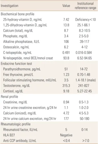
To determine the underlying cause of the fracture, we per- formed additional work-up post-operatively. Routine labo- ratory blood work was within normal range: hemoglobin 15.4 g/dL, white blood cell 8,210/µL, platelet 2,215,000/µL, sodium 138 mmol/L, potassium 4.0 mmol/L, and chloride 104 mmol/L. All biochemical, endocrine, rheumatology, and hematology evaluations were normal, except for severe vi- tamin D deficiency (Table 1). The patients also underwent bone scan and gastro-colonoscopy to rule out cancer le- sions; both tests were normal. His lumbar spine bone min- eral density, which to his knowledge was checked for the first time in his life, was -1.6 indicating osteopenia based on World Health Organization classification.
Fixation was performed in both hips and the patient was directed not to bear weight three weeks postoperatively;
assisted loading with crutches were authorized thereafter.
The patient also received one vitamin D injection (200,000 IU) postoperatively, and was prescribed vitamin D supple- mentation (6,000 IU/day) to take after discharge, based on the American Endocrinology Guideline.[4] At the 6-month follow up, the patient had no problems with ambulation or weight bearing, and an X-ray of the fractured site showed
Table 1. Biochemical, endocrine, renal and rheumatologic values to institutional laboratory reference ranges
Investigation Value Institutional
reference range Biochemical bone profile
25-hydroxy-vitamin D, ng/mL 7.42 Deficiency<10 1,25-dihydroxy-vitamin D, pg/mL 13.8 25.1-66.1
Calcium (total), mg/dL 8.7 8.2-10.5
Phosphate, mg/dL 3.4 2.5-5.0
Alkaline phosphatase, IU/L 106 39-117
Osteocalcin, ng/mL 6.2 4-12
C-telopeptide, ng/mL 0.491 0.016-0.584
N-telopeptide, nmol BCE/mmol creat 93.8 6.52-94.65 Endocrine function test
Parathyroidhormone, pg/mL 51 14-72
Free thyroxine, pmol/L 1.23 0.70-1.48
Follicular stimulating hormone, mIU/mL 3.5 1.4-18.1 (male)
Testosterone, ng/dL 315.3 241-827
Cortisol, ug/dL 9.18 5.27-22.45
Renal profile
Creatinine, mg/dL 0.94 0.5-1.3
24 hr urine creatinine excretion, g/24 hr 1.1 1.0-2.0
Calcium (ionized), mg/dL 4.72 4.5-5.3
24 hr urine calcium excretion, mg/24 hr 177 50-180 Rheumatologic profile
Rheumatoid factor, IU/mL 5 0-14
HLA B27 Negative
Anti CCP antibody, U/mL <0.4 >7.0
nmoL BCE/mmol creat, nmoL of bone collagen equivalents/mmoL creati- nine; HLA, human leukocyte antigen; CCP, cyclic citrullinated peptide.
Fig. 1. (A) Pelvic anteroposterior X-ray demonstrating the transcervical fracture of the neck of the left femur. (B) T2-weighted magnetic resonance imaging of both hips showing increased signal around the femoral necks in keeping with acute bilateral femoral neck fractures.
A B
Atraumatic Bilateral Fracture of the Femoral Neck
https://doi.org/10.11005/jbm.2017.24.3.197 http://e-jbm.org/
199
complete union (Fig. 2). His vitamin D level recovered to normal (25[OH]D, 31.1 ng/mL; 1,25-dihydroxy-vitamin D, 34.5 pg/mL).
DISCUSSION
We presented the unusual case of a 37-year-old man with an atraumatic bilateral hip fracture. Hip fractures following minimal trauma in young patients are usually associated with metabolically altered bones, including chronic renal failure and steroid use, and malabsorption syndromes such as celiac disease.[5-7] None of which are applicable in this case.
The patient’s bone mineral density was calculated via dual energy X-ray absorptiometry to be −1.6; this marked- ly low 25(OH)D value is so low that osteomalacia may fea- sibly occur. Although all other biochemical indices, except vitamin D, were within normal range, osteomalacia in the hip area must be assumed in the clinical scenario of insuf- ficient bilateral femur neck fracture.[8]
Several case reports have described insufficiency fractures in older or pregnant patients that were secondary to vita- min D deficiency.[9-11] To our knowledge, this is the first case of a young male patient with such a severe deficiency of vitamin D that led to insufficiency femur neck fracture.
This case highlights the importance of vitamin D and should not be overlooked by clinicians, even in young adults, and
should be regarded as an independent disease entity.
Vitamin D deficiency is an emerging global health issue, which has been now recognized as a pandemic. Although few data are available regarding the prevalence of vitamin D deficiency among young adults, serum 25(OH)D deficien- cy is more prevalent than previously thought.[12] One study showed that vitamin D deficiency was more prominent in urban clinic-based adolescents.[13] Although the etiology is varied, vitamin D deficiency occurs primarily from lack of natural sunlight, poor diet, and malabsorption. In our case report, the patient was not heavy drinker or smoker, and had none of the typical risk factors for vitamin D deficien- cy.[14] In another study, researchers showed that low body mass index (BMI) was significantly associated with 25(OH) D levels.[13] The only factor we believe could have poten- tially contributed to vitamin D deficiency in our patient was his low BMI (20.6 kg/m2).
In conclusion, we present the case of a 37-year-old man with atraumatic bilateral hip fractures and suspected con- comitant osteomalacia. Although he did not present with any typical of the risk factors, severe 25(OH)D deficiency was evident and was likely to have due to concomitant os- teomalacia. This case report highlights the importance of vitamin D levels in young patients, even in those without any history of disease. Clinicians should also be aware that lower level of vitamin D can lead to femoral neck fractures.
REFERENCES
1. Yoon DS, Lee YK, Ha YC, et al. Inadequate dietary calcium and vitamin D intake in patients with osteoporotic fracture.
J Bone Metab 2016;23:55-61.
2. Holick MF, Chen TC. Vitamin D deficiency: a worldwide prob- lem with health consequences. Am J Clin Nutr 2008;87:
1080s-6s.
3. Kwon YU, Kong GM, Park JH. Femur neck fracture in a young marfan syndrome patient. Hip Pelvis 2016;28:264-8.
4. Holick MF, Binkley NC, Bischoff-Ferrari HA, et al. Evaluation, treatment, and prevention of vitamin D deficiency: an En- docrine Society clinical practice guideline. J Clin Endocri- nol Metab 2011;96:1911-30.
5. Ly TV, Swiontkowski MF. Treatment of femoral neck frac- tures in young adults. J Bone Joint Surg Am 2008;90:2254- 66.
6. Malluche HH, Porter DS, Mawad H, et al. Low-energy frac- Fig. 2. At 6 months postoperatively, the radiograph showed complete
union of the fractures.
Byung-Ho Yoon, et al.
200
http://e-jbm.org/ https://doi.org/10.11005/jbm.2017.24.3.197tures without low T-scores characteristic of osteoporosis:
a possible bone matrix disorder. J Bone Joint Surg Am 2013;
95:e1391-6.
7. Selek O, Memisoglu K, Selek A. Bilateral femoral neck fati- gue fracture due to osteomalacia secondary to celiac dis- ease: Report of three cases. Arch Iran Med 2015;18:542-4.
8. Rader CP, Corsten N, Rolf O. Osteomalacia and vitamin D deficiency. Orthopade 2015;44:695-702.
9. Desai M, Nadkarni S. Spontaneous bilateral fracture neck of femur secondary to vitamin D deficiency: a case report.
Clin Cases Miner Bone Metab 2015;12:282-4.
10. Carter T, Nutt J, Simons A. Bilateral femoral neck insufficien- cy fractures secondary to vitamin D deficiency and con-
current corticosteroid use--a case report. Arch Osteoporos 2014;9:172.
11. Baki ME, Uygun H, Ari B, et al. Bilateral femoral neck insuf- ficiency fractures in pregnancy. Eklem Hastalik Cerrahisi 2014;25:60-2.
12. Hanley DA, Davison KS. Vitamin D insufficiency in North America. J Nutr 2005;135:332-7.
13. Gordon CM, DePeter KC, Feldman HA, et al. Prevalence of vitamin D deficiency among healthy adolescents. Arch Pe- diatr Adolesc Med 2004;158:531-7.
14. Holick MF. The influence of vitamin D on bone health across the life cycle. J Nutr 2005;135:2726s-7s.