• 검색 결과가 없습니다.

False Femoral Neck Fracture Detected during Shaft Nailing: A Mach Band Effect

N/A
N/A
Protected

Academic year: 2022

Share "False Femoral Neck Fracture Detected during Shaft Nailing: A Mach Band Effect"

Copied!
3
0
0

로드 중.... (전체 텍스트 보기)

전체 글

(1)

Yonsei Med J http://www.eymj.org Volume 54 Number 3 May 2013 803

Case Report http://dx.doi.org/10.3349/ymj.2013.54.3.803

pISSN: 0513-5796, eISSN: 1976-2437 Yonsei Med J 54(3):803-805, 2013

False Femoral Neck Fracture Detected during Shaft Nailing:

A Mach Band Effect

Kyu Hyun Yang, 1 Sungjun Kim, 2 and Ji Do Jeong 1

Departments of

1

Orthopaedic Surgery and

2

Radiology, Gangnam Severance Hospital, Yonsei University College of Medicine, Seoul, Korea.

Received: September 4, 2012 Revised: October 9, 2012 Accepted: November 5, 2012

Corresponding author: Dr. Kyu Hyun Yang, Department of Orthopaedic Surgery, Gangnam Severance Hospital, Yonsei University College of Medicine, 211 Eonju-ro, Gangnam-gu, Seoul 135-720, Korea.

Tel: 82-2-2019-3414, Fax: 82-2-573-5393 E-mail: [email protected]

∙ The authors have no financial conflicts of interest.

© Copyright:

Yonsei University College of Medicine 2013 This is an Open Access article distributed under the terms of the Creative Commons Attribution Non- Commercial License (http://creativecommons.org/

licenses/by-nc/3.0) which permits unrestricted non- commercial use, distribution, and reproduction in any medium, provided the original work is properly cited.

Femoral neck fractures are associated with femoral shaft fractures in 1% to 9% of cases. Undisplaced neck fractures are susceptible to displacement during shaft nailing. We report the case of a 57-year-old male patient in whom we performed standard intramedullary nailing for a femoral shaft fracture. In doing so, we identi- fied a vertical radiolucent line at the femoral neck, which was thought to be further displacement of a hidden silent fracture or an iatrogenic fracture that developed during nail insertion. Consequently, we decided to switch to reconstructive femo- ral nailing. Postoperative hip imaging failed to show the femoral neck fracture that we saw in the operating room. Here, we discuss the aforementioned case and re- view the literature concerning this artifact.

Key Words: Pseudofracture, femur neck, nailing, mach band

INTRODUCTION

Femoral neck fractures are associated with femoral shaft fractures in 1% to 9% of cases.

1,2

Some femoral neck fractures are undisplaced and can only be diagnosed by preoperative computed tomography (CT) (i.e., silent neck fractures). Such frac- tures are susceptible to displacement during preparation of the patient for surgery or during reaming or insertion of the nail.

3

During an operation, we encountered what appeared to be a femoral neck fracture, which caused us to change our proce- dure from antegrade femoral nailing to reconstruction nailing. After the operation, however, we determined that it was a false femoral neck fracture, as no fracture could be detected on simple radiogram, fluoroscopy or CT scan.

CASE REPORT

A 57-year-old man sustained a motor vehicle accident and was transferred to our

ER for femoral shaft comminuted fractures on the left side. We planned closed re-

duction and internal fixation with intramedullary nails for the left femoral shaft

fracture. In the operating room, the patient was placed on a fracture table under

general anesthesia. A skin incision was made proximal to the tip of the greater tro-

chanter about 5 centimeters. A 3.2-mm guide pin was inserted at the tip of the

(2)

Kyu Hyun Yang, et al.

Yonsei Med J http://www.eymj.org Volume 54 Number 3 May 2013 804

days after surgery, we took a CT scan of the proximal fe- mur in order to analyze the radiolucent line observed dur- ing intraoperative fluoroscopy; the femoral neck was con- firmed as intact. Reversal of the fluoroscopic image taken in the operating room showed a radiolucent line running from the femoral neck to the medial thigh (Fig. 2).

DISCUSSION

Mach bands are a perceptual phenomenon in which dark and bright lines are observed on the borders of structures of different radiographic densities, resulting in contrast-en- hancing effects in various areas of the body. Pseudo-frac- tures (i.e., false fractures) are among a group of illusory phe- nomena that result from overlapping images, differences in background illumination, subjective contour formation and parallax. Pseudofracture at the base of the dens of the sec- greater trochanter. After trochanteric reaming along the 3.2-

mm guide pin, a ball-tip guide was introduced into the med- ullary cavity of the proximal and distal fragments after re- duction. A femoral nail (greater trochanter starting nail, 12 mm in diameter, 40 cm in length) was introduced after ream- ing a hole to 13 mm. The nail was inserted uneventfully without excessive force. At that time, we identified a vertical radiolucent line at the femoral neck, which was thought to be further displacement of a hidden silent fracture or an iatro- genic fracture that developed during nail insertion (Fig. 1).

Consequently, we decided to perform reconstructive femo- ral nailing. We inserted two 6.0-mm partially threaded can- cellous screws into the femoral neck and head. Afterwards, we observed that the radiolucent line at the femoral neck had decreased in size. Three distal interlocking screws were then inserted, and a nail cap was applied.

Postoperative hip imaging failed to show the femoral neck fracture that we saw in the operating room. Fourteen

Fig. 1. (A) A 3.2-mm guide pin was inserted at the tip of the greater trochanter; the radiolucent line was not visible at that time. Hemostatic forceps were used to control the external rotation of the proximal fragment.

4

(B) A radiolucent line mimicking a femoral neck fracture was visible after nail insertion. (C) A reconstruction nail was inserted for fixation of the femoral neck and shaft fracture; the radiolucent line appeared to decrease in size after insertion of two 6.0-mm partially threaded cancellous screws into the femoral neck and head.

Fig. 2. (A) Postoperative fluoroscopic study failed to reveal the femoral neck fracture in various limb rotations. (B) A CT scan confirmed that the femoral neck was intact. (C) Reversal of the fluoroscopic image taken in the operating room showed a radiolucent line running from the femoral neck to the medial thigh (arrows).

A

A

B

B

C

C

(3)

Mach Band Effect: Femoral Neck Fracture

Yonsei Med J http://www.eymj.org Volume 54 Number 3 May 2013 805

after femoral neck screw fixation; however, we realize that rotation of the limb was different. The lesser trochanter was more prominent in the post-fixation image. We suspect a tiny difference in limb rotation caused the decrease in pseu- dofracture gap on the fluoroscopic image.

5

In conclusion, we recommend that surgeons check the fracture line again in the reversed fluoroscopic image (tracing the adjacent fat plane and its connectivity with the fracture line) before con- verting to reconstructive nailing. However, if the presence of a pseudofracture cannot be definitively ascertained, then antegrade femoral nailing must be converted to reconstruc- tive nailing or another method of fixation. Additionally, all supporting fluoroscopic images should be saved for further evaluation and documentation.

REFERENCES

1. Tornetta P 3rd, Kain MS, Creevy WR. Diagnosis of femoral neck fractures in patients with a femoral shaft fracture. Improvement with a standard protocol. J Bone Joint Surg Am 2007;89:39-43.

2. Riemer BL, Butterfield SL, Ray RL, Daffner RH. Clandestine femoral neck fractures with ipsilateral diaphyseal fractures. J Or- thop Trauma 1993;7:443-9.

3. Yang KH, Han DY, Park HW, Kang HJ, Park JH. Fracture of the ipsilateral neck of the femur in shaft nailing. The role of CT in di- agnosis. J Bone Joint Surg Br 1998;80:673-8.

4. Park J, Yang KH. Correction of malalignment in proximal femoral nailing--reduction technique of displaced proximal fragment. Inju- ry 2010;41:634-8.

5. Daffiner RH. Skeletal pseudofractures. Contemp Diagn Radiol 1992:15:1-5.

ond cervical vertebra is a well-known artifact. Skin fold or fat tissue plane simulating trochanteric fracture as well as pseudofracture of the femoral neck secondary to osteophyte formation on the femoral head and acetabulum have also been reported.

5

In our case, the pseudofracture was not detected during preparation of the entry point, but instead was found after final settlement of the femoral nail. Unfortunately, it is not uncommon to detect displacement of an undisplaced femo- ral neck fracture during antegrade shaft nailing. The pseu- dofracture was vertically oriented in the middle of the fem- oral neck, thus mimicking a typical femoral neck fracture associated with femoral shaft fracture. After the operation, we realized that it was a pseudofracture, so we attempted to determine the cause of the event. We put the patient on the fracture table and recreated the positions that we used for femoral nailing, but we were unable to make the false fem- oral neck fracture reappear. We then reversed the saved im- age that we took during the operation and determined that a radiolucent line was running from the femoral neck to the medial thigh. The boundary of the radiolucent band was a little smoother than that of fracture lines seen in real cases.

Intraoperative CT scan or 3D fluoroscopy may also have al- lowed us to determine that it was a false femoral neck frac- ture; however, this would have been particularly impractical during femoral nailing with the patient positioned on the fracture table.

To our knowledge, this is the first report of a pseudofrac-

ture in the femoral neck associated with femoral shaft nail-

ing. It is true that the pseudofracture line seemed to decrease

수치

Fig. 1. (A) A 3.2-mm guide pin was inserted at the tip of the greater trochanter; the radiolucent line was not visible at that time

참조

관련 문서

In an atypical diaphyseal femoral fracture with marked anterior and lateral bowing, there are some obstacles in fixation of the fracture, such as difficulty in insertion of

After closed reduction and internal fixation with proximal femoral nail for intertrochanteric fracture, some patients com- plained lateral hip pain. We report two cases of lateral

Park BJ, Cho HM, Kim JH, Sin WJ: Excessive sliding of the helical blade and the femoral neck fracture after insertion of proximal femoral nail anti-rotation for type

Purpose: This study examined the outcomes of exchange nailing for the hypertrophic nonunion of femoral shaft fractures treated with intramedullary nailing as well as the

Fractures of the femoral shaft with marked bowing face some obstacles in fixation of the fracture such as difficulty in insertion of the intramedullary nail (IM nail) or

10) Song KJ, Hwang BY and Lee JH: Interlocking intrame- dullary nailing in the treatment of the tibial shaft fracture- comparative study between reamed and unreamed nailing.

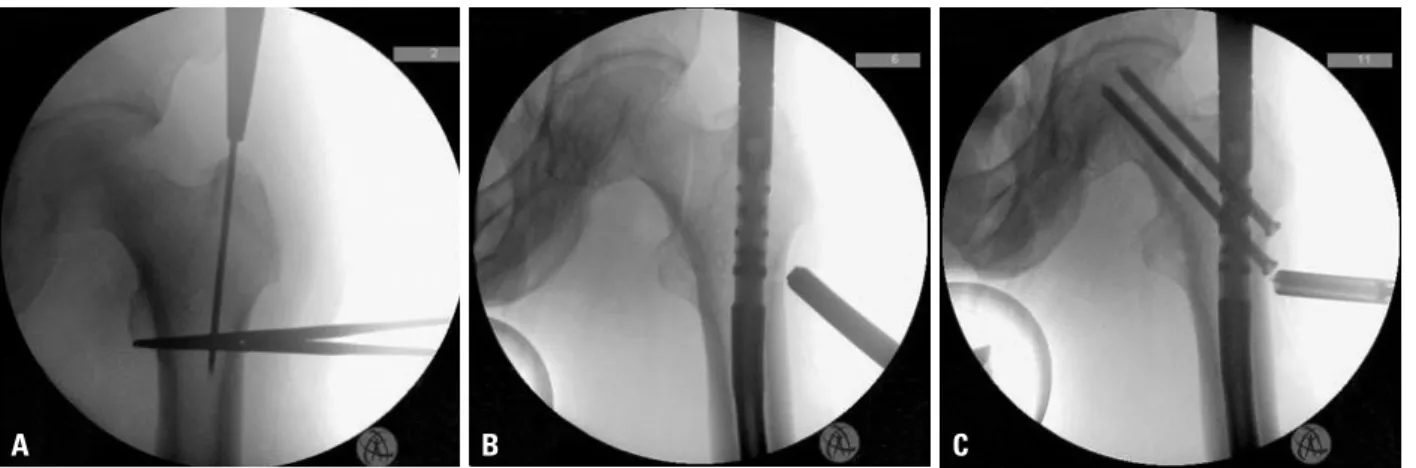
Initial anteroposterior radiograph of the right hip shows compression type stress fracture manifested by sclerosis at inferomedial portion of the femoral neck arrow... Devas 는

Yang et al. 8) and Grimaldi et al. 9) reported superficial femoral arterial ruptures following the insertion of distal interlocking screws with Asian-Pacific type Gamma nail and Gamma