• 검색 결과가 없습니다.

Parameters to Guide Retreatment After Initial Intravenous Immunoglobulin Therapy in Kawasaki Disease

N/A
N/A
Protected

Academic year: 2022

Share "Parameters to Guide Retreatment After Initial Intravenous Immunoglobulin Therapy in Kawasaki Disease"

Copied!
6
0
0

로드 중.... (전체 텍스트 보기)

전체 글

(1)

379

Open Access

Parameters to Guide Retreatment After Initial Intravenous Immunoglobulin Therapy in Kawasaki Disease

Hyun Kwon Kim, MD, Jungeun Oh, MD, Young Mi Hong, MD, and Sejung Sohn, MD Department of Pediatrics, Ewha Womans University School of Medicine, Seoul, Korea

ABSTRACT

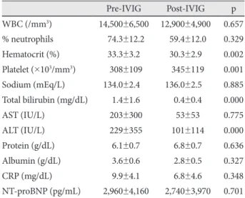
Background and Objectives: We sought to determine parameters to guide the decision of retreatment in patients with Ka- wasaki disease (KD) who remained febrile after initial intravenous immunoglobulin (IVIG). Subjects and Methods: A to- tal of 129 children with KD were studied prospectively. Patients were treated with IVIG 2 to 9 days after the onset of disease.

Laboratory measures, such as white blood cell (WBC), percentage of neutrophils, C-reactive protein (CRP), and N-terminal pro-brain natriuretic peptide (NT-proBNP), were determined before and 48 to 72 hours after IVIG treatment. Patients were classified into IVIG-responsive and IVIG-resistant groups, based on the response to IVIG. Results: Of a total of 129 patients, 107 patients (83%) completely responded to a single IVIG therapy and only 22 patients (17%) required retreatment: 14 had persistent fever and 8 had recrudescent fever. There was no significant difference between the groups in age, gender distri- bution, and duration of fever to IVIG initiation, but coronary artery lesions developed significantly more often in the resis- tant group than in the responsive group (31.8% vs. 2.8%, p=0.000). Compared with pre-IVIG data, post-IVIG levels of WBC, percentage of neutrophils, CRP, and NT-proBNP decreased to within the normal range in the responsive group, whereas they remained high in the resistant group. Multivariate logistic regression indicated that neutrophil counts, CRP, and NT-proBNP were independent parameters of retreatment. Conclusion: Additional therapy at an early stage of the disease should be ad- ministered for febrile patients who have high values of CRP, NT-proBNP, and/or neutrophil counts after IVIG therapy. (Ko- rean Circ J 2011;41:379-384)

KEY WORDS: Kawasaki disease; Intravenous immunoglobulins; Retreatment.

Received: February 22, 2011 Accepted: April 19, 2011

Correspondence: Sejung Sohn, MD, Department of Pediatrics, Ewha Womans University School of Medicine, Mokdong Hospital, 911-1 Mok- dong, Yangcheon-gu, Seoul 158-710, Korea

Tel: 82-2-2650-5579, Fax: 82-2-2653-3718 E-mail: [email protected]

• The authors have no financial conflicts of interest.

cc

This is an Open Access article distributed under the terms of the Cre- ative Commons Attribution Non-Commercial License (http://creativecom- mons.org/licenses/by-nc/3.0) which permits unrestricted non-commer- cial use, distribution, and reproduction in any medium, provided the origi- nal work is properly cited.

Introduction

Intravenous immunoglobulin (IVIG) infusion is an effec- tive therapy for acute Kawasaki disease (KD).

1)

Administra- tion within a few days of illness of a single dose of IVIG, in combination with high-dose aspirin, ceases fever, rapidly re- solves clinical signs and symptoms of inflammation, and re- duces the incidence of coronary artery aneurysms.

1)2)

How-

ever, approximately 10% to 20% of KD patients have persist- ent or recrudescent fever, despite IVIG therapy.

2)3)

When fever persists or recurs with signs of mucocutaneous

inflammation after initial IVIG therapy, retreatment can be

indicated. However, when the patients have fever with few cli-

nical signs of ongoing inflammation, these patients pose a th-

erapeutic dilemma. The KD patients who fail to respond to

initial IVIG treatment should be reassessed to exclude the pos-

sibility of a bacterial infection. Although current diagnostic

criteria for KD require exclusion of infectious diseases before

making a definite diagnosis, there are actually several reports

of patients with typical features of KD, including coronary ar-

tery aneurysm, who also have had a documented infection.

4)5)

In addition, we have sometimes experienced KD patients

who became afebrile with IVIG treatment, but shortly febrile

again due to newly developed viral infection, pharyngoton-

sillitis, gastroenteritis, or urinary tract infection during hos-

pitalization. In such cases, the values of acute phase reactants

were usually within the normal range and fever subsided with-

(2)

out the use of IVIG. In patients who remain febrile despite IVIG therapy, our institutional protocol requires that we al- ways deliberate on the values of acute phase reactants, when a need for additional therapy is recognized. If the patients be- come afebrile after treatment, no laboratory test is perform- ed. Most physicians clinically judge post-IVIG fever to be at- tributable to active KD, rather than to other causes,

2)6)

and tend to retreat this group of patients in the absence of the systemic evaluation required to guide the decision of treatment. We be- lieve that there is a need for objective laboratory measures that identify KD patients who remain febrile after IVIG treat- ment and therefore need additional therapy.

Our goal was to determine from routine laboratory meas- ures the parameters that may serve to guide retreatment in KD patients who remained febrile after initial IVIG therapy.

Subjects and Methods

During the study period between November 2008 and Sep- tember 2010, a total of 129 children who met the diagnostic criteria for KD

7)

were prospectively enrolled. Subjects com- prised 72 boys (55.8%) and 57 girls, aged 2 months to 11 years. All patients were treated with IVIG 2 g/kg for 2 to 9 days after the onset of disease, and were also given aspirin 80 to 100 mg/kg/day until defervescence, and then 5 mg/kg/

day as a single daily dose. When the patients failed to defer- vesce after the first IVIG therapy, they were retreated with a second dose of IVIG (n=14). When they remained still febrile despite a second dose of IVIG therapy, then pulse methyl- prednisolone (30 mg/kg/day for 3 days) was administered (n=8).

We measured white blood cell (WBC), percentage of neu- trophils in WBCs (% neutrophils), hematocrit, platelet counts, serum sodium, total bilirubin, aspartate aminotransferase, alanine aminotransferase (ALT), protein, albumin, C-reac- tive protein (CRP), and N-terminal pro-brain natriuretic pep- tide (NT-proBNP), before and 48 to 72 hours after IVIG tr- eatment. For patients who failed to respond to the second dose of IVIG, laboratory measures were repeated before pulse steroid therapy. Coronary arteries were assessed by echocar- diography performed at the time of diagnosis and at disch- arge, and repeated at weeks 2, 4, and 8 after treatment, and then as needed thereafter. Coronary artery lesions (CALs) were diagnosed according to the criteria of the Japanese Mi- nistry of Health and Welfare.

8)

Patients who presented with CAL before the initial treatment were excluded from the study.

Subjects were divided into 2 groups according to respon- siveness to IVIG treatment: IVIG-responsive patients (n=

107) and IVIG-resistant patients (n=22). Laboratory mea- sures before and after IVIG therapy were compared in each group. We selected the variables that showed a different ch- ange after IVIG therapy between the groups. Pre- and post-

IVIG concentrations of each variable between the groups were compared. The value of the fractional change (FC) of each variable between the groups was also compared, as de- scribed previously.

9)

FC was defined as follows:

FC=(Y-X)/X

In which X represents data before IVIG treatment and Y re- presents data at 48 to 72 hours after IVIG treatment.

This study was approved by the Institutional Review Bo- ard of Ewha Womans University Mokdong Hospital (#ECT 230-2-28) and written informed consents were obtained from the parents of each patient.

Definitions

Duration of fever was defined as the time from the onset of disease to the start of initial IVIG infusion. Persistent fe- ver was defined as temperature ≥38°C that persisted beyond 48 hours after completion of the IVIG infusion. Recrudes- cent fever was defined as temperature <37.5°C for more than 24 hours after IVIG infusion, followed by temperature ≥38°C.

IVIG-responsive patients were defined as patients who had defervescence within 48 hours after initial IVIG infusion.

IVIG-resistant patients were defined as those who required additional rescue therapy, owing to either persistent or recru- descent fever at least 48 hours after the end of initial IVIG infusion.

Data analyses

All data were analyzed with the Statistical Package for the Social Sciences for Windows software (SPSS) version 12.0 (SPSS Inc, Chicago, IL, USA). Data are presented as either mean±SD for continuous variables or as the number (percen- tage) of patients for categorical variables. Intra-group vari- ables were compared using the paired t-test, and inter-group variables and FCs using the Student t-test. Proportions were compared using either the χ

2

test or the Fisher’s exact test. To determine the cutoff value of each parameter found to be significant by FC, receiver operating characteristic (ROC) curves were used. Multivariate logistic regression analysis was used to determine independent parameters of retreat- ment and results were expressed as an odds ratio with a 95%

confidence interval (CI). A 95% CI that did not include 1.0 was interpreted to indicate statistical significance. A level of p<0.05 was considered as statistically significant.

Results

Clinical characteristics of intravenous immunoglobulin-resistant patients

Of a total of 129 patients, 107 patients (83%) completely

responded to a single IVIG therapy and 22 patients (17%)

(3)

required retreatment: 14 had persistent fever and 8 had re- crudescent fever. Of the 14 patients with persistent fever, 7 patients achieved defervescence after the second dose of IVIG. However, the remaining 7 failed to respond to the second IVIG therapy and received pulse methylprednisolone therapy. In contrast, 7 of the 8 patients with recrudescent fe- ver achieved defervescence after the second course of IVIG.

However, according to fever pattern, no significant difference in responsiveness to a second dose of IVIG was found (p=

0.086).

IVIG-resistant patients did not differ from the IVIG-re- sponsive patients with respect to age, gender distribution, and duration of fever measured until IVIG initiation. However, CALs developed significantly more often in the IVIG-resis- tant patients than in IVIG-responsive patients (Table 1).

Changes in laboratory measures after intravenous immunoglobulin therapy in responsive patients

Compared with pre-IVIG data, post-IVIG levels of WBC,

% neutrophils, total bilirubin, CRP, and NT-proBNP decre- ased to within the normal range and protein concentration increased (Table 2).

There were 8 patients who became afebrile but had high values of CRP (4.01 to 8.69 mg/dL) and NT-proBNP (338 to 3,308 pg/mL) after IVIG treatment. We repeated the labora- tory tests and confirmed their normalization.

Changes in laboratory measures after intravenous immunoglobulin therapy in resistant patients

There was no difference between pre- and post-IVIG data in WBC, % neutrophils, protein, albumin, CRP, and NT-pro- BNP. The values of WBC, % neutrophils, CRP, and NT-pro- BNP remained high after IVIG treatment (Table 3). Parado- xically, WBC counts were elevated in 35% of IVIG-resistant children (vs. 9.4% in responsive patients, p=0.001), % neutro- phils in 15% (vs. 4.7% in responsive patients, p=0.15), CRP in 23.8% (vs. 0.9% in responsive patients, p=0.000), and NT- proBNP in 47.4% (vs. 14% in responsive patients, p=0.002) after IVIG therapy, as compared with pre-IVIG data.

In 8 patients who remained febrile after a second dose of IVIG and required pulse steroid therapy, the lab measures before the second dose of IVIG did not significantly differ

from those before steroid therapy: WBC, 14,400±6,100 vs.

14,800±3,600, p=0.885; % neutrophils, 65±9 vs. 62±14, p=

0.684; CRP, 8.8±5.1 vs. 6.8±3.4, p=0.438; and NT-proBNP, 4,800±5,400 vs. 3,100±3,800, p=0.518.

There were 2 patients who remained febrile despite the se- cond dose of IVIG therapy but had normal laboratory mea- sures, including CRP (1.08 mg/dL and 0.71 mg/dL, respecti- vely). We just observed them, without using the steroid, and they achieved defervescence 24 to 33 hours later.

Fractional change of laboratory parameters

We excluded 5 variables that showed a similar change after IVIG treatment between IVIG-responsive and IVIG-resis- tant patients, and selected the remaining 7 variables that sh-

Table 1. Comparison of basic characteristics between IVIG-res- ponsive and IVIG-resistant patients

Responsive (n=107)

Resistant (n=22) p

Age (month) 29.0±20.8 35.9±28.6 0.212

Gender (male/female) 57/50 15/7 0.147

Duration of fever to IVIG (d) 4.3±1.6 3.8±1.8 0.921

CAL, n (%) 3 (2.8) 7 (31.8) 0.000

IVIG: intravenous immunoglobulin, CAL: coronary artery lesion

Table 2. Comparison between pre- and post-IVIG laboratory me- asures in IVIG-responsive patients

Pre-IVIG Post-IVIG p WBC (/mm

3

) 13,800±4,500 8,200±2,800 0.002

% neutrophils 061.6±15.6 33.7±16.3 0.000 Hematocrit (%) 33.3±2.6 32.4±3.00 0.000 Platelet (×10

3

/mm

3

) 338±90 438±132 0.000 Sodium (mEq/L) 136.4±2.50 135.8±10.70 0.874 Total bilirubin (mg/dL) 00.6±0.4 0.2±0.1 0.000

AST (IU/L) 0135±290 135±368 0.491

ALT (IU/L) 0117±208 116±325 0.845

Protein (g/dL) 06.5±0.5 7.8±0.8 0.000

Albumin (g/dL) 03.8±0.3 3.3±0.3 0.001

CRP (mg/dL) 08.6±6.2 1.4±1.3 0.000

NT-proBNP (pg/mL) 01,132±1,651 274±315 0.000 IVIG: intravenous immunoglobulin, WBC: white blood cell, AST: as- partate aminotransferase, ALT: alanine aminotransferase, CRP: C-re- active protein, NT-proBNP: N-terminal pro-brain natriuretic peptide Table 3. Comparison between pre- and post-IVIG laboratory meas- ures in IVIG-resistant patients

Pre-IVIG Post-IVIG p WBC (/mm

3

) 14,500±6,500 12,900±4,900 0.657

% neutrophils 074.3±12.2 059.4±12.0 0.329

Hematocrit (%) 33.3±3.2 30.3±2.9 0.002

Platelet (×10

3

/mm

3

) 0308±109 0345±119 0.001 Sodium (mEq/L) 134.0±2.40 136.0±2.50 0.885 Total bilirubin (mg/dL) 01.4±1.6 00.4±0.4 0.000

AST (IU/L) 0203±300 053±53 0.775

ALT (IU/L) 0229±355 0101±114 0.000

Protein (g/dL) 06.1±0.7 06.8±0.7 0.636 Albumin (g/dL) 03.6±0.6 02.8±0.5 0.327

CRP (mg/dL) 09.9±4.1 06.8±4.6 0.348

NT-proBNP (pg/mL) 02,960±4,160 02,740±3,970 0.701

IVIG: intravenous immunoglobulin, WBC: white blood cell, AST: as-

partate aminotransferase, ALT: alanine aminotransferase, CRP: C-re-

active protein, NT-proBNP: N-terminal pro-brain natriuretic peptide

(4)

owed a different post-IVIG change between the groups: WBC,

% neutrophils, ALT, protein, albumin, CRP, and NT-proBNP.

Of these variables, ALT was also excluded from the further an- alysis because its post-IVIG level in the resistant group decreas- ed toward the normal range.

Pre- and post-IVIG concentrations of the 6 variables were compared between the groups (Table 4). There was no differ- ence between the groups in pre-IVIG levels of WBC, CRP, and NT-proBNP. However, the value of % neutrophils was higher and the concentrations of protein and albumin were lower in the IVIG-resistant patients. With regard to post-IVIG data, all 6 variables showed a significant difference between the groups. To verify the possibility that post-IVIG differences between the groups in % neutrophils, protein, and albumin may be derived from their pre-IVIG differences, FCs of the 6 variables were determined (Table 5). As expected from Table 4, WBC, CRP, and NT-proBNP showed a significant FC dif- ference between the groups. Except for protein and albumin, there was a significant inter-group difference in FC of % neu- trophils, indicating that its post-IVIG difference seen in Table 4 was not derived from the pre-IVIG difference. Therefore, th- ese 4 variables may represent suitable parameters to guide the decision of retreatment in IVIG-resistant KD.

Cutoff values of parameters

The cutoff value of parameters was determined by ROC cur- ves: a WBC cutoff value of 10,850/mm

3

provided a sensitivity of 80% and a specificity of 84%, with an area under the curve

(AUC) of 0.838±0.057 (95% CI, 0.73-0.95, p=0.000); a % neu- trophils cutoff value of 53.4% provided a 75% sensitivity and a 87.7% specificity {AUC=0.896±0.031 (95% CI, 0.84-0.96, p=0.000)}; a CRP cutoff value of 3.68 mg/dL yielded 76.2%

sensitivity and 92.5% specificity {AUC=0.905±0.041 (95%

CI, 0.83-0.99, p=0.000)}; and a NT-proBNP cutoff value of 479 pg/mL yielded 78.9% sensitivity and 86.0% specificity {AUC=0.883±0.05 (95% CI, 0.79-0.98, p=0.000)}.

Independent parameters of retreatment

By multivariate logistic regression analysis, % neutrophils, CRP, and NT-proBNP were identified as independent para- meters of retreatment (Table 6). Patients with post-IVIG CRP

≥3.68 mg/dL, NT-proBNP ≥479 pg/mL, and/or % neutro- phils ≥53.4% were subjected to retreatment, with a sensitivi- ty of 80% and a specificity of 96.3%.

Discussion

Although IVIG plus aspirin therapy are effective for the treat- ment of acute KD,

1)

approximately 10% to 20% of patients ex- perience either persistent or recrudescent fever, despite ini- tial IVIG treatment.

2)3)

These patients are at increased risk of developing coronary artery aneurysm.

2)10)11)

Therefore, it is important to determine the effectiveness of IVIG soon after therapy, especially in resistant cases, to recognize a need for additional therapy, such as a second dose of IVIG, steroids, or infliximab.

12)13)

However, no evaluation system currently ex- ists to determine the effectiveness of IVIG therapy. Both fever resulting from cytokine release and increased CRP levels me- diated by interleukin-6 are clinically useful measures of sys- temic inflammation in KD. Post-IVIG fever, however, may ar- ise from other causes (e.g., intercurrent infection), as well as from KD itself during hospitalization. Thus other adjuvant in- flammatory markers may be required to more precisely assess the ongoing inflammation arising from KD and to support the decision for retreatment in patients who remain febrile af- ter IVIG therapy. We investigated routine laboratory meas- ures and propose herein CRP, NT-proBNP, and % neutro- phils as independent parameters to guide the decision of retreat- ment in KD patients who are still febrile after initial IVIG th- Table 4. Comparison of pre- and post-IVIG laboratory measures between IVIG-responsive and IVIG-resistant patients

Pre-IVIG

p Post-IVIG

Responsive Resistant Responsive Resistant p

WBC 13,800±4,500 14,500±6,500 0.688 8,200±2,800 12,900±4,900 0.000

% neutrophils 061.6±15.6 074.3±12.2 0.000 33.7±16.3 059.4±12.0 0.000

Protein 06.5±0.5 06.1±0.7 0.025 7.8±0.8 06.8±0.7 0.003

Albumin 03.8±0.3 03.6±0.6 0.004 3.3±0.3 02.8±0.5 0.046

CRP 08.6±6.2 09.9±4.1 0.337 1.4±1.3 06.8±4.6 0.000

NT-proBNP 01,132±1,651 02,960±4,160 0.096 274±315 02,740±3,970 0.013

IVIG: intravenous immunoglobulin, WBC: white blood cell, CRP: C-reactive protein, NT-proBNP: N-terminal pro-brain natriuretic peptide Table 5. Fractional changes of laboratory parameters

FC* p

Responsive Resistant

WBC -0.36±0.29 -0.01±0.60 0.011

% neutrophils -0.45±0.25 -0.18±0.22 0.000

Protein -0.16±0.21 -0.13±0.17 0.777

Albumin -0.14±0.08 -0.20±0.19 0.362

CRP -0.76±0.75 -0.27±0.60 0.005

NT-proBNP -0.51±0.61 -0.99±3.10 0.045

*FC=(Data at 48 to 72 hours after IVIG)-(Data before IVIG)/Data

before IVIG. WBC: white blood cell, CRP: C-reactive protein, NT-

proBNP: N-terminal pro-brain natriuretic peptide, FC: fractional

changes

(5)

erapy.

In this study, IVIG-resistance occurred in 17% of KD pa- tients. The clinical indications for retreatment were persistent fever in 14 patients and recrudescent fever in 8 patients. Of these 22 patients retreated with a second dose of IVIG, 14 patients (63.6%) achieved defervescence with this treatment.

This finding was similar to results of other studies in which ap- proximately two thirds of the initial non-responders eventual- ly responded to a second dose of IVIG.

3)6)

In addition, alth- ough there was no statistical significance, persistent fever was likely to cease either by a second dose of IVIG or by steroid th- erapy, whereas recrudescent fever was more likely to cease by a second dose of IVIG only.

The optimal period to begin IVIG administration remains controversial. Some investigators reported that early treat- ment, i.e., before day 5 of illness, is associated with increased need for additional IVIG therapy.

14)

Contrarily, others advo- cated that earlier treatment would be better.

15)

In this study, we found no difference between the IVIG-responsive and IVIG-resistant group in the proportion of patients who were treated before day 5 of illness (62.0% vs. 77.3%, p=0.215) and also in the mean duration of fever until IVIG initiation. Thus neither group was affected by the time of IVIG administration in this study.

The patients who failed to defervesce after IVIG therapy were characterized by significantly higher values of post-IV- IG WBC, % neutrophils, CRP, and NT-proBNP, compared with IVIG-responsive patients. Thus, we demonstrated that WBC, % neutrophils, CRP, and NT-proBNP may be suitable parameters of retreatment after initial IVIG therapy. When assessed by multivariate analysis, the neutrophil counts and serum concentrations of CRP and NT-proBNP were found to be independent parameters of the response to IVIG therapy.

Both the sensitivity and specificity of these 3 independent pa- rameters, as defined in this study, may allow clinicians to iden- tify patients who remain febrile and need additional therapy after initial IVIG treatment. We are able to justify retreatment more reliably by measuring not only CRP, but also NT-proB- NP and % neutrophils, rather than CRP alone.

Our results were similar to the findings of Mori et al.

9)

who demonstrated that increased WBC, neutrophil counts, and CRP after IVIG treatment are markers of treatment efficacy and also useful predictors of CALs. The role of neutrophils

in acute KD has been elucidated. Neutrophil activation can occur by stimulation of increased granulocyte colony-stim- ulating factor and cytokines that are involved in coronary ar- tery dilation.

16)17)

In the acute phase of disease, enhanced neu- trophil function may contribute to the pathogenesis of coro- nary vascular endothelial injury through either production of elastase or expression of vascular endothelial growth factor.

17)18)

This was demonstrated in a pathologic study showing early infiltration of neutrophils in the CALs of patients who died 10 days after the onset of disease.

19)

Thus, it is natural that neu- trophil is a marker of systemic inflammation. As a parameter in the multivariate analysis, the neutrophil count is interpret- ed to be more significant than WBC.

We proposed a CRP cutoff value of 3.68 mg/dL as a marker of ongoing inflammation after IVIG therapy. This value is si- milar to 4 mg/dL calculated as the upper limit of post-IVIG CRP (mean+2 SDs) in the responsive group, as shown in Table 4, but is still a little higher than the value of 3 mg/dL, proposed by the American Heart Association as a criterion for the evalu- ation of suspected incomplete KD.

20)

We reported previously that NT-proBNP increases during the acute phase of KD, i.e., before IVIG treatment, and decrea- ses to within the normal range in the convalescent phase.

21)

The change in NT-proBNP according to the phase of KD is equi- valent to that in CRP, suggesting that NT-proBNP may be used as a marker of systemic inflammation, as well as myo- cardial involvement, in acute KD. This finding is consistent with that of the other study.

22)

Moreover, the fact that, after IVIG administration, NT-proBNP was paradoxically elevat- ed in nearly half of IVIG-resistant patients suggests that NT- proBNP may be a useful marker of ongoing inflammation after IVIG treatment as well. Thus we put an emphasis on the clinical significance of NT-proBNP as a marker of retreat- ment. A NT-proBNP cutoff value of 479 pg/mL, indicated as a marker of retreatment in the present study, was higher than both 260 pg/mL and 170 pg/mL proposed as a diagnostic test of acute KD in our previous study,

21)

and in the other study,

22)

respectively.

Using the defined parameters, here are our suggested guide- lines of retreatment for KD patients who receive IVIG treat- ment: 1) Do necessarily retreat patients who remain febrile despite IVIG therapy and have high values of CRP, NT-proB- NP and/or neutrophil counts; 2) When the patients become Table 6. Multivariate logistic analysis for parameters of retreatment

Logistic coefficient (β) SE Odds ratio 95% CI

Lower Upper

WBC (10,850/mm

3

) 0.93 0.99 02.54 0.36 17.69

Neutrophils (53.4%) 3.11 1.26 22.32 1.88 264.94

CRP (3.68 mg/dL) 2.16 1.03 08.68 1.14 65.75

NT-proBNP (479 pg/mL) 2.67 1.24 14.43 1.27 163.34

WBC: white blood cell, CRP: C-reactive protein, NT-proBNP: N-terminal pro-brain natriuretic peptide

(6)

febrile and show normal lab measures (i.e., parameters), do not retreat for KD and observe for 1 to 2 days, with anticipa- tion of defervescence. In these cases, fever is less likely to be caused by KD; 3) When the patients become afebrile and show abnormal lab values/parameters, do not retreat them and re- peat laboratory tests; 4) Do not retreat patients who become afebrile and have normal parameters.

In conclusion, our study shows that, in KD patients who re- main febrile despite IVIG treatment, additional therapy can be facilitated early by measuring CRP in combination with NT-proBNP and/or % neutrophils. Further study in a larger sample of patients is warranted to clarify our guidelines of retreatment for KD patients.

REFERENCES

1) Newburger JW, Takahashi M, Burns JC, et al. The treatment of Kawa- saki syndrome with intravenous gamma globulin. N Engl J Med 1986;

315:341-7.

2) Burns JC, Capparelli EV, Brown JA, Newburger JW, Glode MP. Intra- venous gamma-globulin treatment and retreatment in Kawasaki disease US/Canadian Kawasaki Syndrome Study Group. Pediatr Infect Dis J 1998;17:1144-8.

3) Wallace CA, French JW, Kahn SJ, Sherry DD. Initial intravenous gammaglobulin treatment failure in Kawasaki disease. Pediatrics 2000;105:E78.

4) Reller M, DeCristofaro J, Schwartz DC. Coronary aneurysms in a pa- tient with atypical Kawasaki syndrome and a streptococcal infection.

Pediatr Cardiol 1984;5:205-7.

5) Johnson D, Azimi P. Kawasaki disease associated with Klebsiella pneumoniae bacteremia and parainfluenza type 3 virus infection. Pe- diatr Infect Dis 1985;4:100.

6) Sundel RP, Burns JC, Baker A, Beiser AS, Newburger JW. Gamma globulin re-treatment in Kawasaki disease. J Pediatr 1993;123:657-9.

7) Dajani AS, Taubert KA, Gerber MA, et al. Diagnosis and therapy of Kawasaki disease in children. Circulation 1993;87:1776-80.

8) Research committee on Kawasaki disease. Report of subcommittee on standardization of diagnostic criteria and reporting of coronary ar- tery lesions in Kawasaki disease. Tokyo: Ministry of Health and Wel- fare;1984.

9) Mori M, Imagawa T, Yasui K, Kanaya A, Yokota S. Predictors of co- ronary artery lesions after intravenous gamma-globulin treatment in Kawasaki disease. J Pediatr 2000;137:177-80.

10) Fukunishi M, Kikkawa M, Hamana K, et al. Prediction of non-respon- siveness to intravenous high-dose gamma-globulin therapy in patients with Kawasaki disease at onset. J Pediatr 2000;137:172-6.

11) Kobayashi T, Inoue Y, Takeuchi K, et al. Prediction of intravenous im- munoglobulin unresponsiveness in patients with Kawasaki disease.

Circulation 2006;113:2606-12.

12) Burns JC, Mason WH, Hauger SB, et al. Infliximab treatment for re- fractory Kawasaki syndrome. J Pediatr 2005;146:662-7.

13) Song MS, Lee SB, Sohn S, et al. Infliximab treatment for refractory Kawasaki disease in Korean children. Korean Circ J 2010;40:334-8.

14) Muta H, Ishii M, Egami K, et al. Early intravenous gamma-globulin treatment for Kawasaki disease: the nationwide surveys in Japan. J Pediatr 2004;144:496-9.

15) Tse SM, Silverman ED, McCrindle BW, Yeung RS. Early treatment with intravenous immunoglobulin in patients with Kawasaki disease.

J Pediatr 2002;140:450-5.

16) Suzuki H, Noda E, Miyawaki M, Tacheuchi T, Uemura S, Koike M.

Serum levels of neutrophil activation cytokines in Kawasaki disease.

Pediatr Int 2001;43:115-9.

17) Samada K, Igarashi H, Shiraishi H, Hatake K, Momoi MY. Increased serum granulocyte colony-stimulating factor correlates with coronary artery dilatation in Kawasaki disease. Eur J Pediatr 2002;161:538-41.

18) Hamamichi Y, Ichida F, Yu X, et al. Neutrophils and mononuclear cells express vascular endothelial growth factor in acute Kawasaki disease:

its possible role in progression of coronary artery lesions. Pediatr Res 2001;49:74-80.

19) Takahashi K, Oharaseki T, Naoe S, Wakayama M, Yokouchi Y. Neutro- philic involvement in the damage to coronary arteries in acute stage of Kawasaki disease. Pediatr Int 2005;47:305-10.

20) Newburger JW, Takahashi M, Gerber MA, et al. Diagnosis, treatment, and long-term management of Kawasaki disease: a statement for he- alth professionals from the Committee on Rheumatic Fever, Endocar- ditis and Kawasaki Disease, Council on Cardiovascular Disease in the Young, American Heart Association. Circulation 2004;110:2747-71.

21) Lee H, Kim H, Kim HS, Sohn S. NT-pro BNP: a new diagnostic sc- reening tool for Kawasaki disease. Korean J Pediatr 2006;49:539-44.

22) Dahdah N, Siles A, Fournier A, et al. Natriuretic peptide as an adjunc-

tive diagnostic test in the acute phase of Kawasaki disease. Pediatr Car-

diol 2009;30:810-7.

수치

Table 2. Comparison between pre- and post-IVIG laboratory me- me-asures in IVIG-responsive patients

참조

관련 문서

This proportion was similar to the mean pro- portion of patients with KD (1.48%) in a recent na- tionwide survey. 15) Therefore, the authors think that the diagnosis of KD in

One hundred and ten patients out of 216 patients were treat- ed intravenously with 0.3 mg/kg of dexamethasone per day (0.1 mg/kg, 3 times a day) for three consecutive days, begin-