Received: 2013.8.22. Revised: 2013.10.18. Accepted: 2013.11.25.
Corresponding author: Ki Hyung Kim
Department of Obstetrics and Gynecology, Pusan National University School of Medicine, 179 Gudeok-ro, Seo-gu, Busan 602-739, Korea
Tel: +82-51-240-7287 Fax: +82-51-248-2384 E-mail: [email protected]
Articles published in Obstet Gynecol Sci are open-access, distributed under the terms of the Creative Commons Attribution Non-Commercial License (http://creativecommons.
org/licenses/by-nc/3.0/) which permits unrestricted non-commercial use, distribution, and reproduction in any medium, provided the original work is properly cited.
Copyright © 2014 Korean Society of Obstetrics and Gynecology
Introduction
Endometrial carcinoma is the most common malignancy of the female genital tract in the United States. In Korea, the incidence of endometrial cancer has been increasing in recent years; the age standardized incidence rate per 100,000 dur- ing 2010 was 5.0 [1]. The recent increasing prevalence of risk factors such as obesity and diabetes will result in further increases in the incidence of endometrial carcinoma.
Patients with endometrial cancer generally have a good prognosis due to early presentation with postmenopausal bleeding. In addition, early stage cancer does not spread be- yond the uterus. However, recurrent or metastatic endometrial cancers still have a poor prognosis. The improvement of clini- cal outcomes will require a much better understanding of the
processes that inhibit and stimulate cancer progression [2].
Several investigations have assessed different biological
S100 expression in dendritic cells is inversely correlated with tumor grade in endometrial carcinoma
Young Joo Lee 1 , Sun Young Kang 1 , Moo Sung Jo 1 , Dong Soo Suh 1,2 , Ki Hyung Kim 1,2 , Man Soo Yoon 1,2
1
Department of Obstetrics and Gynecology, Pusan National University School of Medicine,
2Biomedical Research Institute and Pusan Cancer Center, Pusan National University Hospital, Busan, Korea
Objective
The aim of this study was to determine the expression of S100 positive dendritic cells (DCs) and the relationship with clinicopathologic factors in endometrial carcinoma.
Methods
Samples were collected from 89 patients with endometrial endometrioid adenocarcinoma treated in Pusan National University Hospital from 2004 to 2011. Normal endometrial tissues were obtained from 30 hysterectomized women with benign adnexal masses and served as controls. Paraffin-embedded sections were immunohistochemically stained for S100 was performed, and the number of positive DCs was counted. The relationship of these cells to the stage, histological grade, myometrial invasion, and lymph node metastasis was analyzed.
Results
The proportion of S100-positive DCs in the endometrial endometrioid adenocarcinoma was 31.5% (28/89), which was significantly higher (P<0.05) than in the control group. The proportion of S100-positive DC expression was negatively correlated with the histologic grade, but was not associated with the stage, myometrial invasion, or lymph node metastasis.
Conclusion
High DC density was inversely correlated with histologic grade in endometrial carcinoma. Tumor-infiltrating S100+ DCs may be used as pathologic marker in endometrial carcinoma.
Keywords: Dendritic cells; Endometrial neoplasms; S100 http://dx.doi.org/10.5468/ogs.2014.57.3.201
pISSN 2287-8572 · eISSN 2287-8580
variables in tissue and serum from endometrial carcinoma patients to detect biomarkers predicting the clinical outcome.
These biomarkers could be used for the stratification of pa- tients for better tailored treatment [3].
Recently, an influencing factor in the clinical outcome in human cancers has been found to be linked with the fight between host immunity and the tumor. One component in the tumor microenvironment that plays central role in antitumor immunity is the dendritic cells (DCs). DCs are recognized as the strongest antigen-presenting cells and are potent in the ability to activate initial T-lymphocytes to initiate immune re- sponses [4]. The presence of a large number of DCs in tumor tissues may therefore suggest a favorable prognosis.
A positive association between tumor-infiltrating DCs and clinical prognosis has been reported in a variety of human solid tumors [5]. The S100+ DCs, in particular, represent one of the important factors reflecting the immune system’s ability to in- hibit tumor growth. Available evidence indicates that high num- bers of infiltrating immune cells in the tumor microenvironment correlate with an improved prognosis for cancer patients [6].
In the present study, we analyzed the tumor infiltration of S100+ DCs to determine whether the presence of DCs was associated with known prognostic factors in endometrial car- cinoma. Our data demonstrated that a high rate of infiltration of S100+ DCs was negatively correlated with histologic grade.
Materials and methods
1. Patients and tissue samples
A total of 89 patients with endometrial endometrioid car- cinoma who underwent surgery and were diagnosed from 2004 to 2011 were selected from the archives of the Pusan National University Hospital in this study. All patients under- went a total abdominal hysterectomy and a bilateral salpingo- oophorectomy. More extensive treatment with pelvic and/
or para-aortic lymph node dissection was performed in case of more advanced disease (stage II and higher) or unfavor- able features (grade 2 and higher). H&E stained sections were reviewed and reclassified by World Health Organization guidelines [7]. The following parameters had been evaluated in the tissue materials: histologic type, grade of differentiation, stage, depth of myometrial invasion. This retrospective study was approved by the Ethical Review Committee of Pusan Na- tional University Hospital.
2. Immunohistochemistry
The tissue specimens were fixed in 10% formalin and embed- ded in paraffin. Sections, 4 μm in thickness, were deparaf- finized in xylene and rehydrated through a series of graded ethanol. Endogenous peroxidase activity was blocked by incubation with 3% hydrogen peroxide in methanol for 10 minutes. Antigen retrieval was performed by microwaving the slides in citrate buffer (pH 6.0). The sections were then incu- bated at 4°C overnight with anti-S100 antibody (rabbit poly- clonal, Z0311, 1:400; Dako-Cytomation, Glostrup, Denmark).
Immunoreactivity was visualized using 3,3’-diaminobenzidine (Dako-Cytomation). Slides were counterstained with Meyer’s hematoxylin. Human Schwannoma tissue was used as a posi- tive control and phosphate-buffered saline without the pri- mary antibody served as a negative control.
3. Evaluation of staining
Each slide was evaluated independently by two pathologists who were blinded to clinical and outcome data. Condensed staining areas in the tumor tissue were selected for observa- tion. Areas with maximally positive cells were observed under a microscope at high magnification (×400) and the numbers of S100-positive DCs were counted. Five fields were selected for every section, and the cell numbers were averaged. The patients were divided into 2 groups based on the median S100+ cell counts. DC numbers 10 or greater per high power fields (HPF) were considered a positive group, whereas the one less than 10/HPF was considered a negative group [8].
4. Statistical analysis
For statistical analysis, SPSS ver. 15.0 (SPSS Inc., Chicago, IL, USA) was used. The χ
2test and Fisher’s exact test were used to evaluate the correlation between the expression of S100 and the clinicopathologic parameters. P-values of <0.05 were considered statistically significant.
Results
1. Patients’ characteristics
The clinicopathological characteristics of the 89 patients are
presented in Table 1. The mean age of the patients was 61
years (range, 40–78 years). Patients with endometrial cancer
included 12 with premenopausal status and 77 with post-
menopausal status. Clinical follow-up was available for all
cases. The median follow-up period was 64 months (range, 8–84 months). Overall, 86.5% of the patients had early-stage disease (I and II) and 13.5% had advanced-stage disease. In addition, 40.4% had well-differentiated cancer (G1), 32.65%
had moderately differentiated cancer (G2), and 26.9% had poorly differentiated cancer (G3). None of the patients had undergone radiation or chemotherapy before surgery. A to- tal of 30.3% underwent total hysterectomy and a bilateral salpingo-oophorectomy (BSO) alone, while 69.7% underwent total hysterectomy and BSO and pelvic and/or para-aortic lymph node dissection. No patients had remaining macro- scopic tumors at the time of surgery.
2. S100+ dendritic cell infiltration into endometrial carcinoma tissues
S100 antigens were stained in endometrial carcinoma tissues.
Expression of S100 protein was noted in the nucleus and/or in the cytoplasm of DCs, appearing as brown granules. Based on the analysis of the DCs, samples were divided into groups;
one with less than 10 DCs per mm
2and the other with 10 or more DCs per mm
2.
Positive DCs were sparse in normal endometrial tissue.
S100+ DCs were observed more frequently in the stroma sur- rounding tumors than within the tumors. The proportion of S100+ DCs in the endometrial adenocarcinoma was 31.5%
(28/89), which was significantly higher (P<0.001) than in the normal endometrial tissues.
3. S100+ dendritic cells infiltration and clinicopathological factors
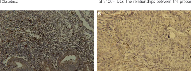
Fig. 1 shows a representative case of endometrioid endo- metrial carcinoma that illustrates the distribution pattern of S100+ DCs. The relationships between the proportion of
Fig. 1. Immunohistochemical staining of dendritic cell infiltration in endometrial carcinoma. Representative patient tissues were highly stained for S100 (A). (B) was patient tissues with low S100 cell count (×400).
A B
Table 1. Clinicopathological characteristics of the patients with endometrial carcinoma
No. of patients (%) Age
≤60 57 (64.0)
>60 32 (36.0)
Menopause
Pre 12 (13.5)
Post 77 (86.5)
BMI
≤25 27 (30.3)
>25 62 (69.7)
FIGO grade
G1 36 (40.0)
G2 29 (32.6)
G3 24 (27.4)
FIGO stage
I 64 (71.9)
II 13 (14.6)
III 10 (11.2)
IV 2 (2.3)
BMI, body mass index; FIGO, International Federation of Gynaecol-
ogy and Obstetrics.
S100+ DCs and clinicopathological variables are assessed and summarized in Table 2. Tumor grade (P<0.001) was significantly associated with the rate of S100+ DCs. Stage (P=0.094), myometrial invasion (P=0.879), and lymph node metastasis ( P=0.427) were not significantly associated with the proportion of S100+ DCs. Lymph node positive group
are biopsy proven positive cases while lymph node negative group include biopsy proven negative cases and cases with no lymphadenectomy and no significant lymphadenopathy on magnetic resonance imaging.
4. Correlation with survival rate
The correlation between the number of S100+ cells and survival rate is shown in Fig. 2. In the Kaplan-Meier plot for progression free survival and overall survival, a slight tendency towards a higher progression free and overall survival rates for endometrial carcinoma patients with S100 positive DCs, although these were not statistically significant ( P=0.092, P=0.151, respectively).
Discussion
This study examined the prognostic significance of the tumor infiltrating DCs in endometrial carcinoma patients. The present study showed that the proportion of S100-positive DCs was negatively correlated with the histologic grade.
DCs are recognized as the most potent antigen-presenting cells and are useful markers for immune responses because they are capable of inducing cytotoxic T lymphocytes from native T cells. T and B cell’ activation by DCs occurs by pre- sentation of antigens on major histocompatibility complex class I and II molecules. Thus, DCs play a central role in the Table 2. Association of tumor-infiltrating dendritic cells with clini-
copathological parameters
S100 P-value
a)Positive Negative LN metastasis
Negative 27 (33.3) 54 (66.7) 0.427
b)Positive 1 (1.3) 7 (98.7)
Myometrial invasion
<1/2 20 (32.8) 41 (67.2) 0.879
>1/2 8 (28.6) 20 (71.4)
FIGO grade
G1 24 (66.7) 12 (33.3) <0.001
G2, G3 4 (8.2) 49 (91.8)
FIGO stage
I–II 27 (35.1) 50 (64.9) 0.094
b)III–IV 1 (8.3) 11 (91.7)
Values are presented as number (%).
FIGO, International Federation of Gynecology and Obstetrics.
a)