The Melanoma Antigen Gene as a Surveillance
Marker for the Detection of Circulating Tumor Cells in Patients with Breast Carcinoma
Soim Kwon,
M.S.1Seok Hyung Kang,
M.D.1Jungsil Ro,
M.D.1Chang-Ho Jeon,
M.D.2Jong-Wook Park,
M.D.3Eun Sook Lee,
M.D., Ph.D.11
Research Institute and Hospital, National Cancer Center, Goyang, Gyeonggi, South Korea.
2
Laboratory Medicine, Catholic Medical Center of Daegu, Daegu, South Korea.
3
The Institute of Medical Science, School of Med- icine, Keimyung University, Daegu, South Korea.
Address for reprints: Eun Sook Lee, M.D., Ph.D., Center for Breast Cancer, National Cancer Center, Madu-1-dong 809, Ilsan-gu, Goyang-si, Gyeonggi- do 411-769, South Korea; Fax: (011) 82 319201948; E-mail: [email protected]
Received November 30, 2004; revision received March 6, 2005; accepted March 10, 2005.
BACKGROUND. Circulating occult tumors cells could be used for the surveillance of metastases after primary breast carcinoma therapy, but their detection is limited by the lack of specific molecular markers. Melanoma antigen genes (MAGEs), which are expressed in malignant tissues but not in normal tissues (except for placenta and testis), might provide such a marker. To date, however, the use of MAGEs in the detection of occult tumor cells using reverse transcription-poly- merase chain reaction (RT-PCR) has been limited because of the heterogeneity and low expression of individual MAGEs in tumor tissues.
METHODS. We developed multiple MAGE-recognizing primers (MMRPs) that were capable of binding to the cyclic DNA of 6 MAGE-A gene subtypes (MAGE-A1–
MAGE-A6). We assessed the ability of the MMRPs to detect the expression of MAGE-A gene subtypes in peripheral blood obtained from patients with benign or malignant breast disease.
RESULTS. MAGE-A gene expression was not detected in 32 patients with benign disease but was detected in 1 of 31 patients (3%) patients with negative lymph node breast carcinoma, in 10 of 52 patients (19%) with 1–3 positive lymph nodes, in 11 of 53 patients (21%) with ⱖ 4 positive lymph nodes, and in 20 of 52 patients (39%) with metastatic disease. The results were statistically significant (P ⬍ 0.0001;
chi-square test for linear-by-linear association). The results also showed that the detection of MAGE-A gene expression in the blood predicted tumor progression or recurrence.
CONCLUSIONS. The results suggested that MAGE-A gene expression may be used for the surveillance of circulating breast carcinoma cells after primary therapy by RT-nested PCR using MMRPs. Cancer 2005;104:251– 6.
© 2005 American Cancer Society.
KEYWORDS: breast carcinoma, melanoma antigen gene, surveillance marker, re- verse transcriptase-nested polymerase chain reaction, peripheral blood.
B reast carcinoma cells frequently express several well character- ized, tumor-associated antigens, such as carcinoembryonic anti- gen (CEA), MUC-1, and Her-2/neu.1– 4Because these antigens also are expressed in normal epithelial and blood cells, however, their utility for monitoring disease progression or recurrence is limited.
5,6
The human melanoma antigen gene (MAGE) family encodes tumor- specific antigens that are recognized by autologous cytotoxic T lympho- cytes.
3,7,8MAGEs have been identified in a number of neoplasms, in- cluding testicular germ cell tumors and carcinomas of the liver, lung, and breast.
9 –13The functions of MAGEs are not known, although they may play a role in embryonal development and tumor transformation or in certain aspects of tumor progression.
14,15The most relevant fact is that MAGEs are expressed in malignant cells but not in normal cells (except for testis and placenta).
16,17Thus, MAGEs would be ideal markers for
© 2005 American Cancer Society DOI 10.1002/cncr.21162
Published online 3 June 2005 in Wiley InterScience (www.interscience.wiley.com).
occult tumor cells that could be detected in blood by a simple reverse transcription-polymerase chain reaction (RT-PCR) analysis.
Twenty-five MAGEs have been cloned since 1991.
7,15,18Among the 3 most studied MAGE families (MAGE-A, MAGE-B, and MAGE-C), MAGE-A, which has 12 subtypes (MAGE-A1–MAGE-A12), has been identified the most in tumors and has been characterized the best.
17,19 –22Although various malignancies express MAGE-A genes, individual subtypes are expressed too sporadically and too weakly to be suitable for tumor cell detection.
7,23,24Most carcinomas, however, express of at least one MAGE-A gene. Therefore, we developed mul- tiple MAGE-recognizing primers (MMRPs) that can si- multaneously detect 6 MAGE-A gene subtypes (MAGE- A1–MAGE-A6).
16,25In this study, we investigated whether the MMRPs could detect the corresponding mRNAs expressed by circulating tumor cells in patients with breast carcinoma. We found that MMRPs did detect MAGE-A gene expression and that the frequency of ex- pression was correlated significantly with tumor pro- gression and metastases.
MATERIALS AND METHODS Patients and Clinical Data
The patients studied were 32 women with benign breast disease and 188 women with breast carcinoma (Table 1) (mean age, 47 years; age range, 27–79 years) who were seen in our clinic from October 2001 to December 2003. The patients with benign disease showed no evidence of acute infection and had no history of carcinoma, diabetes, heart disease, chronic bronchitis, or thrombotic events during the previous year. Institutional Review Board approval was ob- tained for the use of human blood specimens. All patients provided written, informed consent. We col- lected the following data retrospectively: menopausal status, disease stage, tumor size, number of axillary lymph node metastases, HER-2/neu expression, and estrogen and progesterone receptor status.
RNA Preparation and cyclic DNA Synthesis
We extracted total RNA from peripheral blood samples (2 mL collected in ethylenediamine tetraacetic acid tubes) using TRIzol LS reagent (Invitrogen Inc., Carlsbad, CA) according to the supplier’s protocol and treated the extract with DNase (Gentra Inc., Minneapolis, MN). We calculated total RNA and assessed its purity and quality by ultraviolet spectrophotometry. We synthesized cyclic DNA by incubating 3 g RNA in a 20-L reaction mixture that contained 50 mM Tris-HCl, 75 mM KCl, 2.5 mM MgCl
2, 10 mM dithiothreitol, 250 M deoxyadenosine triphosphate (dATP), 250 M deoxycytidine triphos- phate (dCTP), 250 M deoxythymidine triphosphate (dTTP), 250 M deoxyguanine triphosphate (dGTP), RNase inhibitor (2 U/ L), Maloney murine leukemia virus RT (5 U/ L), and 5 M random hexamer. We incubated the reaction mixture at room temperature for 10 minutes, at 42 °C for 60 minutes, at 95 °C for 5 minutes, and at 5 °C for 5 minutes, and then stored it at
⫺ 80 °C until further use.
Nested PCR
We performed the first PCR reactions in 20 L reaction mixture that contained 10 mM Tris-HCl, 50 mM KCl, 1.5 mM MgCl
2, 200 M dATP, 200 M dCTP, 200 M dTTP, 200 M dGTP, 0.6 U Taq DNA polymerase, 0.5
M sense primer, 0.5 M antisense primer, and 2 L of the reaction products. Denaturation was initiated at 95 °C for 5 minutes followed by 30 cycles at 95 °C for 30 seconds, at 60 °C for 45 seconds, and at 72 °C for 45 seconds. The final extension was performed at 72 °C for 10 minutes. We used 1 L of the first PCR product as the template for the second (nested) PCR and the same conditions. We used a combination of sense and antisense primers for simultaneous detection of MAGE-A1–MAGE-A6 gene expression. Glyceraldehyde 3-phosphate dehydrogenase (GAPDH) served as an internal control. Samples were amplified for 25 cycles at 94 °C for 30 seconds, at 62 °C for 30 seconds, and at 72 °C for 45 seconds. We used an MJ Research PTC-200 Peltier Thermal Cycler (MJ Research, Inc., Waltham, MA) for all reactions and analyzed the PCR products in 1% agarose gels. The MAGE-A gene products ampli- fied were ⬇ 469–493 base pairs.
The primer pairs were as follows: for MMRP1 (first PCR), 5 ⬘-CTGAAGGAGAAGATCTGCC-3⬘ and 5⬘-CTC- CAGGTAGTTTTCCTGCAC-3 ⬘; for MMRP2 (nested PCR), 5 ⬘-CTGAAGGAGAAGATCTGCCWGTG-3⬘ (W is A or T) and 5 ⬘-CCAGCATTTCTGCCTTTGTGA-3⬘; and, for GAPDH, 5 ⬘-TGATGACATCAAGAAGGTGGTGAAG-3⬘ and 5 ⬘-TCCTTGGAGGCCATGTGGGCCAT-3⬘. In a previous study, we verified that this method could detect a few malignant cells among millions of normal cells.
TABLE 1
Melanoma Antigen Gene-A Expression in Peripheral Blood from Patients with Benign or Malignant Breast Tumors
Type of breast disease
No. of patients
No. (%) positive
MAGE-A expression P valuea
Benign 32 0 (0)
Malignant
Lymph node negative 31 1 (3) ⬍ 0.0001
1–3 Positive lymph nodes 52 10 (19) ⱖ 4 Positive lymph nodes 53 11 (21)
Metastatic 52 20 (39)
MAGE: melanoma antigen gene.
aChi-square test (linear by linear association).
Statistical Analysis
We used a chi-square test for linear-by-linear associ- ation to determine whether the association between pathologic status and frequency of MAGE-A gene ex- pression was significant. We used SPSS software (ver- sion 9.0; SPSS Inc., Chicago, IL) and considered P values ⬍ 0.05 statistically significant.
RESULTS
The occult tumor cells from 2 mL of peripheral blood were detected by RT-nested PCR using with MMRPs.
To determine its sensitivity, the total RNA isolated from gastric carcinoma cells (SNU484), a MAGE-A gene-positive cell line, was used to detect MAGE-A1–
MAGE-16 in our previous study. An in vitro model was assessed in which serially diluted (1 ⫻ 10
6) SNU484 cells were mixed with 10
7normal donor SNU638 cells.
The results showed that 20 PCR cycles of nested PCR were enough to detect the MAGE-A gene expression of 1–5 SNU484 cells in a background of 10
7SNU638 cells (Fig. 1).
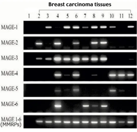
25MMRPs could detect six subtypes of the MAGE-A gene in one step. In 11 samples of breast carcinoma tissues, the RT-nested PCR products that were de- tected with each specific MAGE subtype primer varied with the sample, but MMRPs detected MAGE expres- sion in all samples except negative controls (Fig. 2, Lane 1). These results indicate that MMRPs overcame the problem of heterogeneity and low expression lev- els of individual MAGEs in the detection of tumor cells.
MAGE-A gene expression was not detected in 32 patients with benign breast disease but was detected in 1 of 31 patients (3%) with lymph node-negative breast carcinoma, in 10 of 51 patients (19%) with 1–3 positive lymph nodes, in 11 of 53 patients (21%) with ⱖ 4 positive lymph nodes, and in 20 of 52 patients (39%) with metastatic disease (Fig. 3). The frequency of MAGE-A gene expression correlated significantly with advancement of breast carcinoma (P ⬍ 0.0001) (Table 1). Moreover, expression frequency increased
significantly with tumor size (P ⫽ 0.003), the number of axillary lymph node metastases (P ⫽ 0.043), and disease ⱖ Stage IIA (P ⫽ 0.001). MAGE-A gene expres- sion also was associated with positive estrogen recep- tor status (P ⫽ 0.002) and progesterone receptor status (P ⫽ 0.02) but not with menopausal status or Her2/
neu expression (Table 2).
DISCUSSION
The current study on the detection by RT-nested PCR of circulating tumor cells in patients with breast car- cinoma revealed several new findings. First, the use of MMRPs overcame the problems of heterogeneity and low expression levels of individual MAGE-A gene sub- types. Second, the technique detected no MAGE ex- pression in benign breast disease, and the rate of detection was correlated significantly with tumor size, lymph node status, and disease stage. Third, the find- ing that the frequency of detection was significantly greater in advanced or metastatic breast carcinoma, like bone marrow micrometastases, may have prog- nostic impact. MAGE expression may be useful for the monitoring of therapeutic efficacy in the adjuvant set- ting in patients with no measurable disease, and it may be helpful in predicting disease recurrence in patients with initially negative status who turn out to be positive at the time of recurrence.
The recurrence rate among patients with breast carcinoma who show no evidence of metastases at diagnosis is as high as 30 – 40% over 5–20 years,
26 –28presumably as a consequence of undetected micro- metastases that occurred before diagnosis or treat- ment. Clinical studies also have demonstrated that distant metastases do not increase significantly in pa- tients who do not have their lymph nodes removed until they develop clinical evidence of disease.
8,29Lymphovascular invasion and bone marrow metasta- ses are the important prognostic factors in patients with breast carcinoma. The most recent studies con- sistently reported that the presence of disseminated tumor cells in bone marrow had a strong impact on FIGURE 1. Melanoma antigen gene (MAGE) detection sensitivity for MAGE-A subtype 1– 6 assays. SNU484 gastric carcinoma cells were mixed with normal donor SNU638 cells at various rates, and the total RNA (4 g) isolated from mixed cells was used for MAGE 1– 6 assays.
One microliter of the first reverse tran-
scriptase-polymerase chain reaction
(PCR) products diluted with distilled wa-
ter (100-fold) was used for the second
PCR.
FIGURE 2. Electrophoretic analysis showed expression of melanoma anti- gen gene A subtypes 1– 6 (MAGE-1–
MAGE-6) detected by reverse transcrip- tase-nested polymerase chain reaction analysis. These images show the MAGE- A subtype mRNAs detected by individual MAGE primers and by multiple MAGE- recognizing primers (MMRPs) in RNA ex- tracted from breast carcinoma tissues.
Lane 1, negative control; lanes 2–12, breast carcinoma tissues from different patients.
FIGURE 3. Melanoma antigen gene A (MAGE-A) expression in peripheral blood samples. MAGE-A gene expression was not detected in 32 patients with benign breast disease but was detected in 1 of 31 patients (3%) who had lymph node- negative breast carcinoma, in 10 of 52 patients (19%) who had breast carci- noma with 1–3 positive lymph nodes, in 11 of 53 patients (21%) who had breast carcinoma with ⱖ 4 positive lymph nodes, and in 20 of 52 patients (39%) with metastatic breast carcinoma.
GAPDH: glyceraldehyde 3-phosphate
dehydrogenase (internal control).
patient survival.
30However, the prognostic signifi- cance of circulating tumor cells is much less clear than the significance of disseminated tumor cells in bone marrow. It is not known whether a significant propor- tion of circulating tumor cells survive and, subse- quently, are capable of forming detectable metasta- sis.
31–36Mehes and colleagues reported that a large proportion of circulating tumor cells in patients with breast carcinoma were apoptotic.
37However, several other groups reported that, in patients with breast carcinoma, the presence of circulating tumor cells, as detected by immunohistochemical or molecular methods, was correlated with tumor stage and disease progression.
38 – 40If the circulating tumor cells can be detected successfully, then peripheral blood should
provide easy assess for the detection of disseminated tumor cells.
17Our findings that MAGE-A genes were not detected in blood from patients with benign dis- ease and that the detection rate was correlated signif- icantly with disease progression strongly suggested this possibility. There also was a study showing that the number of circulating tumor cells before treat- ment was an independent prognostic predictor of dis- ease-free and overall survival.
41Although our method could detect 1–5 tumor cells in a background of 10
7normal cells, the intensity of expression could not predict the number of circulating tumor cells.
25We partially would quantify our product compared with the internal control (GAPDH). It is known that quan- tification produces more interobserver variances. Our objective is to develop a simple and easy method for the detection of circulating tumor cells. If we verify more about the clinical significance of circulating tu- mor cells in future study, then our method may prove to be useful as a simple surveillance marker, like CEA in colon carcinoma.
In the current study, we expected to find a higher rate of MAGE expression in metastatic disease than what we observed. We speculated that most blood samples from patients with metastatic disease were collected after intensive systemic therapy, which may have affected the frequency of MAGE expression. The other interesting finding was that MAGE-A gene ex- pression was detected with significantly greater fre- quency in patients who had estrogen receptor-positive tumors. We expected the opposite, because estrogen receptor-negative tumors are more malignant, but we have too little information to speculate on the signif- icance of those data.
It is too early to assess the clinical importance of our findings. In a future study, we intend to compare MAGE- A gene expression in the same patients before and after treatment. Long-term clinical follow-up of patients with circulating occult tumors cells will determine the prog- nostic significance of the technique. In addition, corre- lation of sequential tests with other clinical data during follow-up will determine its value in the prediction of recurrence after primary treatment. Detecting circulat- ing tumor cells with MMRPs potentially offers a practi- cal, safe, and cost-effective method for assessing prog- nosis and risk of recurrence among patients with MAGE- positive breast carcinoma.
REFERENCES
1. Bostick PJ, Chatterjee S, Chi DD, et al. Limitations of specific reverse-transcriptase polymerase chain reaction markers in the detection of metastases in the lymph nodes and blood of breast cancer patients. J Clin Oncol. 1998;16:2632–2640.
TABLE 2
Correlation between Clinicopathologic Parameters and MAGE-A Gene Expression in Peripheral Blood from 188 Patients with Breast Tumors
aClinical factor
No. of patients
No. (%) positive for
MAGE-A P value
Tumor size
ⱕ 2 mm 53 4 (7.5) 0.003
ⱖ 2 mm to 5 mm 69 15 (21.7)
⬎ 5 mm 9 3 (33.3)
No. of lymph node metastases
0 31 1 (3.2) 0.043
ⱕ 1 to ⬍ 4 52 10 (19.2)
ⱖ 4 53 11 (20.8)
Disease stage
ⱕ Stage IIA 43 3 (7.0) 0.001
ⱖ Stage IIB 93 19 (20.4)
Metastatic 52 20 (38.5)
Receptor status Estrogen receptor
Negative 63 5 (7.9) 0.002
bPositive 115 32 (27.8)
Unknown 10 5
Progesterone receptor
Negative 88 12 (13.6) 0.02
bPositive 90 25 (27.8)
Unknown 10 5
Menopausal status
Premenopausal 110 28 (22.5) 0.223
Postmenopausal 78 14 (17.9)
Her-2/neu expression
Low 82 17 (20.7) 0.972
bIntermediate 59 12 (20.3)
High 32 6 (18.8)
Unknown 15 7
MAGE-A: melanoma antigen gene A.
aThe frequency of MAGE-A gene expression was correlated significantly with tumor size, lymph node status, and pathologic stage. Higher expression frequencies were observed in patients with large tumors (P⫽ 0.003), axillary lymph node metastases (P ⫽ 0.043), and advanced disease (P ⫽ 0.001). MAGE-A gene expression also was correlated significantly with hormone receptor status but not with meno- pausal status or Her2/neu gene expression.
bExcludes patients with unknown status.